Original Article
miR-21
expression predicts prognosis in diffuse large
B-cell lymphoma
Jing Li1,2, Rong Fu1, Liping Yang3, Weiping Tu2
1Department of Hematology, Tianjin Medical University General Hospital, Tianjin 300052, China; 2Department
of Hematology, Shangyu People’s Hospital, Shangyu 312300, Shaoxing, Zhejiang, China; 3Department of
Hematology, Yuebei People’s Hospital, Shaoguan 512000, Guangdong, China
Received May 28, 2015; Accepted June 29, 2015; Epub November 1, 2015; Published November 15, 2015
Abstract: Background: Expression patterns of microRNAs in serum are involved in potentially biomarkers for various diseases. The aim of the study was to investigate the expression level of miR-21 in diffuse large B cell lymphoma (DLBCL) and its prognostic value. Methods: Real-time quantitative polymerase chain reaction (qRT-PCR) was used to measure miR-21 levels in serum samples from 112 patients with DLBCL as well as in serum samples from 45 healthy controls. The associations between miR-21 expression and clinicopathologic parameters and overall surviv-al of the patients, were ansurviv-alyzed by chi-square test and Kaplan-Meier method. The Cox proportionsurviv-al hazards regres-sion analyses were performed to estimate the prognostic values for patient survival prediction. Results: We found that serum miR-21 expression was markedly upregulated in patients with DLBCL than healthy controls. Increased
miR-21 expression was significantly correlated with B symptoms, IPI score, CHOP-like treatment and Rituximab (all
Ps<0.05). Moreover, DLBCL patients with miR-21 higher expression have shown significantly worse overall survival than those with lower miR-21 expression. And miR-21 expression was an independent prognostic marker of overall survival in a multivariate analysis (P=0.001, HR: 4.404, 95% CI: 1.770-10.956). Conclusion: The results of the pres-ent study suggested miR-21 expression level could be a novel potential biomarker for DLBCL prognosis.
Keywords:MicroRNA-21, diffuse large B-cell lymphoma, prognosis
Introduction
Diffuse large B-cell lymphoma (DLBCL) is one of the most lethal malignancies and is becom-ing one of most deadly threat to human health and life worldwide [1]. DLBCL is the most com-mon lymphoma worldwide, accounting for near-ly 30 to 40% of non-Hodgkin’s near-lymphoma cases and is highly heterogeneous from both morpho-logical and clinical standpoints [2]. The patho-genic mechanism contributing to the malignant biological characteristics in DLBCL urgently remains to be clarified by the reason of lacking of specific clinical manifestations and respond-ing poor to existrespond-ing treatment [3, 4]. Although there are latest advancements in diagnostic and therapeutic techniques, a large number of DLBCL patients still have an unfavorable prog-nosis every year. In addition, despite several biomarkers emerged to better classify and pre-dict outcome at diagnosis, there are not yet routinely used in clinical practice [5]. Thus,
exploring more molecular biomarkers involved in DLBCL pathogenesis may novel provide effective therapeutic opportunity.
MicroRNAs (miRNAs) are a class of small, natu-rally occurring, noncoding and single-stranded RNA molecules (18, 22 nucleotides) that func-tion as post-transcripfunc-tional regulators by direct-ly cleaving target messenger RNA (mRNA) or translational repression [6]. A growing number of both direct and indirect evidence suggests a relationship between differential miRNA expres-sion and cancer [7, 8]. However, some miRNAs were found to act as tumor suppressors, where-as others acted where-as oncogenes, depending on the targets of the miRNAs, which may provide insights into the functional detection of human malignancies [9].
most commonly over-expressed miRNA in can-cers. It has been identified as the only miRNA commonly over-expressed in various solid tumors, including lung, breast, stomach, pros-tate, colon, brain, head and neck, esophagus, and pancreas, as well as in chronic lymphocytic leukemia, uterine leiomyomas, and malignant hepatocytes [9, 11-15]. In addition, a correla-tion between miR-21 expression and the carci-nogenesis of DLBCL has also been reported [16]. However, the underlying mechanism is not completely clear. Thus, further analyses are needed to clarify the role of miR-21 in DLBCL prognosis based on clinicopathologic stage. In the present study, serum miR-21 expression levels in DLBCL were examined, and the clinico-pathologic significance and potential prognos-tic value for DLBCL were assessed.
Methods and materials
Patients and serum samples
Serum samples were obtained from 112 pati- ents who were diagnosed with DLBCL enrolled at the Tianjin Medical University General Hospital at the time of diagnosis. All of these patients have undergone molecular and pheno-typic classification with available clinical data. 45 serum samples from healthy individuals. Blood samples of all patients and healthy con-trol patients were collected. The samples were allowed to stand at room temperature for 30
min and then centrifuged at 3,000 rpm for 15 min at 4°C. To remove cellular contaminants, serum samples were subjected to additional centrifugation at 12,000 rpm for 10 min. The supernatants to be used for RNA extraction were snap-frozen and then stored at -80°C. The study was approved by the Ethics Committee of Tianjin Medical University General Hospital, and ethical permission and informed consent were obtained from all participants.
Isolation of total RNA and real-time quantita-tive PCR analysis (qRT-PCR)
MiR-21 expression in serum samples from 112 patients with DLBCL and 45 healthy controls was measured by reverse transcription and real-time PCR (RT-PCR). Total RNA was isolated from frozen samples using Trizol reagent (Invitrogen, CA, USA) according to the manufac-turer’s protocol. The TaqMan microRNA assay and TaqMan universal PCR master mix were used to detect the expression of miR-21, and the U6 gene was used as an internal control to normalize variances. Relative quantification of target miRNA expression was evaluated using the comparative cycle threshold (CT) method. Each sample was examined in triplicate and the raw data were presented as the relative quan-tity of target miRNA, normalized with respect to U6. RT-PCR primers: miR-21: F: 5’-GCGGGT- AGCTTATCAGACTG-3’; R: 5’-GTGCAGGGTCCGA- GGT-3’; U6: F: 5’-GCGCGTCGTGAAGCGTTC-3’; R: 5’-GTGCAGGGTCCGAGGT-3.
Statistical analysis
[image:2.629.101.294.81.233.2]All statistical calculations were performed using SPSS 18.0 for Windows (SPSS Inc, IL, USA) and GraphPad Prism 5 (GraphPad Software Inc., CA, USA). miR-21 expression lev-els in serum samples were shown by mean and standard deviation (mean ± SD) and compared using Student’s t-test. The two-tailed Chi-squared test was employed to explore the cor-relation between miR-21 expression and clini-cal pathologiclini-cal features. Survival rates were calculated according to the Kaplan-Meier method and survival curves were plotted; sta-tistical differences were analyzed using the log-rank test. Multivariate analysis of the prognos-tic factors was performed with Cox regression model. P<0.05 was considered statistically significant.
Results
Serum miR-21 is significantly up-regulated in patients with DLBCL
We analyzed the expression levels of miR-21 in serum samples from 112 DLBCL patients and 45 healthy individuals. As revealed by qRT-PCR analysis, miR-21 expression was significantly higher in serum samples from DLBCL patients than that from healthy controls (P<0.05, Figure 1).
Association between miR-21 expression and the clinicopathological features of DLBCL
For better understanding of the clinical rele-vance of miR-21 expression in DLBCL, we
divid-high miR-21 expression was an independent prognostic factor for patients with DLBCL (Table 2) (P=0.001, HR: 4.404, 95% CI: 1.770- 10.956).
Discussion
[image:3.629.100.373.95.476.2]Recently, miRNAs have been demonstrated to play a key role in tumorigenesis. miRNAs may offer a new regulatory model of gene expres-sion, and miRNA expression levels correlate closely with specific clinical features of cancer, so that they can be used to classify normal and cancerous tissues, as well as for prognosis [17-19]. It has been reported that miR-21 plays a role in the development of tumor via regulating the expression of the tumor suppressor, such as PDCD4, PTEN, and TPM1. Suppression of
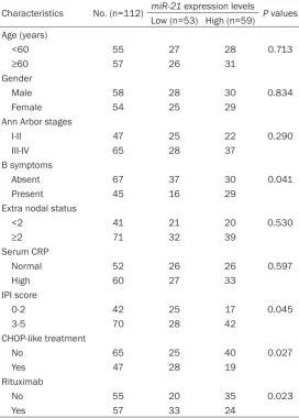
Table 1.miR-21 expression and clinicopathological features Characteristics No. (n=112) miR-21 expression levels P values
Low (n=53) High (n=59) Age (years)
<60 55 27 28 0.713
≥60 57 26 31
Gender
Male 58 28 30 0.834
Female 54 25 29
Ann Arbor stages
I-II 47 25 22 0.290
III-IV 65 28 37
B symptoms
Absent 67 37 30 0.041
Present 45 16 29
Extra nodal status
<2 41 21 20 0.530
≥2 71 32 39
Serum CRP
Normal 52 26 26 0.597
High 60 27 33
IPI score
0-2 42 25 17 0.045
3-5 70 28 42
CHOP-like treatment
No 65 25 40 0.027
Yes 47 28 19
Rituximab
No 55 20 35 0.023
Yes 57 33 24
IPI: International Prognostic Index; CHOP-like refers to CHOP (cyclophosphamide, adriamycin, vincristine and prednisone) or a CHOP-like regimen.
ed the 112 DLBCL patients into a high expression group (n=53) and a low expression group (n=59), according to the expres-sion level of miR-21 in all sam-ples. And the relationship of the
miR-21 with various clinical fea-tures of DLBCL was analyzed and is summarized in Table 1. As shown in the results, miR-21
expression was closely associ-ated with B symptoms, IPI score, CHOP-like treatment and Ritu- ximab (all Ps<0.05). However, but there was no relationship with other characteristics, such as age, gender, Ann Arbor stag-es, extra nodal status, and serum CRP (all Ps>0.05).
Prognostic values of miR-21 expression in serum samples from DLBCL patients
The correlation between miR-21
miR-21 can inhibit tumor growth, which could indicate miR-21 functions as an oncogene [20-22]. It has been reported that the dysregulation of miR-21 performed an important function in different types of cancer [9, 11-15]. However, few studies are available on its expression and functions in DLBCL.
In this study, we investigated the expression of
miR-21 in serum samples from DLBCL patients and healthy controls by qRT-PCR for the first time. Based on the relative expression level analysis, it was investigated that the associa-tion of miR-21 with clinicopathological factors and prognosis of patients with DLBCL. Results showed that the serum level of miR-21 was up-regulation in DLBCL patients compared with that in healthy controls (P<0.05). The
expres-sion pattern of miR-21 found in our study is in line with previous findings that the expression of miR-21 was increased in DLBCL cells and was used as an indepen-dent prognostic indicator for DLBCL patients by Lawrie’s studies [23]. Fang et al. found that miR-21 were significantly elevated in DLBCL serum when compared with normal controls [24]. Wang et al.
reported that miR-21 expression was sig-nificantly higher in hepatocellular carcino-ma tissues compared with norcarcino-mal adja-cent liver tissues [25]. Alexander et al.
showed that miR-21 showed significantly increased levels in the cerebrospinal fluid of patients with primary central nervous system lymphoma compared with the cerebrospinal fluid of control patients [26]. Charles et al. revealed that miR-21 levels were higher in DLBCL patient than healthy control serum [27]. Chen et al. reported that expression of miR-21 was increased in DLBCL cell lines (Ly1, Ly3, OCI-Ly4, OCI-Ly7, OCI-Ly8, OCI-Ly10, OCI-Ly18, OCI-Ly19, and HBL) [28].
In addition, it was also proved that the rel-ative expression level of miR-21 was close-ly associated with B symptoms, IPI score, CHOP-like treatment and Rituximab. However, miR-21 expression was not asso-ciated with age, gender, Ann Arbor stages, extra nodal status, and serum CRP. More importantly, we proved that miR-21
expression was significantly associated with overall survival by Kaplan-Meier
anal-Table 2. Multivariate analyses of prognostic variables of overall survival in DLBCL patients
Variables HR 95% CI P Values
MiR-21 expression 4.404 1.770-10.956 0.001
B symptoms 1.137 0.551-2.348 0.729
IPI score 1.128 0.498-2.556 0.773
CHOP-like treatment 1.036 0.484-2.217 0.928
[image:4.629.98.332.77.314.2]Rituximab 1.709 0.831-3.514 0.145
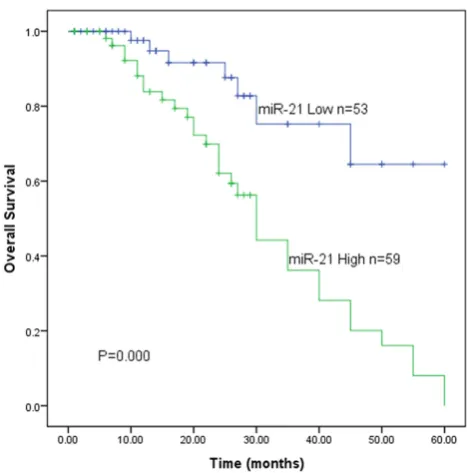
Figure 2. Overexpression of miR-21 is associated with poor overall survival in patients with DLBCL. Kaplan-Meier analysis of overall survival was analyzed according to miR-21 expres-sion levels (P=0.000).
ysis and log-rank test. Patients with high levels of miR-21 expression had worse overall surviv-al compared with those with low levels of miR-21 expression. By a Cox proportional hazards model adjusted for factors related to survival of DLBCL, miR-21 up-regulation could be an inde-pendent prognostic marker in patients with DLBCL. These data indicated that miR-21
expression play a crucial role in tumorigenesis, and progression of DLBCL. Mao et al. revealed that serum miR-21 was used as an indepen-dent and powerful predictor of overall survival for primary central nervous system lymphoma [29]. Go et al. reported that overexpression of
[image:4.629.100.332.410.494.2]rituximab-com-bined chemotherapy [16]. MG Narducci et al.
reported that miR-21 was upregulated in cuta-neous T-cell lymphoma and could discriminate patients with unfavorable and favorable out-come [30].
In conclusion, the results suggest that miR-21
is a novel biomarker and a prognostic target for DLBCL in future. Because there are only a few studies on the relationship between miR-21
expression and the prognosis of DLBCL, further study is required to elucidate the exact molecu-lar mechanisms to verify our conclusions.
Disclosure of conflict of interest
None.
Address correspondence to: Dr. Rong Fu, Depart- ment of Hematology, Tianjin Medical University General Hospital, Tianjin 300052, China. Tel: +86-22-60362205; E-mail: [email protected]
References
[1] Hunt KE and Reichard KK. Diffuse large B-cell lymphoma. Arch Pathol Lab Med 2008; 132: 118-124.
[2] Harris NL, Jaffe ES, Stein H, Banks PM, Chan JK, Cleary ML, Delsol G, De Wolf-Peeters C, Falini B, Gatter KC, et al. A revised European-American classification of lymphoid neo-plasms: a proposal from the International Lym-phoma Study Group. Blood 1994; 84: 1361-1392.
[3] Kuppers R. Mechanisms of B-cell lymphoma pathogenesis. Nat Rev Cancer 2005; 5: 251-262.
[4] Rosenwald A, Wright G, Chan WC, Connors JM, Campo E, Fisher RI, Gascoyne RD, Muller-Her-melink HK, Smeland EB, Giltnane JM, Hurt EM, Zhao H, Averett L, Yang L, Wilson WH, Jaffe ES, Simon R, Klausner RD, Powell J, Duffey PL, Longo DL, Greiner TC, Weisenburger DD, Sanger WG, Dave BJ, Lynch JC, Vose J, Armit-age JO, Montserrat E, Lopez-Guillermo A, Gro-gan TM, Miller TP, LeBlanc M, Ott G, Kvaloy S, Delabie J, Holte H, Krajci P, Stokke T and Staudt LM. The use of molecular profiling to predict survival after chemotherapy for diffuse large-B-cell lymphoma. N Engl J Med 2002; 346: 1937-1947.
[5] Berglund M, Thunberg U, Amini RM, Book M, Roos G, Erlanson M, Linderoth J, Dictor M, Jerkeman M, Cavallin-Stahl E, Sundstrom C, Rehn-Eriksson S, Backlin C, Hagberg H, Rosen-quist R and Enblad G. Evaluation of immuno-phenotype in diffuse large B-cell lymphoma
and its impact on prognosis. Mod Pathol 2005; 18: 1113-1120.
[6] Bartel DP. MicroRNAs: genomics, biogenesis, mechanism, and function. Cell 2004; 116: 281-297.
[7] Fabbri M, Croce CM and Calin GA. MicroRNAs. Cancer J 2008; 14: 1-6.
[8] He L and Hannon GJ. MicroRNAs: small RNAs with a big role in gene regulation. Nat Rev Gen-et 2004; 5: 522-531.
[9] Volinia S, Calin GA, Liu CG, Ambs S, Cimmino A, Petrocca F, Visone R, Iorio M, Roldo C, Ferracin M, Prueitt RL, Yanaihara N, Lanza G, Scarpa A, Vecchione A, Negrini M, Harris CC and Croce CM. A microRNA expression signature of hu-man solid tumors defines cancer gene targets. Proc Natl Acad Sci U S A 2006; 103: 2257-2261.
[10] Roehle A, Hoefig KP, Repsilber D, Thorns C, Ziepert M, Wesche KO, Thiere M, Loeffler M, Klapper W, Pfreundschuh M, Matolcsy A, Ber-nd HW, Reiniger L, Merz H aBer-nd Feller AC. Mi-croRNA signatures characterize diffuse large B-cell lymphomas and follicular lymphomas. Br J Haematol 2008; 142: 732-744.
[11] Shibuya H, Iinuma H, Shimada R, Horiuchi A and Watanabe T. Clinicopathological and prog-nostic value of microRNA-21 and microR-NA-155 in colorectal cancer. Oncology 2010; 79: 313-320.
[12] Gong C, Yao Y, Wang Y, Liu B, Wu W, Chen J, Su F, Yao H and Song E. Up-regulation of miR-21 mediates resistance to trastuzumab therapy for breast cancer. J Biol Chem 2011; 286: 19127-19137.
[13] Tomimaru Y, Eguchi H, Nagano H, Wada H, To-mokuni A, Kobayashi S, Marubashi S, Takeda Y, Tanemura M, Umeshita K, Doki Y and Mori M. MicroRNA-21 induces resistance to the an-ti-tumour effect of interferon-alpha/5-fluoro-uracil in hepatocellular carcinoma cells. Br J Cancer 2010; 103: 1617-1626.
[14] Kwak HJ, Kim YJ, Chun KR, Woo YM, Park SJ, Jeong JA, Jo SH, Kim TH, Min HS, Chae JS, Choi EJ, Kim G, Shin SH, Gwak HS, Kim SK, Hong EK, Lee GK, Choi KH, Kim JH, Yoo H, Park JB and Lee SH. Downregulation of Spry2 by miR-21 triggers malignancy in human gliomas. On-cogene 2011; 30: 2433-2442.
[16] Go H, Jang JY, Kim PJ, Kim YG, Nam SJ, Paik JH, Kim TM, Heo DS, Kim CW and Jeon YK. MicroR-NA-21 plays an oncogenic role by targeting FOXO1 and activating the PI3K/AKT pathway in diffuse large B-cell lymphoma. Oncotarget 2015; 6: 15035-49.
[17] Huang Y, Shen XJ, Zou Q, Wang SP, Tang SM and Zhang GZ. Biological functions of microR-NAs: a review. J Physiol Biochem 2011; 67: 129-139.
[18] Perera RJ and Ray A. MicroRNAs in the search for understanding human diseases. BioDrugs 2007; 21: 97-104.
[19] Calin GA and Croce CM. MicroRNA signatures in human cancers. Nat Rev Cancer 2006; 6: 857-866.
[20] Lu Z, Liu M, Stribinskis V, Klinge CM, Ramos KS, Colburn NH and Li Y. MicroRNA-21 motes cell transformation by targeting the pro-grammed cell death 4 gene. Oncogene 2008; 27: 4373-4379.
[21] Zhu S, Si ML, Wu H and Mo YY. MicroRNA-21 targets the tumor suppressor gene tropomyo-sin 1 (TPM1). J Biol Chem 2007; 282: 14328-14336.
[22] Lou Y, Yang X, Wang F, Cui Z and Huang Y. Mi-croRNA-21 promotes the cell proliferation, in-vasion and migration abilities in ovarian epi-thelial carcinomas through inhibiting the expression of PTEN protein. Int J Mol Med 2010; 26: 819-827.
[23] Lawrie CH, Soneji S, Marafioti T, Cooper CD, Palazzo S, Paterson JC, Cattan H, Enver T, Mag-er R, Boultwood J, Wainscoat JS and Hatton CS. MicroRNA expression distinguishes be-tween germinal center B cell-like and activated B cell-like subtypes of diffuse large B cell lym-phoma. Int J Cancer 2007; 121: 1156-1161. [24] Fang C, Zhu DX, Dong HJ, Zhou ZJ, Wang YH,
Liu L, Fan L, Miao KR, Liu P, Xu W and Li JY. Serum microRNAs are promising novel bio-markers for diffuse large B cell lymphoma. Ann Hematol 2012; 91: 553-559.
[25] Wang WY, Zhang HF, Wang L, Ma YP, Gao F, Zhang SJ and Wang LC. miR-21 expression pre-dicts prognosis in hepatocellular carcinoma. Clin Res Hepatol Gastroenterol 2014; 38: 715-719.
[26] Baraniskin A, Kuhnhenn J, Schlegel U, Chan A, Deckert M, Gold R, Maghnouj A, Zollner H, Rei-nacher-Schick A, Schmiegel W, Hahn SA and Schroers R. Identification of microRNAs in the cerebrospinal fluid as marker for primary dif-fuse large B-cell lymphoma of the central ner-vous system. Blood 2011; 117: 3140-3146. [27] Lawrie CH, Gal S, Dunlop HM, Pushkaran B,
Liggins AP, Pulford K, Banham AH, Pezzella F, Boultwood J, Wainscoat JS, Hatton CS and Har-ris AL. Detection of elevated levels of tumour-associated microRNAs in serum of patients with diffuse large B-cell lymphoma. Br J Hae-matol 2008; 141: 672-675.
[28] Chen W, Wang H, Chen H, Liu S, Lu H, Kong D, Huang X, Kong Q and Lu Z. Clinical significance and detection of microRNA-21 in serum of pa-tients with diffuse large B-cell lymphoma in Chinese population. Eur J Haematol 2014; 92: 407-412.
[29] Mao X, Sun Y and Tang J. Serum miR-21 is a diagnostic and prognostic marker of primary central nervous system lymphoma. Neurol Sci 2014; 35: 233-238.