DUNCAN S. OWEN,
JR.,
M. D., F.
A. C.
P.
Associate Professor of Medicine, Division of Connective Tissue
Diseases, Medical College of Virginia, Health Sciences Division of
Virginia Commonwealth University, Richmond
Synovial fluid analysis is a frequently ignored examination except in suspected cases of septic con-ditions. It has been shown that it is an extremely valuable procedure in making rapid and accurate diagnoses in many types of joint diseases.
Table 1 illustrates the findings in ten separate joint states. The gross appearance, "wet-prep" microscopic examination, leukocyte count and sugar content are procedures that are extremely important and can be performed with a paucity of equipment. We have not found the mucin-clot test or protein content to be very helpful. Of course, if infection is a possibility, Gram's stain and appropriate cultures should be instituted. It should be remembered, how-ever, that only 25-30% of the cases of gonococcal arthritis will be associated with a positive Gram's stain or culture. The culture yield may possibly improve with the increasing use of Thayer-Martin media. The anticoagulant should be either heparin or EDT A instead of oxalate because the examiner may confuse oxalate crystals with urate or pyro-phosphate crystals.
Cell Counting. The white blood cell count is of particular importance in suspected septic conditions. Methods have been outlined in detail in an excel-lent book by Ropes and Bauer. The fluid should .be collected in an anticoagulant tube. The diluent should be··physiologic saline because acetic acid will cause precipitation of mucin and make an accurate count practically impossible. The addition of methylene blue to the diluent will help differentiate the cell types. If the fluid is hemorrhagic, a
hypo-*
Presented by Dr. Owen at the 45th Annual McGuire Lecture Series, November 8, 1973, at the Medical College of Virginia, Richmond.MCV QUARTERLY 10(1): 13·17, 1974
tonic diluent of 0.3% sodium chloride can be tried which theoretically will disrupt the red blood cells but not the nucleated cells.
A differential white count using Wright's stain is performed in the same manner as a peripheral blood smear. If the total white blood count in the synovial fluid is less than 5,000/mm3, it is best to centrifuge the fluid at 2,000-3,000 rpm for ten minutes. The supernatant is then removed arid physiologic saline added to the sediment until. the original volume is obtained. The solution should then be recentrifuged which will remove most of the mucin. The sediment can then be easily smeared on a slide, air dried, and stained. In cases of sys-temic lupus erythematosus (SLE), the.LE cell may be seen.
Glucose. The glucose value of normal synovial fluid usually parallels that of serum. The results are of greater value if the patient has been fasting for about six hours. It is even possible for normal synovial fluid sugar to be higher than that of the serum for several hours postprandially.
In any suspected septic joint condition, a glu-cose determination should always be performed. We have found the glucose levels to be low in almost all cases of septic arthritis observed at the Medical College of Virginia during the past seven years. It is important to have the glucose level determined immediately after aspiration. Because of the glu-colytic action of white cells, falsely low levels may be observed in cases of rheumatoid arthritis and gout, for example, if the white cell count of the fluid is high and the fluid is not tested for one or more hours. The falsely low sugar level can also be prevented by placing the fluid to be tested in a sodium fluoride tube. A synovial glucose of 50 mg %
TABLE 1
SYNOVIANAL YSIS
Mucin Leukocytes
Disease Appearance Clot Viscosity per mm3
Normal Clear, Good High <200
amber <25% polys
Gouty Milky or Poor Low 15,000+
Arthritis yellow 70% polys
Pseudogout Turbid, Fair to Fair or 25,000± amber, or Poor Low 90% polys± yellow
Rheumatoid Turbid, Poor Low 15,000+
Arthritis amber to 70% polys
light green
Septic Very turbid, Poor Low 70,000±
Arthritis gray 90% + polys
Tuberculous Turbid, Poor Low 30,000±
.(\rthritis amber, or 60%± polys
yellow
Osteoarthritis Clear, Good High 1000±
amber 10% polys
Acute Bloody or Good High <2000
Traumatic turbid <25% polys
Arthritis
Systemic Slightly Good High 3000±
Lupus turbid, <10% polys
Erythematosus amber
Rheumatic Slightly Good Fair 15,000
Fever turbid, to Low 50% polys
amber
less than fasting serum should be considered as strongly indicating an infectious process until 'proved otherwise.
"Wet Drop" Preparation Studies. The micro-scopic examination of a drop of fresh or anticoagu-lated synovial fluid may yield diagnostic information, especially in cases of crystal deposition diseases (gout and pseudogout). This examination is per-formed by placing a small drop of synovial fluid on a glass slide, covering with a cover slip and sealing the edges with clear fingernail polish. Only a small drop is used to prevent individual cells from
Sugar:
serum-synovial difference
(mg/lOOml) Crystals Comment
<10 None
10± Sodium Strongly negative bire-urate fringent intra- and
extra-cellular urate crystals.
10± Calcium Few-to-many weakly pyrophosphate positive intra- and
extra-cellular calcium pyro-phosphate crystals.
25 Occasional WBC inclusions fre-cholesterol quently present.
Rheumatoid factor usually present.
70 None Culture positive in only 20-30% of GC cases.
so+ None Biopsy frequently helpful.
<10 None Cartilage fibrils may be present.
<10 None Occasional cartilage fibril. Many RBC's.
10± None Examine for LE cells.
10± None Occasional WBC
inclusion
moving across the field of view. Sealing the slide prevents the formation of artifacts while drying and
the compensated polarizing microscope is an ex-pensive piece of equipment, which is not readily available in the office of most physicians and it is infrequently found in general hospitals.
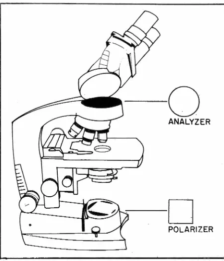
Simple Polarization. A simple polarizing micro-scope can be made from an ordinary micromicro-scope by inserting a polarizing filter below the condenser, usually on top of the light source (the polarizer), and using one filter above the objective, usually in the barrel or above the eye piece (the analyzer). The polarizer is then rotated until the darkest field possible is obtained. The monosodium urate (MSU) crystals usually appear as bright needles against this black background. The calcium pyrophosphate dihydrate ( CPPD) crystals usually appear in bright monoclinic, triclinic, rectangular or rhomboid forms, and may be quite difficult to differentiate from MSU crystals in synovial fluid. Figure 1 illustrates the placement of simple plastic polarizing filters in a laboratory microscope.
Compensated Polarization. The compensated polarizing microscope utilizes a first-order red filter, the compensator, which is inserted between the objective and the analyzer. The compensator retards red light and the polarized background becomes red instead of black. The axis of the line of slow vibration of the compensator is then defined. The MSU and CPPD crystals will be either yellow or blue, depending upon their respective positions to this axis. The MSU crystal is brightly yellow when parallel to the axis and indicates that it is strongly negatively birefringent. The CPPD crystal is faintly blue when in this position, indicating weakly posi-tive birefringence.
On occasion, both MSU and CPPD crystals are noted in the same fluid indicating coexisting disease. Previously injected adrenocorticosteroids, oxalate anticoagulant, cholesterol crystals, and scratches on the slide or coverslip should not be confused with either MSU or CPPD crystals.
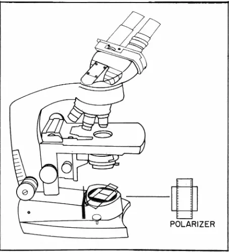
New and Simple Technique. We have found a very adequate substitute for the first-order red filter which can be used with an ordinary light micro-scope which has been adapted with simple polarizing filters. One needs only a clean microscopic glass slide and simple transparent cellophane tape. One piece of the cellophane tape is carefully applied to the top side of the slide and then another piece of tape is applied over this. Wide tape is preferable but two pieces of narrow tape carefully applied
Fig. I-Illustration of simple plastic polarizing filters in barrel of microscope (analyzer) and above light source
(polarizer) .
beside two other pieces is satisfactory. The scope is then adapted for simple polarizing micro-scopy as previously described. (One may first wish to focus on the material to be examined.) The cellophane-taped slide is then placed over the polarizer and carefully rotated until the background is quite red. We have discovered that the long axis of the taped slide substitutes amazingly well for the axis of slow vibration of the first-order red compensator. The examiner can theh mentally pro-ject the axis of the taped slide on the stage. Figure 2 illustrates the celiophane-taped slide on top of the polarizer. With the stage arranged to permit free movement of the "wet-prep" slide, the examiner can then rotate the "wet-prep" slide and define whether the observed crystals are negatively or positively birefringent.
a blue background. The interested examiner may have to purchase a few rolls of tape until a satis-factory one is found.
The improvised compensated polarizing micro-scope techniques described above have been a tremendous help in making a rapid and accurate diagnosis of crystal-induced synovitis. Faculty, housestaff, and students are encouraged to carry polarizing filters and a cellophane-taped microscope slide with them. A great deal of enthusiasm has been expressed. For the first time, individuals are really learning the true meaning of the planes of bire-fringence. In the past, many individuals memorized, in preparation for written examination, which crys-tals were negatively or positively birefringent. It is hoped that others will discover these techniques to be helpful to them. Examiners will be greatly helped in establishing theii: own personal techniques by smearing some MSU crystals from a tophus on a clean microscopic slide. They can then experiment with the filters until the simple maneuvers are mastered.
Ragocytes (RA cells, inclusion body cells, raisin seed cells) in syriovial fluid of patients with rheumatoid arthritis were originally described by Dr. Joseph Hollander and associates. These cells
-a
POLARIZER Fig. 2-Illustration of "cellophane tape compensator" above the polarizer.are leukocytes containing inclusion bodies. They appear as dark granules under regular light micro-scopy at high dry magnification and as clear vacuo~ lated areas with phase microscopy. It was assumed that these bodies were phagocytosed rheumatoid factor and they were noted in from 5-95% of the total leukocyte population of rheumatoid synovial fluid. It was believed that phagocytosis of rheuma-toid factor played a role in the pathogenesis of rheumatoid arthritis in a similar fashion to the phagocytosis of sodium utate crystals in the patho-genesis of gouty arthritis. This concept was supported with the production of acute synovitis by the . in-jection of autologous yG-globulin into inactive joints of patients with rheumatoid arthritis. Subsequent studies have shown that the findings of these leuko-cyte inclusions are not specific for rheumatoid ar-thritis. They may be found in other types of inflam-matory joint disease and may be scavengers which have phagocytosed products of inflammation includ-ing y-globulins.
Osteoarthritis fluid may reveal
a
few-to-many cartilage fibrils and fragments. These may also be seen in cases of pseudogout.Cholesterol crystals are noted rarely in the fluid of cases of chronic rheumatoid arthritis. These crystals are large, rhomboid, have punched-out corners and are birefringent.
An inexperienced examiner may confuse pre-viously injected corticosteroid ester crystals with urate or pyrophosphate crystals. The steroid crystals may be phagocytosed by white cells and apparently can precipitate a "postinjection flare" of the in-jected joint. The crystals are usually negatively birefringent.
Reiter's syndrome synovial fluid has been re-ported to show large mononuclear cells containing many vacuoles. Some of these cells appear to con-tain polymorphonuclear cells. The specificity of this is unclear.
Table 2 illustrates the complement (C') levels in various disease entities. It is most helpful, how-ever, to know the normal values and type of com-plement determination for each laboratory. For example, if total complement is determined on a viscous synovial fluid and if a diffusion technique is used, poor diffusion may occur due to viscosity and erroneously low level results.
TABLE 2
COMPLEMENT
Rheumatoid arthritis Systemic lupus erythematosus Reiter's
Septic Crystal disease Spondylitis & psoriasis
Serum
N (rarely l)
l
N
N (occ. i)
N N
Synovial Fluid
l
l ii
t
Nori Nori
definitive diagnosis of one of the various types of arthritis, especially gout, pseudogout and septic arthritis. Unfortunately, it is one of the most fre-quently ignored tests. It should be remembered that only a few drops of fluid are needed for a "wet drop" examination for crystals, ragocytes, Gram's stain and a Wright's stain.
BIBLIOGRAPHY
COHEN AS (ed): Laboratory diagnostic procedures in the
rheumatic diseases. Boston, Little, ~rown & Co., 1967.
HOLLANDER JL, McCARTY DJ Jr, ASTROGA G, CASTRO-MURILLO E: Studies on the pathogenesis of rheumatoid joint inflammation. I. The RA cell and a work-ing hypothesis. Ann Intern Med 62:271, 1965.
HOLLANDER JL, REGINATO A, TORRALBA TP:
Ex-amination of synovial fluid as a diagnostic aid in arthritis.
Med Clin North Am 50:1281, 1966.
KAHN CB, HOLLANDER JL, SCHUMACHER HR: Corticosteroid crystals in synovial fluid. JAMA 21 I :807,
1970.
McCARTY DJ Jr: A basic guide to arthrocentesis. Hosp
Med 4:77, 1968.
McCARTY DJ Jr: On the crystal deposition diseases.
Dis-ease-a-Month, pp 1-31, March, 1970.
McCARTY DJ Jr, HOLLANDER JL: Identification of urate crystals in gouty synovial fluid. Ann Intern Med 54:452, 1960.
McCARTY DJ Jr, KOHN NN, FAIRES JS: The signifi-cance of calcium phosphate crystals in the synovial fluid of arthritic patients. The "pseudogout syndrome." I. Clinical aspects. Ann Intern Med 56:711, 1962.
MOSKOWJTZ RW, KATZ D: Chondrocalcinosis and chonqrocalsynovitis ( pseudogout syndrome). Am J Med
43:322, 1967.
OWEN DS Jr, COOKE CL, TOONE E: Practical synovial fluid examination. Va Med Month 97:88, 1970.
PEKIN JJ, MALININ IL, ZV AIFLER NJ: Unusual
syno-vial fluid findings in Reiter's syndrome. Ann Intern Med 66:677, 1967.
PHELPS P, STEELE AD, McCARTY DJ Jr: Compensated polarized light microscopy. Identification of crystals in synovial fluids from gout and pseuqogout. JAMA 203:508,
1968.
ROPES MW, BAUER W: Synovial fluid changes in joint disease. Cambridge, Harvard University Press, 1953.
SCHUMACHER HR: Intracellular crystals in synovial fluid anticoagulated with oxalate. N Engl J M,ed 274:1372, 1966.
SKINNER M, COHEN AS: Calcium pyrophosphate dihy-drate crystal deposition disease. Arch Intern Med 123:636, 1969.
VAUGHAN JH, BARNETT EV, SOBEL MV, JACOX RF: Jntracytoplasmic inclusions of immunoglobulins in rheumatoid arthritis and other diseases. Arth Rheum 11: 125, 1968.
VAUGHAN JH, JACOX RF, NOELL P: Relationship of intracytoplasmic inclusions in joint fluid leukocytes to anti-'YG-globulins. A rth Rheum 11: 135, 1968.
ZUCKNER J, UDDIN J, GANTNER GE, DORNER RW: Cholesterol crystals in synovial fluid. Ann Intern Med 60: 436, 1964.