R E S E A R C H
Open Access
HIV induces expression of complement
component C3 in astrocytes by NF-
κ
B-dependent activation of interleukin-6
synthesis
Jadwiga Nitkiewicz
1,4, Alejandra Borjabad
1, Susan Morgello
2, Jacinta Murray
2, Wei Chao
1, Luni Emdad
3,
Paul B. Fisher
3, Mary Jane Potash
1and David J. Volsky
1,5*Abstract
Background:Abnormal activation of the complement system contributes to some central nervous system diseases but the role of complement in HIV-associated neurocognitive disorder (HAND) is unclear.
Methods:We used real-time PCR and immunohistochemistry to detect complement expression in postmortem brain tissue from HAND patients and controls. To further investigate the basis for viral induction of gene expression in the brain, we studied the effect of HIV on C3 expression by astrocytes, innate immune effector cells, and targets of HIV. Human fetal astrocytes (HFA) were infected with HIV in culture and cellular pathways and factors involved in signaling to C3 expression were elucidated using pharmacological pathway inhibitors, antisense RNA, promoter mutational analysis, and fluorescence microscopy.
Results:We found significantly increased expression of complement components including C3 in brain tissues from patients with HAND and C3 was identified by immunocytochemistry in astrocytes and neurons. Exposure of HFA to HIV in culture-induced C3 promoter activity, mRNA expression, and protein production. Use of
pharmacological inhibitors indicated that induction of C3 expression by HIV requires NF-κB and protein kinase signaling. The relevance of NF-κB regulation to C3 induction was confirmed through detection of NF-κB translocation into nuclei and inhibition through overexpression of the physiological NF-κB inhibitor, I-κBα. C3 promoter mutation analysis revealed that the NF-κB and SP binding sites are dispensable for the induction by HIV, while the proximal IL-1β/IL-6 responsive element is essential. HIV-treated HFA secreted IL-6, exogenous IL-6 activated the C3 promoter, and anti-IL-6 antibodies blocked HIV activation of the C3 promoter. The activation of IL-6 transcription by HIV was dependent upon an NF-κB element within the IL-6 promoter.
Conclusions:These results suggest that HIV activates C3 expression in primary astrocytes indirectly, through NF-κ B-dependent induction of IL-6, which in turn activates the C3 promoter. HIV induction of C3 and IL-6 in astrocytes may contribute to HIV-mediated inflammation in the brain and cognitive dysfunction.
Keywords:HIV, HAND, Complement component C3, Astrocytes, IL-6, Neurodegeneration, NF-κB, HIV-associated dementia, Brain tissue
* Correspondence:[email protected]
1
Department of Medicine, Division of Infectious Diseases, Icahn School of Medicine at Mount Sinai, New York 10029, NY, USA
5Department of Medicine, Division of Infectious Diseases, 1468 Madison
Avenue, Annenberg Building, 21st Floor, Room 42, New York 10029, NY, USA Full list of author information is available at the end of the article
Background
HIV-infected patients are at high risk of central nervous system diseases termed HIV-associated neurocognitive disorders (HAND), which include in a decreasing order of severity HIV dementia (HAD), mild neurocognitive dis-order (MND), and asymptomatic neurocognitive impair-ment (ANI) [1]. HAD is a severe neurodegenerative brain disease which is often accompanied by the pathological manifestation of encephalitis (HIVE) and which presents with cognitive, motor, and behavioral symptoms [2]. ANI and MND are milder, chronic cognitive dysfunctions that generally do not progress to dementia [3] and are diag-nosed solely by the extent of neurocognitive impairment (NCI) in neuropsychological tests [1, 4]. Wide access to antiretroviral therapies (ART) diminished the prevalence of HAD but had little effect on the milder forms of NCI which are now the predominant HIV brain dysfunctions seen in about 50% of patients on ART [5–8].
The HIV role in HAND is subject of intense research. Although HIV generally does not target neurons [9], the virus was shown to cause neuropathogenesis through production of neurotoxic viral proteins and cellular me-diators secreted by HIV-infected cells, primarily macro-phages, microglia, and astrocytes (reviewed in [10]). Some potential mediators of neuropathogenesis, includ-ing CCL-2, IL-8, and IL-6, can also be produced by as-trocytes exposed to HIV, the HIV proteins Tat and gp120, or SIV [11–14]. Because astrocytes are the most numerous cells in the brain [15], their capacity to amp-lify neuropathogenic effects of HIV-infected macro-phages and microglia in this manner may be significant.
In our investigation of activation of primary human fetal astrocytes (HFA) by HIV, we found that HIV bind-ing is sufficient to induce transcription and secretion of IL-6 and IL-8 [16]. IL-6 is one of the cytokines elevated in the brain of individuals with HAND and it is consid-ered a predictive marker of neuropathogenesis of SIV-infected macaques [17, 18] and in HIV-SIV-infected humans [19]. IL-6 may also be involved in synaptic function and brain pathologies [20–22]. To identify other potential pathogenic products of astrocytes, we investigated the global responses of these cells to HIV exposure [22] and to productive infection in culture by gene expression profiling [23]. Among the most highly induced cellular transcripts in infected HFA is complement component C3, the pivotal protein in the classical complement cas-cade, and other complement components. Precedents exist for the synthesis of C3 by astrocytes after HIV acti-vation. Speth and colleagues reported induction of C3 by exposure of astrocytic cell lines to HIV as well as some of its proteins and investigated the mechanism of its in-duction [24–26].
Complement has been described as an important fac-tor in the pathogenesis of many central nervous system
diseases including infectious, autoimmune and degen-erative disorders, and lately has been implicated in neuropsychiatric diseases [27–29]. Complement overex-pression is associated with acute brain injury and chronic neurodegenerative diseases including Alzhei-mer’s disease [30–32] and Huntington’s disease [33]. Furthermore, C3 serves as a stage-biomarker of Alzhei-mer’s disease in CSF [34]. Elevated complement expres-sion was also found in the brains of patients with HIV dementia [35], cerebrospinal fluid of patients with HAND [36–38], in the brain tissues of macaques with SIV encephalitis [39], and in the brain of mice infected with chimeric HIV [40], indicating that induction of complement may be part of the neuroinflammatory in-sult associated with HIV and SIV neuropathogenesis.
The role of elevated complement in HAND is un-known. One mechanism through which C3 may impair neuronal function has been suggested by studies show-ing that C1q and C3 are required for synaptic remodel-ing durremodel-ing brain development [41]. The authors suggested that aberrant expression of complement com-ponents in the adult brain might mediate inappropriate synaptic elimination, impairing neuronal function [41]. Consistent with this view, it is noteworthy that SIV-infected macaques display C3 deposition on neuronal membranes in the brain [42] and that antiretroviral treatment of macaques reduces the SIV-associated ex-pression of complement components in the brain [43]. Recent studies in this regard suggest that the disruption of microglia CR3/C3 signaling results in sustained defi-cits in synaptic connectivity [44] and that complement and microglia mediate synaptic pruning and remodeling of synaptic connectivity in the brain [31, 45]. The synap-tic loss mediated by complement and microglia was de-scribed in a model of frontotemporal dementia [46] and in a model of Alzheimer’s disease [47]. It also was asso-ciated to synaptic changes in multiple sclerosis [48] and schizophrenia [29, 49]. Vasek and colleagues described a similar process during synaptic loss in memory impair-ment after virus infection. Infection with West Nile virus induces complement-mediated elimination of presynap-tic terminals [50]. The parpresynap-ticipation of astrocytes in this pathogenic process is indicated by findings that C3 se-creted from astrocytes interacts with microglial C3a re-ceptor (C3aR) to alter cognitive function in Alzheimer’s disease [51]. These findings suggest that complement is an important mechanism in synaptic remodeling in neuropathology and that astrocytes may play an import-ant role in the process.
cellular pathway through which activation occurs. We find that the initial response of primary astrocytes to HIV involves multiple protein kinases and NF-κ B-dependent induction of IL-6; in turn, IL-6 appears to promulgate the response by activating expression of C3. Intervening at pivotal sites in this network can interrupt the spread of pathogenic responses from cell-to-cell and reduce HIV neuropathogenesis.
Methods
Patients and brain samples
Adult human brain specimens were provided by the Manhattan HIV Brain Bank, a member of the National NeuroAIDS Tissue Consortium (U24MH100931) under an Institutional Review Board-approved protocol at Icahn School of Medicine at Mount Sinai. HIV and gene expres-sion analyses were conducted on coded brain samples without subject identifiers under an “exempt” status ap-proved by an Institutional Review Board of St. Luke’ s-Roo-sevelt Hospital Center (presently Mount Sinai). Samples from four HIV-positive and three HIV-negative subjects were used in this study. All HIV-positive patients had evi-dence of HIVE; HIV-negative subjects (controls) had nor-mal neurological function and unremarkable brain histology. Ages ranged from 30 to 63 years; five were male and two were female; and three were white, two hispanic, and two black. All samples used for analyses were from a similar location within gray matter of the frontal lobe.
Analysis of brain samples
RNA was extracted using the RNeasy Mini Kit (QIAGEN, Valencia, CA). cDNA was prepared from individual brain samples using the WT-Ovation™RNA Amplification Sys-tem (NuGEN Technologies, Inc., San Carlos, CA). QPCR was conducted using TaqMan chemistry and using probes from the Universal Probe Library (Roche Diagnostics, In-dianapolis, IN) and primers designed with the online Pro-beFinder software (https://lifescience.roche.com/en_us/ brands/universal-probe-library.html). In the PCR reaction, 5μl of cDNA obtained as previously described was com-bined with 10 μl of 2X Universal Master Mix (Thermo Fisher Scientific, Waltham, MA), 0.2 μl of each forward and reverse primers at 200 nM, 0.2μl of probe at 100 nM, and RNAse/DNAase-free water. All reactions were per-formed in duplicate and were run in a 7500 real-time PCR system (Thermo Fisher Scientific). Data were normalized using glyceraldehyde-3-phosphate dehydrogenase (GAPDH). For immunohistochemical analysis, formalin-fixed blocks were used to construct tissue microarrays with three punches (diameter of 1 mm) from each block. Five-micrometer sections were cut and immunohisto-chemistry performed using a rabbit polyclonal anti-C3c complement antibody (1:3000, DakoCytomation) and ei-ther mouse anti-GFAP or anti-CD68 as the primary
reagents and diaminobenzidine or amino ethyl carbazole as the chromogens.
Cell culture, HIV preparation, and infection
HFA were isolated from second trimester human fetal brains obtained from elective abortions in full compli-ance with NIH guidelines as previously described [52]. Highly enriched populations of astrocytes were obtained by high-density cultures condition in the absence of growth factors in F12 Dulbecco’s modified Eagle’s medium (Thermo Fisher Scientific), containing 10% fetal bovine serum (Hyclone, Piscataway, NJ) antibiotics and fungizone. Cells were incubated at 37 °C in 5% CO2/95% air. HIV-1 molecular clones used were NL4-3 (clone pNL4-3; M19921) from Dr. Malcolm Martin’s laboratory [53] that expresses all HIV-1 proteins. HIV/NL4-3 stocks were prepared by transfection of 293T cells. Cells were transfected by calcium phosphate co-precipitation method [54] and then purified by sedimentation as de-scribed; mock virus was sedimented in parallel from su-pernatants of 293T cells [52]. Residual plasmid DNA was removed post-transfection using DNAse I digestion (Sigma, St. Louis, MO). Early passages of HFA were cul-tured for 5–7 days until 75% confluence; the cells were washed in warm PBS and infected with cell-free HIV-1 at the indicated number of picograms per cell or com-parable dilutions of mock virus for 2 h at 37 °C and were washed three times to remove the virus. For more de-tailed protocol, see [55] and [16].
Detection and quantification of IL-6 or C3 by ELISA
Cell supernatants were collected as indicated and the levels of IL-6 were measured by ELISA (Raybiotech, Norcross, GA). To detect C3, a (indirect) sandwich ELISA was con-ducted using goat anti-human C3 polyclonal antibody (Sigma) as the antibody in solid phase, followed by cell su-pernatants. Bound C3 was detected using rabbit anti-human C3c complement antibody (Dako, Carpinteria, CA), as the detection antibody followed by peroxidase conjugated-monoclonal anti-rabbit IgG (γ-chain specific) (Sigma). The concentration of C3 protein was measured using complement C3 protein from human serum (Sigma) as a standard (in serial dilution).
Luciferase reporter plasmids containing C3 promoter or mutant promoter constructs
was performed in a buffer containing 10 mM Tris-HCl, 50 mM KCl, 0.1% Triton X-100, 200 μM each dNTP, 2.5 mM MgCL2, 4 units Taq polymerase (Promega), 0.5 μg DNA, and 0.6 μM each primer, in total reaction of 50 μL. The cycling parameters were 94 °C for 1 min, 55 °C for 1 min, and 72 °C for 2 min, for a total 25 cycles for both stages. Six C3 promoter deletion mutants were constructed by PCR amplification using sense primers containing aNheI cleavage site as follows:
C3P50: 5′GCGCGCTAGCTGGGGGAAAGGCAGG AGCCAGATAA 3′
C3P100: 5′GCGCGCTAGCCTGGGGCAGCCCCA AAAGGGGAGAGG 3′
C3P200: 5′GCGCGCTAGCAGCTGCATTCATGCT GCTGGGGAAC 3′
C3P300: 5′GCGCGCTAGCCTCCAGACCTTAGTG TTCTTCCACTAC 3′
C3P600: 5′GCGCGCTAGCCAGGAAGTTTTCCCT GACCCTCCAAG 3′
C3P875: 5′GCGCGCTAGCGATCAATATGAATAT ATTATACACACAG 3′
and an antisense primer containing aXhoI cleavage site
(5′ GCGCCTCGAGAGCAGCGCCTGCTGGAGCTG
GCTTTTTATC 3′). Six replacement mutations were constructed in specific response elements within the C3 promoter by PCR. The table contains the mutagenic primers and sequences altered. Reaction products were inserted into the pGL3 basic vector.
IL-6 promoter constructs
Dr. Gail Bishop of the University of Iowa (Iowa City, IA) kindly provided plasmids encoding luciferase whose ex-pression is driven by the intact murine IL-6 promoter or a mutated promoter lacking the NF-κB recognition element [56]. Their function in HFA was assessed as described for constructs encoding C3 promoter-dependent luciferase expression.
QPCR for detection of C3 gene expression in HFA, and other cellular gene expression in human brain tissue
RNA was isolated from astrocytes with RNeasy kits (QIAGEN), and cDNA was synthesized using the Super-script kit (Thermo Fisher Scientific), all according to manufacturer’s instructions. Primers for C3 and GAPDH were purchased from Applied Biosystems (Thermo Fisher Scientific). Primers for other human genes were designed using the Roche Universal Probe Library Assay Center and were purchased from Thermo Fisher Scien-tific; the Universal Probe Library (Roche) was used to provide probes. C3 gene expression in HFA was evalu-ated by fold change of Relative Quantification. In the hu-man brain tissue study, QPCR reactions were prepared as described in [35]. All reactions were performed in du-plicate. Raw data was analyzed using the 7900 System SDS Software (Thermo Fisher Scientific). Data was nor-malized using GAPDH expression values. Relative quan-tification employed the comparative threshold cycle method. Student’sttest was used to test significant ver-suscontrol groups.
Analysis of promoter function by luciferase activity
HFA were grown to 80% confluence in 12-well plates and transfected with plasmid DNA as follows: 1.5μg C3 or IL-6 promoter driving firefly luciferase and 0.5 μg of Renillaluciferase vector, after 2.5 h of transfection using lipofectamine 2000 (Thermo Fisher Scientific), cells were washed and incubated 48 h with various stimuli, then lysed and both luciferase activities were measured using the Promega Dual Luciferase Assay kit according to the manufacturer’s instructions, firefly luciferase is reported as relative light units (RLU), normalized toRenilla lucif-erase activity.
Inhibitors of signal transduction pathways
Astrocytes were preincubated for 6 h with one of pharma-cological inhibitors (EMD Chemicals, Gibbstown, NJ) of signal transduction pathways or with vehicle as indicated:
Mutants Primer Sequence Position/response element
M1 5′GGAAATGGTATTGGAGGATCTGGGGCAGCC 3′ 5′GGCTGCCCCAGATCCTCCAATACCATTTCC 3′
ATTGAGAAATCTGGGGCAG; ATTGGAGGATCTGGGGCAG
−109–(−90); IL-1β/IL-6
M2 5′GGAAATGGTATCAGGAAATCTGGGGCAGCC 3′ 5′GGCTGCCCCAGATTTCCTGATACCATTTCC 3′
TGAGAAA;CAGGAAA −106–(−100); IL-6
M3 5′GAAAAGCTTAGGGGGTGGTATTGAGAAATC 3′ 5′GATTTCTCAATACCACCCCCTAAGCTTTTC 3′
GAAATGGT——ATTGAGAA;
GAAAAGCT——ATTGAGAA −
116–(−100); IFN-γ
M4 5′CATTCATGCTGCTAAAGAACATGCCCTCAG 3′ 5′CTGAGGGCATGTTCTTTAGCAGCATGAATG 3′
TGGGGAA;
TAAAGAA −
181–(−175); IL-6
M5 5′CCCATCTGAAATGCTTCCTCCTACAGGAAG 3′ 5′CTTCCTGGTAGGAGGAAGCATTTCAGATGGG 3′
GGGGACATTTCA;
GGAAGCATTCA −
615–(−604); NF-κB
M6 5′CTCTAGAAATGAAAGCTTTCCTCAGTGATG 3′ 5′CATCACTGAGGAAAGCTTTCATTTCTAGAG 3′
GAGGAC-TTTCC;
GAAAGC-TTTCC −
AG17 2μg/ml AG18 10μg/ml, CAPE 0.5μg/ml, genistein 25 μg/ml, JNK inhibitor II 1 μg/ml, PDTC 5 μM, SB 202190 10μM, SB 203580 10μM, U0126 10μM, and wort-mannin 0.1μg/ml. After preincubation with inhibitor, cells were washed and then were cultured in 7.5% FBS DMEM with/or without inhibitor, followed by HIV infection or mock control. Alternatively, HFA were infected with adeno-virus control or an adenoadeno-virus expressing super-repressor I-κBmt32 as described [57]; cells were then transfected with the C3-luciferase construct, followed by HIV or mock in-fection and luciferase activity measured.
Detection and quantification of NF-κB
For quantitation of nuclear content of NF-κB, nuclei were isolated using the Panomics Nuclear Extraction Kit and protein was measured using the Transbinding TM NF-κB Assay Kit according to the manufacturer’s in-structions. Alternatively, astrocytes were cultured on two-well chamber slides, fixed with 4% formaldehyde, permeabilized with 0.1% Triton X-100 and after blocking nonspecific binding with 1% bovine serum albumin, stained with anti-p65 antibody (1:100; Santa Cruz Bio-technology, Santa Cruz, CA) overnight at 4 °C. Cells were then rinsed three times for 5 min each in PBS and incubated with Alexa488-conjugated anti-rabbit IgG (Thermo Fisher Scientific) for 1 h at room temperature. After three rinses for 5 min each in PBS, cells were mounted in Vectashield fluorescence mounting medium containing 4.6-diamidino-2-phenylindole (Vector La-boratories, Burlingame, CA). Images were taken with a Confocal Laser Scanning Microscope LSM Multiphoton 510 (Zeiss, Thornwood, NY).
Statistics
Student’sttest was used to test significant differences in between two groups with asterisk indicating p< 0.05 (Figs. 1 and 8b). For all other studies (Figs. 2, 3, 4, 5, 6, 7, and 8a), one-way analysis of variance (ANOVA) was used to assess significant differences among the groups. Dunnett’s multiple comparison post hoc test was per-formed generally to compare all the groups to untreated mock-infected or untreated medium or in Fig. 6 to com-pare differences in values between the intact C3 pro-moter and deletion or point mutations in HIV infected. Significance is indicated by asterisk (p< 0.05). All the ANOVA and Dunnett’s statistical comparisons and values are included in Additional file 1: Table S1.
Results
Expression of C3 protein in HFA and in acute phase reactants in the brain from HIV-infected patients
Our results and observations by other investigators indi-cate that HIV exposure of transformed or primary human
astrocytes in culture leads to induction of C3 [23–26]. To address the physiological significance of these findings in HIV neuropathogenesis, we measured the relative levels of the transcripts of C1qa, C1qb, C3, and C4a in brain tissue obtained at autopsy from individual patients with HIVE in comparison to brain tissue from uninfected individuals (Fig. 1A). C3, C1qb, and C4a transcripts, but not C1qa, were significantly upregulated in the brains of HIVE pa-tients compared to controls. Other markers of inflamma-tion, some previously implicated in HAD [23, 35, 58, 59], were also transcriptionally induced in the brains of HIVE patients including interferon-related genes IFIT1 and STAT1 and the macrophage inflammatory protein 2-alpha(MIP2-α), also known as the chemokine CXCL2.
To identify the cell types producing C3, we performed two-label immunohistochemistry staining on paraffin sections from control and HIVE brains for C3c and ei-ther GFAP for astrocytes or CD68 for microglia/macro-phages (Fig. 1B). C3c was detected with AEC-conjugated antibody (red) and GFAP and CD68 with DAB (brown/ black). Brain sections from an HIV-negative subject showed no C3c immunoreactivity indicating no baseline complement activation (Fig. 1Ba). In contrast, brain sec-tions from a patient with HIVE (Fig. 1Bb–d) showed strong C3c staining, particularly in neurons (dark red cells in Fig. 1Bb and Bd) and some astrocytes (light red). Double staining for C3 and GFAP (Fig. 1Bc) confirmed the astrocytic expression of C3 during HIV infection in patients (red C3 staining in cell body and brown GFAP staining in cell processes; arrows). There was limited cel-lular co-localization of C3c and CD68 markers (Fig. 1Bd; arrow) suggesting that in this tissue, macrophages/ microglia were not the major source of C3. These find-ings indicate that the environment of the HIV-infected brain generates signals to neurons and astrocytes to pro-duce elevated C3. The remainder of this study is dedi-cated to identifying the specific triggers to this innate immune response.
HIV activates the C3 locus in primary astrocytes in an NF-κB-dependent pathway
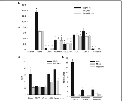
As a first step to understand the mechanism of C3 ac-tivation, we began investigation of the protein kinase sig-naling pathways in HFA required for C3 induction. HFA were pretreated with pharmacological inhibitors of spe-cific protein kinases, then were infected by HIV/NL4-3, and transfected with the C3 promoter-luciferase con-struct to assay the activity of the C3 promoter (Fig. 3). NF-κB inhibitors (CAPE and PDTC) as well as inhibitors
of p38 (SB203580, SB202190), protein tyrosine kinase (Genistein), JNK, platelet-derived growth factor receptor tyrosine kinase (AG17), and epidermal growth factor re-ceptor tyrosine kinase (AG18) blocked the HIV activa-tion of transcripactiva-tion from the C3 promoter. Based upon the maintenance of activation of the C3 promoter in the presence of specific inhibitors, PI3K and MEK are not involved in induction of C3 by HIV in astrocytes. Earlier
Fig. 1Upregulation of C3 and identification of producer cells in HIV-infected brain.acDNA was prepared from the brain tissue from four patients
who died with HIV dementia or three HIV-negative patients and subjected to QPCR amplifying the designated transcripts, data are expressed as fold change relative to uninfected tissue. The significance of gene induction of HIV-infected vs. uninfected was tested by Student’sttest with
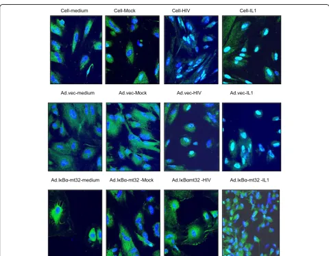
studies using astrocytic cell lines implicated cyclic AMP, protein kinase C, and C/EBPδbut not NF-κB in the acti-vation of C3 expression in astrocytes by HIV [24]. To supplement the evidence obtained using pharmaco-logical inhibitors (Fig. 3), we performed three comple-mentary assays to investigate the state of activation of NF-κB after exposure of HFA to HIV. First, nuclear localization of p50 monomer of NF-κB in astrocytes was measured by ELISA after various stimuli (Fig. 4a). Like the positive control IL-1β, HIV induced the entry of NF-κB into the nucleus and this event was inhibited by CAPE. Second, we employed an adenovirus vector of super-repressor I-κBmt32 with the expectation that the complex of NF-κB and I-κBmt32 will resist degradation and NF-κB will be retained in the cytoplasm and cannot function in transcription. HFA were infected by control adenovirus or adenovirus encoding I-κBmt32 and then infected with HIV and induction of the C3 promoter was measured by luciferase assay (Fig. 4b). Like the ex-periment shown in Fig. 2, HIV exposure activated tran-scription from the C3 promoter; the control adenovirus vector had no effect upon this activation. In contrast, ex-pression of I-κBmt32 and inhibition of activation of NF-κB completely blocked C3 promoter induction by HIV in HFA (Fig. 4b). Finally, to unambiguously demonstrate that HIV activates NF-κB in astrocytes, we localized NF-κB in HFA by confocal microscopy. Cells were activated by various stimuli in the presence of control adenovirus or adenovirus encoding I-κBmt32 super-repressor of NF-κB. Cells were fixed and stained for the p65 chain of NF-κB, nuclei were visualized with DAPI (Fig. 5). In the absence of stimuli, NF-κB is mainly cytoplasmic in HFA;
however, both HIV and IL-1β induced the translocation of p65 into the nucleus (upper row). In contrast, the expression of the super-repressor I-κBmt32 arrested NF-κB nuclear localization induced either by HIV or by IL-1β(lower row); control adenovirus had no effect (middle row). The findings in Figs. 3, 4, and 5 demonstrate un-equivocally that HFA respond to HIV by NF-κB-linked C3 synthesis. Further studies dissect the elements in the C3 promoter that control HIV-induced C3 responses.
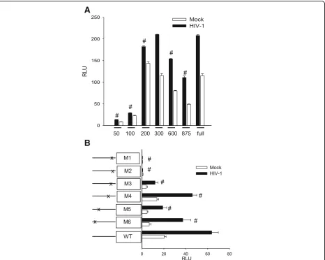
We prepared a series of deletion and point mutations in the C3 promoter in the luciferase construct and tested their response during activation of HFA by HIV (Fig. 6). The minimal promoters consisting of only 50 or 100 base pairs upstream of the initiation codon were in-active; restoring the promoter to 200 base pairs restored the response to HIV (Fig. 6a). The constitutive activity of this construct was somewhat higher than that of the intact promoter, suggesting that there is a negative regu-latory element upstream. To identify essential sites within the promoter recognizing transcriptional modula-tors, we prepared five mutations (Fig. 6b). Mutation of the proximal IL-1β/IL-6 site (M1 or M2) abolished both constitutive and HIV-induced transcription directed by the C3 promoter. In contrast, mutation of the interferon-γresponse element (M3), the more distal IL-6 element (M4), or either of two NF-κB sites (M5 or M6) had only modest effects upon the C3 promoter response to HIV by HFA. Studies in the previous section demon-strated that HIV-induced C3 through NF-κB, but here it is clear that the NF-κB sites in the C3 promoter are dispensable for the response to HIV. This apparent paradox can be resolved if NF-κB is required for
A
B
C
Fig. 2HIV activates the C3 promoter inducing transcription and protein production by HFA. HFA were exposed to NL4-3 at 0.5, 1.0, and 2.5 pg per cell
induction of expression of an intermediate protein that itself induces C3.
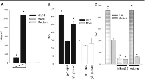
We propose that HIV activates IL-1βor IL-6 expres-sion through NF-κB and one or both of these cytokines then initiates a program to activate the C3 promoter in HFA. As shown in Fig. 1, both IL-1βand IL-6 are prom-inently expressed in the HIV-infected brain, consistent with the premise that these cytokines trigger C3 produc-tion by astrocytes. We have previously shown that HIV can induce IL-6 transcription and secretion [16] and Fig. 7a illustrates dose-dependent induction of IL-6 in HFA by HIV. Because HFA do not produce IL-1βunder the same conditions (not shown), we confined further studies to activities of IL-6 upon HFA. To definitively link the production of IL-6 to the activation of C3
expression, we employed anti-IL-6 neutralizing antibody. HFA expressing the C3 promoter-luciferase construct were exposed to HIV in the presence of anti-IL-6 or control antibody (Fig. 7b). The ability of HIV to activate the C3 promoter was reduced by neutralization of IL-6 to background levels, indicating that IL-6 is an essential intermediate in C3 induction by HIV. To determine whether IL-6 employs NF-κB to activate the C3 pro-moter, we exposed cells to exogenous IL-6 and inhibited NF-κB through transduction of the super-repressor, I-κBmt32 (Fig. 7c). IL-6 increased the activity of the C3 promoter in HFA but infection with the adenovirus vec-tor of I-κBmt32 entirely blunted this response; infection with control adenovirus had no effect upon the C3 pro-moter response. These findings strongly implicate
Fig. 3HIV induces multiple elements for activation of the C3 promoter in HFA. HFA were treated with pharmacological agents as indicated prior
Fig. 5HIV induces nuclear translocation of NF-κB in HFA. HFA were treated as indicated and subjected to confocal microscopy, staining for DNA (blue) and the p65 chain of NF-κB (green). Please see the“Methods”section for details
A
B
Fig. 4C3 induction by HIV in HFA requires activation of NF-κB. HFA were treated as indicated andaassayed for translocation of NF-κB into the
endogenously produced IL-6 in the pathway of HIV acti-vation of C3 by HFA. To determine the transcriptional pathway employed in HIV activation of IL-6 synthesis in HFA, we tested the induction of the IL-6 promoter by pharmacological inhibition of NF-κB or by mutagen-esis. An IL-6 promoter construct directing synthesis of luciferase was tested for activation by HIV in the pres-ence or abspres-ence of CAPE (Fig. 8a) or an intact IL-6 promoter, and a mutant in theκB site driving luciferase expression were used (Fig. 8b). HIV required NF-κB ac-tivity to activate the IL-6 promoter, the presence of CAPE or a mutated κB element abolished promoter function. These findings, coupled with those in Fig. 7, strongly suggest that HIV activates a circuit in astro-cytes, first inducing NF-κB-dependent IL-6 synthesis; extracellular IL-6 then initiates a secondary signaling cascade resulting in C3 transcription and protein expression.
Discussion
The major finding of this work is that HIV initiates an auto-stimulatory pathway in cultured HFA in which NF-κB is activated to induce IL-6 production and IL-6 fur-ther signals to induce C3 transcription and protein pro-duction. IL-6 and C3 are also elevated in the brains of patients with HAND; double staining indicated that C3 was expressed by astrocytes. In culture, these responses involve multiple protein kinases and NF-κB, which are characteristic of the complexity of induction of other acute phase reactants, like serum amyloid A [60].
Several approaches have previously shown that HIV and SIV neuropathogenesis correlates with elevated ex-pression of complement proteins in the central nervous system [32, 35, 36, 39, 61]. Immunohistochemistry stain-ing in SIV-infected macaque brain tissues localized ele-vated expression of complement component proteins C3 and C1q to astrocytes, neurons, and myeloid cells [42].
A
B
Fig. 6Elements in the C3 promoter essential for activation of HFA by HIV. HFA were exposed to HIV or mock virus and then transfected with
We show by immunostaining here that complement C3c can also be overexpressed in astrocytes and cells with neuronal morphology in the brain tissues from patients with HAD/HIVE; these tissues also had elevated levels of C3, C4a, but not C1qa transcripts and, consistent with other studies [62], increased expression of inflammatory effector genes IL-1βand IL-6 (Fig. 1). At present, there is less information about potentially aberrant comple-ment expression in the CNS of patients with mild HIV cognitive disease [63]. However, CSF and brain analyses from these patients revealed markers of increased neuro-inflammation [64, 65] and complement activation is part of the inflammatory response to HIV infection [63]. Re-cent research also shows that HIV persists in astrocytes during presymptomatic stage of HIV infection [66], sup-porting the notion that HIV infection of these cells and potentially complement activation by the virus may im-pact the course of HIV neuropathogenesis.
Consistent with previous studies in astrocytic cell lines [24, 26], exposure of HFA to HIV in culture activates C3 promoter in a virus dose-dependent manner resulting in increased C3 mRNA and production of secretable C3 protein (Fig. 2). Using pharmacological inhibitors, we found that several protein kinases are necessary for the intracellular signal transmission after HIV exposure of
HFA, but their order of activation requires further study (Figs. 3, 4, and 5). The activation by HIV requires, among others, the NF-κB and PTK signaling pathways; however, surprisingly, the C3 core promoter lacks NF-κB and SP binding sites. The requirement for functional NF-κB for the HIV effect on C3 was confirmed by over-expression of the NF-κB inhibitor I-κBα in astrocytes; adenovirus-mediated transduction of I-κBα into HFA, but not of control adenovirus, blocked nuclear transloca-tion of NF-κB and prevented induction of the C3 promoter and mRNA. However, activation of the C3 promoter by HIV was absolutely dependent on the presence of the IL-6/IL-1β responsive element at −109 to −90 and its sub-domain −106 to −100. The neutralization of IL-6 by anti-IL-6 antibody abolished C3 promoter induction revealing an essential role for IL-6 in increased C3 protein synthesis in HFA upon HIV ex-posure. Earlier studies using astrocytic cell lines impli-cated cyclic AMP, protein kinase C, the IL-6/IL-1β responsive element in the C3 promoter, and transcrip-tion factor C/EBPδ, but not NF-κB or IL-6, in induction of C3 in transformed astrocytes by HIV [24, 26]. Our re-sults demonstrate a novel auto-stimulatory pathway in the scheme of complement activation by HIV in primary astrocytes by indicating that HIV activates C3 in these
A
B
C
κ
Fig. 7The induction of IL-6 is essential for the induction of C3 by HIV in HFA.aHFA were exposed to HIV at 0.5 and 2.5 pg per cell or mock virus and
cells indirectly through NF-κB-dependent induction of IL-6, which in turn acts as a second messenger in this pathway through induction of C3 transcription via the
−109 to −90 IL-6/IL-1β responsive element in the C3 promoter.
Our findings from in vitro studies of HIV activation of C3 expression by HFA through IL-6 gain biological significance from observations that IL-6 is overex-pressed in the brains of patients with HAND and the requirement for IL-1β/IL-6 element in the C3 pro-moter for its activation by HIV [26]. Independent studies of IL-6 activity show that when mice are gen-etically engineered to express IL-6 restricted to astro-cytes through the GFAP promoter, C3 is upregulated in the brain but not in the peripheral tissues [67]. As-trocytes are now recognized as one element of the in-nate immune system, responding to brain injury not only by cytoskeletal changes to restrict neuronal injury but also by synthesis of effector proteins including cytokines, interferon-related proteins, and complement components [30]. Furthermore, astrocytes are increasingly recognized as an important HIV res-ervoir in patients at the presymptomatic stage of in-fection [66] and in SIV-infected macaques throughout the course of SIV disease [68]. What has proven particularly striking are findings in the last decade from multiple systems that decoration of neurons with complement components is a pivotal feature of synaptic pruning by microglia that occurs during nor-mal development and that can underlie functional
changes observed during distinct neuropathological disorders (reviewed in [31, 69]). C1q and C3 knock-out mice exhibit impaired synapse elimination during brain development [41]; indeed without C1q, mice are susceptible to both spontaneous and induced epileptic seizures associated with excess excitatory synapses [70]. Synaptic loss in a mouse model of Alzheimer’s disease has been linked to aberrant expression of C1q which was further shown essential for soluble β amyl-oid synaptic toxicity [47]; in mice, West Nile virus infection of neurons drives their expression of complement components leading to pruning of pre-synaptic termini, a disease state absent in C1q or C3 knockout mice [50], and remarkably, C3 knockout mice failed to display even synaptic loss leading to neuronal loss and defects in memory observed during normal aging [71]. These observations suggest that overexpression of complement proteins in the brain, independent of other known neuropathogenic media-tors such as amyloid plaques in AD or HIV Tat pro-tein in HAD, may directly contribute to neuronal injury in diseased brain. Studies in the present work, along with previous reports [24–26], suggest that as-trocytes may serve as an important source of neuro-pathogenic complement components in HIV-infected brain. Our discovery that IL-6 expression is central to C3 induction by HIV in astrocytes (Figs. 7 and 8) may provide an avenue to new therapeutic investiga-tions in this common pathway to diverse brain dis-eases plaguing mankind [72, 73].
Fig. 8HIV activates the IL-6 promoter through NF-κB.aHFA transfected with the IL-6 promoter driving luciferase were exposed to HIV at 0.5 or 2.5 pg
p24 per cell in the presence or absence of CAPE. Luciferase activity was measured and the difference in values between mock-infected and the other experimental groupsa. Luciferase activity was measured for HFA transfected with the IL-6 promoter driving luciferase were exposed to HIV at 0.5 or 2.5 pg per cell or mock virus in the presence or absence of CAPEaand IL-6 promoter and a mutant in theκB siteb. The differences in values between mock-infected and HIV-infected cells were tested byaone-way ANOVA and Dunnett’s post hoc analysis withasteriskindicatingp< 0.05 and by Student’s
Conclusions
The study presented here indicates that HIV induces C3 expression in primary human astrocytes indirectly, through NF-κB dependent induction of IL-6, which in turn activates the C3 promoter. HIV induction of C3 and IL-6 in astrocytes may contribute to HIV-mediated inflammation in the brain and cognitive dysfunction.
Additional file
Additional file 1: Table S1.Supplementary Statistical Results. Result
tables of statistical results for one-way ANOVA and Dunnett’s post hoc analysis. Each sheet in the Excel file contains the corresponding figure analyzed by this approach. (XLS 59 kb)
Acknowledgements
We thank Dr. Gail Bishop of the University of Iowa for her generous gift of constructs of the IL-6 promoter driving luciferase. We thank Galina Bentsman for the technical assistance and Ilene Totillo for the help in manuscript preparation. PBF holds the Thelma Newmeyer Corman Chair in Cancer Research in the MCC.
Funding
Funding was supplied by PHS grants to DJV: R21 MH 086372, R01 MH 083627, R01 DA 017618, R01 DA 037611, R01 MH 104145 and to SM: U01 MH 100931.
Availability of data and materials
All data generated or analyzed during the current study are available from the corresponding author on reasonable request.
Authors’contributions
JN designed the study, performed and analyzed all the HFA in vitro studies, and drafted the manuscript. AB performed and analyzed the human studies and contributed to writing the manuscript. SM provided the human brain tissues and supervised the human studies. JM performed the immunohistochemical analysis. WC collaborated in the in vitro studies and performed part of the promoter analysis. LE and PBF designed and helped with immunostaining study for detection p65 subunits of NF-κB and contributed in the preparation of the manuscript. MJP was a major contributor in writing the manuscript. DJV supervised the design and analysis of the study and contribute to write the manuscript. All authors read and approved the final manuscript.
Competing interests
The authors declare that they have no competing interests.
Consent for publication
Not applicable.
Ethics approval and consent to participate
Adult human brain specimens were provided by the Manhattan HIV Brain Bank (U24MH100931) under an Institutional Review Board-approved protocol at Mount Sinai School of Medicine.
Author details
1Department of Medicine, Division of Infectious Diseases, Icahn School of
Medicine at Mount Sinai, New York 10029, NY, USA.2Manhattan HIV Brain
Bank, Department of Neurology, Icahn School of Medicine at Mount Sinai, New York 10029, NY, USA.3Department of Human and Molecular Genetics,
VCU Massey Cancer Center, School of Medicine, VCU Institute of Molecular Medicine, Virginia Commonwealth UniversitySchool of Medicine, Richmond 23298, VA, USA.4Present Address: PSI-CRO, Wisniowy Business Park C, 1 Sierpnia 6A, 02-134 Warsaw, Poland.5Department of Medicine, Division of
Infectious Diseases, 1468 Madison Avenue, Annenberg Building, 21st Floor, Room 42, New York 10029, NY, USA.
Received: 23 August 2016 Accepted: 10 January 2017
References
1. Antinori A, Arendt G, Becker JT, Brew BJ, Byrd DA, Cherner M, Clifford DB, Cinque P, Epstein LG, Goodkin K, et al. Updated research nosology for HIV-associated neurocognitive disorders. Neurology. 2007;69:1789–99. 2. Price RW, Spudich SS, Peterson J, Joseph S, Fuchs D, Zetterberg H, Gisslen
M, Swanstrom R. Evolving character of chronic central nervous system HIV infection. Semin Neurol. 2014;34:7–13.
3. Brouillette M-J, Yuen T, Fellows LK, Cysique LA, Heaton RK, Mayo NE. Identifying neurocognitive decline at 36 months among HIV-positive participants in the CHARTER Cohort using group-based trajectory analysis. Plos One. 2016;11:e0155766.
4. Saylor D, Dickens AM, Sacktor N, Haughey N, Slusher B, Pletnikov M, Mankowski JL, Brown A, Volsky DJ, McArthur JC. HIV-associated neurocognitive disorder—pathogenesis and prospects for treatment. Nat Rev Neurol. 2016;12:234–48.
5. Harezlak J, Buchthal S, Taylor M, Schifitto G, Zhong J, Daar E, Alger J, Singer E, Campbell T, Yiannoutsos C, et al. Persistence of HIV-associated cognitive impairment, inflammation, and neuronal injury in era of highly active antiretroviral treatment. AIDS. 2011;25:625–33.
6. Heaton RK, Franklin DR, Ellis RJ, McCutchan JA, Letendre SL, Leblanc S, Corkran SH, Duarte NA, Clifford DB, Woods SP, et al. HIV-associated neurocognitive disorders before and during the era of combination antiretroviral therapy: differences in rates, nature, and predictors. J Neurovirol. 2011;17:3–16.
7. Mateen FJ, Shinohara RT, Carone M, Miller EN, McArthur JC, Jacobson LP, Sacktor N. Neurologic disorders incidence in HIV+ vs HIV−men: Multicenter AIDS Cohort Study, 1996–2011. Neurology. 2012;79:1873–80.
8. Robertson KR, Smurzynski M, Parsons TD, Wu K, Bosch RJ, Wu J, McArthur JC, Collier AC, Evans SR, Ellis RJ. The prevalence and incidence of neurocognitive impairment in the HAART era. AIDS. 2007;21:1915–21. 9. Takahashi K, Wesselingh SL, Griffin DE, McArthur JC, Johnson RT, Glass JD.
Localization of HIV-1 in human brain using polymerase chain reaction/in situ hybridization and immunocytochemistry. Ann Neurol. 1996;39:705–11. 10. Spudich S, González-Scarano F. HIV-1-related central nervous system
disease: current issues in pathogenesis, diagnosis, and treatment. Cold Spring Harb Perspect Med. 2012;2:a007120.
11. Conant K, Garzino-Demo A, Nath A, McArthur JC, Halliday W, Power C, Gallo RC, Major EO. Induction of monocyte chemoattractant protein-1 in HIV-1 Tat-stimulated astrocytes and elevation in AIDS. Proc Natl Acad Sci U S A. 1998;95:3117–21.
12. Fitting S, Zou S, Chen W, Vo P, Hauser KF, Knapp PE. Regional heterogeneity and diversity in cytokine and chemokine production by astroglia: differential responses to HIV-1 Tat, gp120, and morphine revealed by multiplex analysis. J Proteome Res. 2010;9:1795–804.
13. Nookala AR, Kumar A. Molecular mechanisms involved in HIV-1 Tat-mediated induction of IL-6 and IL-8 in astrocytes. J Neuroinflammation. 2014;11:214.
14. Zink MC, Clements JE. A novel simian immunodeficiency virus model that provides insight into mechanisms of human immunodeficiency virus central nervous system disease. J Neurovirol. 2002;8 Suppl 2:42–8.
15. Chen Y, Swanson RA. Astrocytes and brain injury. J Cereb Blood Flow Metab. 2003;23:137–49.
16. Li J, Bentsman G, Potash MJ, Volsky DJ. Human immunodeficiency virus type 1 efficiently binds to human fetal astrocytes and induces neuroinflammatory responses independent of infection. BMC Neurosci. 2007;8:31.
17. Roberts ES, Burudi EME, Flynn C, Madden LJ, Roinick KL, Watry DD, Zandonatti MA, Taffe MA, Fox HS. Acute SIV infection of the brain leads to upregulation of IL6 and interferon-regulated genes: expression patterns throughout disease progression and impact on neuroAIDS. J Neuroimmunol. 2004;157:81–92.
18. Mankowski JL, Queen SE, Clements JE, Zink MC. Cerebrospinal fluid markers that predict SIV CNS disease. J Neuroimmunol. 2004;157:66–70.
20. Quintana A, Giralt M, Molinero A, Campbell IL, Penkowa M, Hidalgo J. Analysis of the cerebral transcriptome in mice subjected to traumatic brain injury: importance of IL-6. Neuroimmunomodulation. 2007;14:139–43. 21. Sordillo PP, Sordillo LA, Helson L. Bifunctional role of pro-inflammatory
cytokines after traumatic brain injury. Brain Inj. 2016;1–11:e0149451. 22. Su Z-Z, Kang D-C, Chen Y, Pekarskaya O, Chao W, Volsky DJ, Fisher PB.
Identification and cloning of human astrocyte genes displaying elevated expression after infection with HIV-1 or exposure to HIV-1 envelope glycoprotein by rapid subtraction hybridization, RaSH. Oncogene. 2002;21:3592–602. 23. Kim S-Y, Li J, Bentsman G, Brooks AI, Volsky DJ. Microarray analysis of changes
in cellular gene expression induced by productive infection of primary human astrocytes: implications for HAD. J Neuroimmunol. 2004;157:17–26.
24. Speth C, Schabetsberger T, Mohsenipour I, Stöckl G, Würzner R, Stoiber H, Lass-Flörl C, Dierich MP. Mechanism of human immunodeficiency virus-induced complement expression in astrocytes and neurons. J Virol. 2002;76:3179–88. 25. Speth C, Stöckl G, Mohsenipour I, Würzner R, Stoiber H, Lass-Flörl C, Dierich
MP. Human immunodeficiency virus type 1 induces expression of complement factors in human astrocytes. J Virol. 2001;75:2604–15. 26. Bruder C, Hagleitner M, Darlington G, Mohsenipour I, Würzner R, Höllmüller
I, Stoiber H, Lass-Flörl C, Dierich MP, Speth C. HIV-1 induces complement factor C3 synthesis in astrocytes and neurons by modulation of promoter activity. Mol Immunol. 2003;40:949–61.
27. Morgan BP. The role of complement in neurological and neuropsychiatric diseases. Expert Rev Clin Immunol. 2015;11:1109–19.
28. Orsini F, De Blasio D, Zangari R, Zanier ER, De Simoni MG. Versatility of the complement system in neuroinflammation, neurodegeneration and brain homeostasis. Front Cell Neurosci. 2014;8:380.
29. Sekar A, Bialas AR, de Rivera H, Davis A, Hammond TR, Kamitaki N, Tooley K, Presumey J, Baum M, Van Doren V, et al. Schizophrenia risk from complex variation of complement component 4. Nature. 2016;530:177–83. 30. Farina C, Aloisi F, Meinl E. Astrocytes are active players in cerebral innate
immunity. Trends Immunol. 2007;28:138–45.
31. Hong S, Dissing-Olesen L, Stevens B. New insights on the role of microglia in synaptic pruning in health and disease. Curr Opin Neurobiol. 2016;36: 128–34.
32. Komotar RJ, Kim GH, Otten ML, Hassid B, Mocco J, Sughrue ME, Starke RM, Mack WJ, Ducruet AF, Merkow MB, et al. The role of complement in stroke therapy. Adv Exp Med Biol. 2008;632:23–33.
33. Singhrao SK, Neal JW, Morgan BP, Gasque P. Increased complement biosynthesis by microglia and complement activation on neurons in Huntington’s disease. Exp Neurol. 1999;159:362–76.
34. Hu WT, Watts KD, Tailor P, Nguyen TP, Howell JC, Lee RC, Seyfried NT, Gearing M, Hales CM, Levey AI, et al. CSF complement 3 and factor H are staging biomarkers in Alzheimer’s disease. Acta Neuropathol Commun. 2016;4:14.
35. Borjabad A, Morgello S, Chao W, Kim S-Y, Brooks AI, Murray J, Potash MJ, Volsky DJ. Significant effects of antiretroviral therapy on global gene expression in brain tissues of patients with HIV-1-associated neurocognitive disorders. Plos Pathog. 2011;7:e1002213.
36. Jongen PJ, Doesburg WH, Ibrahim-Stappers JL, Lemmens WA, Hommes OR, Lamers KJ. Cerebrospinal fluid C3 and C4 indexes in immunological disorders of the central nervous system. Acta Neurol Scand. 2000;101:116–21. 37. Reboul J, Schuller E, Pialoux G, Rey MA, Lebon P, Allinquant B, Brun-Vezinet
F. Immunoglobulins and complement components in 37 patients infected by HIV-1 virus: comparison of general (systemic) and intrathecal immunity. J Neurol Sci. 1989;89:243–52.
38. Rozek W, Ricardo-Dukelow M, Holloway S, Gendelman HE, Wojna V, Melendez LM, Ciborowski P. Cerebrospinal fluid proteomic profiling of HIV-1-infected patients with cognitive impairment. J Proteome Res. 2007;6: 4189–99.
39. Roberts ES, Zandonatti MA, Watry DD, Madden LJ, Henriksen SJ, Taffe MA, Fox HS. Induction of pathogenic sets of genes in macrophages and neurons in NeuroAIDS. Am J Pathol. 2003;162:2041–57.
40. Potash MJ, Chao W, Bentsman G, Paris N, Saini M, Nitkiewicz J, Belem P, Sharer L, Brooks AI, Volsky DJ. A mouse model for study of systemic HIV-1 infection, antiviral immune responses, and neuroinvasiveness. Proc Natl Acad Sci U S A. 2005;102:3760–5.
41. Stevens B, Allen NJ, Vazquez LE, Howell GR, Christopherson KS, Nouri N, Micheva KD, Mehalow AK, Huberman AD, Stafford B, et al. The classical complement cascade mediates CNS synapse elimination. Cell. 2007;131: 1164–78.
42. Speth C, Williams K, Hagleitner M, Westmoreland S, Rambach G, Mohsenipour I, Schmitz J, Würzner R, Lass-Flörl C, Stoiber H, et al. Complement synthesis and activation in the brain of SIV-infected monkeys. J Neuroimmunol. 2004;151:45–54.
43. Depboylu C, Schäfer MK, Schwaeble WJ, Reinhart TA, Maeda H, Mitsuya H, Damadzic R, Rausch DM, Eiden LE, Weihe E. Increase of C1q biosynthesis in brain microglia and macrophages during lentivirus infection in the rhesus macaque is sensitive to antiretroviral treatment with 6-chloro-2’,3’ -dideoxyguanosine. Neurobiol Dis. 2005;20:12–26.
44. Schafer DP, Lehrman EK, Kautzman AG, Koyama R, Mardinly AR, Yamasaki R, Ransohoff RM, Greenberg ME, Barres BA, Stevens B. Microglia sculpt postnatal neural circuits in an activity and complement-dependent manner. Neuron. 2012;74:691–705.
45. Paolicelli RC, Bolasco G, Pagani F, Maggi L, Scianni M, Panzanelli P, Giustetto M, Ferreira TA, Guiducci E, Dumas L, et al. Synaptic pruning by microglia is necessary for normal brain development. Science. 2011;333:1456–8. 46. Lui H, Zhang J, Makinson SR, Cahill MK, Kelley KW, Huang HY, Shang Y,
Oldham MC, Martens LH, Gao F, et al. Progranulin deficiency promotes circuit-specific synaptic pruning by microglia via complement activation. Cell. 2016;165:921–35.
47. Hong S, Beja-Glasser VF, Nfonoyim BM, Frouin A, Li S, Ramakrishnan S, Merry KM, Shi Q, Rosenthal A, Barres BA, et al. Complement and microglia mediate early synapse loss in Alzheimer mouse models. Science. 2016;352:712–6. 48. Michailidou I, Willems JG, Kooi EJ, van Eden C, Gold SM, Geurts JJ, Baas F,
Huitinga I, Ramaglia V. Complement C1q-C3-associated synaptic changes in multiple sclerosis hippocampus. Ann Neurol. 2015;77:1007–26.
49. Mayilyan KR, Weinberger DR, Sim RB. The complement system in schizophrenia. Drug News Perspect. 2008;21:200–10.
50. Vasek MJ, Garber C, Dorsey D, Durrant DM, Bollman B, Soung A, Yu J, Perez-Torres C, Frouin A, Wilton DK, et al. A complement-microglial axis drives synapse loss during virus-induced memory impairment. Nature. 2016;534:538–43. 51. Lian H, Litvinchuk A, Chiang AC, Aithmitti N, Jankowsky JL, Zheng H.
Astrocyte-microglia cross talk through complement activation modulates amyloid pathology in mouse models of Alzheimer’s disease. J Neurosci. 2016;36:577–89.
52. Canki M, Thai JN, Chao W, Ghorpade A, Potash MJ, Volsky DJ. Highly productive infection with pseudotyped human immunodeficiency virus type 1 (HIV-1) indicates no intracellular restrictions to HIV-1 replication in primary human astrocytes. J Virol. 2001;75:7925–33.
53. Adachi A, Gendelman HE, Koenig S, Folks T, Willey R, Rabson A, Martin MA. Production of acquired immunodeficiency syndrome-associated retrovirus in human and nonhuman cells transfected with an infectious molecular clone. J Virol. 1986;59:284–91.
54. Sambrook J, Russell DW. Molecular cloning: a laboratory manual. 3rd edition edn. Cold Spring Harbor, New York: Cold Spring Harbor Laboratory Press; 2001. 55. Kim SY, Chao W, Choi SY, Volsky DJ. Cloning and characterization of the 3’
-untranslated region of the human excitatory amino acid transporter 2 transcript. J Neurochem. 2003;86:1458–67.
56. Baccam M, Woo S-Y, Vinson C, Bishop GA. CD40-mediated transcriptional regulation of the IL-6 gene in B lymphocytes: involvement of NF-kB, AP-1, and C/EBP. J Immunol. 2003;170:3099–108.
57. Emdad L, Sarkar D, Su ZZ, Randolph A, Boukerche H, Valerie K, Fisher PB. Activation of the nuclear factor kappaB pathway by astrocyte elevated gene-1: implications for tumor progression and metastasis. Cancer Res. 2006;66:1509–16.
58. Chaudhuri A, Yang B, Gendelman HE, Persidsky Y, Kanmogne GD. STAT1 signaling modulates HIV-1-induced inflammatory responses and leukocyte transmigration across the blood-brain barrier. Blood. 2008;111:2062–72. 59. Winkler JM, Chaudhuri AD, Fox HS. Translating the brain transcriptome in
neuroAIDS: from non-human primates to humans. J Neuroimmune Pharmacol. 2012;7:372–9.
60. Blatteis CM, Li S, Li Z, Perlik V, Feleder C. Signaling the brain in systemic inflammation: the role of complement. Front Biosci. 2004;9:915–31. 61. Pendyala G, Trauger SA, Kalisiak E, Ellis RJ, Siuzdak G, Fox HS. Cerebrospinal
fluid proteomics reveals potential pathogenic changes in the brains of SIV-infected monkeys. J Proteome Res. 2009;8:2253–60.
62. Achim CL, Heyes MP, Wiley CA. Quantitation of human immunodeficiency virus, immune activation factors, and quinolinic acid in AIDS brains. J Clin Invest. 1993;91:2769–75.
64. Abassi M, Morawski BM, Nakigozi G, Nakasujja N, Kong X, Meya DB, Robertson K, Gray R, Wawer MJ, Sacktor N, Boulware DR. Cerebrospinal fluid biomarkers and HIV-associated neurocognitive disorders in HIV-infected individuals in Rakai, Uganda. J Neurovirol. 2016. doi:10.1007/s13365-016-0505-9.
65. Hong S, Banks WA. Role of the immune system in HIV-associated neuroinflammation and neurocognitive implications. Brain Behav Immun. 2015;45:1–12.
66. Thompson KA, Cherry CL, Bell JE, Mclean CA. Brain cell reservoirs of latent virus in presymptomatic HIV-infected individuals. Am J Pathol. 2011;179: 1623–9.
67. Barnum SR, Jones JL, Müller-Ladner U, Samimi A, Campbell IL. Chronic complement C3 gene expression in the CNS of transgenic mice with astrocyte-targeted interleukin-6 expression. Glia. 1996;18:107–17. 68. Clements JE, Babas T, Mankowski JL, Suryanarayana K, Piatak Jr M, Tarwater
PM, Lifson JD, Zink MC. The central nervous system as a reservoir for simian immunodeficiency virus (SIV): steady-state levels of SIV DNA in brain from acute through asymptomatic infection. J Infect Dis. 2002;186:905–13. 69. Stephan AH, Barres BA, Stevens B. The complement system: an unexpected
role in synaptic pruning during development and disease. Annu Rev Neurosci. 2012;35:369–89.
70. Chu Y, Jin X, Parada I, Pesic A, Stevens B, Barres B, Prince DA. Enhanced synaptic connectivity and epilepsy in C1q knockout mice. Proc Natl Acad Sci U S A. 2010;107:7975–80.
71. Shi Q, Colodner KJ, Matousek SB, Merry K, Hong S, Kenison JE, Frost JL, Le KX, Li S, Dodart JC, et al. Complement C3-deficient mice fail to display age-related hippocampal decline. J Neurosci. 2015;35:13029–42.
72. Mastellos DC, Reis ES, Yancopoulou D, Hajishengallis G, Ricklin D, Lambris JD. From orphan drugs to adopted therapies: advancing C3-targeted intervention to the clinical stage. Immunobiology. 2016;221(10):1046–57. 73. Ruseva MM, Ramaglia V, Morgan BP, Harris CL. An anticomplement agent
that homes to the damaged brain and promotes recovery after traumatic brain injury in mice. Proc Natl Acad Sci U S A. 2015;112:14319–24.
• We accept pre-submission inquiries
• Our selector tool helps you to find the most relevant journal
• We provide round the clock customer support
• Convenient online submission
• Thorough peer review
• Inclusion in PubMed and all major indexing services
• Maximum visibility for your research
Submit your manuscript at www.biomedcentral.com/submit