International Educational Applied Research Journal
Volume: 2 | Issue: 1 | Jan 2018
1
INTERNATIONAL EDUCATIONAL APPLIED RESEARCH JOURNAL
Comparison study: two methods for determination of urinary citrate
SAAD AL-FAWAEIR (PhD)
11
Department of Medical Biochemistry, King Hussein Medical Center, Amman, Jordan.
_____________________________________________________________________________________________________________________________________________
Abstract -
Objective:
Measurements of urinary citrate is considered as an indicator of the risk for kidney stone
formation, in this study we compared two methods of urinary citrate assessment, and these methods are colorimetric
method and enzymatic method.
Methods:
urinary citrate levels were determined by a colorimetric method and a commercially enzymatic method in
patients with urolithiasis (n= 60) and in healthy controls (n=40).performance characteristics of both methods were
compared.
Results:
In comparison with the urinary citrate levels of healthy controls the urinary citrate levels in patients with
urolithiasis were lower. The difference between patients and controls was insignificant by enzymatic method, were it
was significant by colorimetric method. Low, intermediate and high citrate containing urinary pool within- run
imprecision for colorimetric method were,2.0,2.75and 3.25, and 3.1,3.4 and 4.5for enzymatic method, run to run
imprecision studies for colorimetric method were 5.0, 6.0 and 6.75 and 7.0,7.75and 8.75 for enzymatic method.
Enzymatic method was linear up to 5mmol/L while colorimetric method was linear up to 10 m mol/L. The detection of
limit of colorimetric method and enzymatic method was 0.09 μ mol/L and 0.375μ mol/L; mean recoveries were
93%and 87% respectively. There is a positive, strong (good) and statistically significant correlation between enzymatic
and calorimetric measures (r=0.853, p<0.001).
Conclusion:
according to the pervious results the colorimetric method has superiority in comparison with enzymatic
method.
Keywords :
Citrate, Urolithiasis, Colorimetric And Enzymatic.
___________________________________________________________________________________________________________________________________________
Introduction
Citric acid is an important intermediate in metabolism. In humans, citrate is both metabolized and excreted by the kidney (1), and its presence in urine contributes to the inhibitory potential against crystallization of calcium salt: Hypocitraturia is seen in a substantial number of patients with calcium nephrolithiasis(2,3). Citrate acts both through surface-controlled mechanism to hamper crystal growth and aggregation (4) and through the formation of stable soluble complexes with calcium (5). Therefore, citrate determination has become an important tool in the assessment of urine super saturation with respect to calcium oxalate and phosphate (6).
There are many reasons that have effects on the balance of inhibiting of urinary stones (7). Urinary citrate concentration plays an important role in formation of calcium phosphate and calcium phosphate stones in urinary tract. Citrate present in the urine forms soluble complexes with calcium leading in decrease of supersaturating of calcium oxalate and calcium phosphate. It is through that inhibitory effect of citrate has a role in prevention of formation of urinary stones (8).
Therefore, urine citrate levels draw interest in urinary tract stone disease. It has been shown in many studies that the citrate levels in patients with urinary tract stone are significantly low (9).
Recently, urinary citrate levels usually determined by enzymatic methods with a rare determination by colorimetric methods. In this study we aimed to compare an enzymatic method with colorimetric method in order to assess performance characteristics of both methods.
Methods and materials
Patient and control groups
Patients group consists of 60 persons with urolithiasis. Control group consists of 40 healthy volunteers. From both control and patients group ,24 hour urine samples were
collected and stored at -80 Co until analysis.
1. Enzymatic method:
International Educational Applied Research Journal
Volume: 2 | Issue: 1 | Jan 2018
2
INTERNATIONAL EDUCATIONAL APPLIED RESEARCH JOURNAL
product,pyruvate) in the presence of LDH(lactate dehydrogenase), MDH(malate dehydrogenase) and NADH. The intensity of the UV-color at this wavelength is proportional to the concentration of citric acid in the tested sample.
Procedure
20 ml of buffer were added to working reagent and mixed gently till dissolution.
Then a 1000μl from this mixture 25μl distilled water was added to prepare the blank, to prepare sample to 1000μl from mixture 25μl sample was added followed by mixing
well and incubation for 3 minutes at 37C0 then the samples
were read against distilled water, then 25μl of working starter was added followed by incubation for 3 minutes at
37C0 then waiting the end of reaction then the absorbance
was read. Citric acid levels were measured using the following formula:
Citric acid =1.28 x the difference of absorbance.
2.
Colorimetric
method:
Colorimetric
Measurements of Citrate
This method was first developed by Millan (6) with a
subsequent modification of Lewis (7) and Mezzour (8) and Seker (9); in this study we used the last modification by Mezzour with changes in this method.
The principle of this method rests on the quantification of the yellow complex formed with iron chloride and urinary citrate, measurable at 390 nm in acidic media.
In alkaline pH phosphates in the urine were precipitated
after reaction with MgCl2 and citrate forms a yellow colored
complex then this complex can be determined spectrophotometerically at 390 nm. Citric acid -monohydrate was used as a standard in this experiment.
Equipments:
Cintra 303 UV-Visible spectrophotometer (GBC Scientific Equipments Ltd, Australia) was used in this study
Reagents:
A Solution of 25% NH4OH (E.Merck. Darmstadt, Germany)
was used in our study while 30%NH4OH was used in some
studies.
0.2 M Magnesium Chloride solution ( E.Merck. Darmstadt, Germany) and Hydrochloric Acid (HCl) solution (E.Merck. Darmstadt, Germany) 10, 1 and 0.0.1M.
Ferric Chloride (Sigma.Aldirch.Germany) FeCl3 18 mM
solution was prepared freshly in deionized water instead of HCl.
Citrate –monohydrate (C6H8O7.H2O) (E.Merck. Darmstadt,
Germany) was used as standard in our study.
Procedure:
Urine samples were kept in (-80) refrigerator until analysis. in analysis day the samples were brought to the room
temperature , to 4 ml urine sample 0.1 ml NH4OH(25%) was
added followed by a well mixing , then 0.9 ml MgCl2 was
added and mixed in a vortex mixer then the mixture was
centrifuged at 4000g for ten minutes in order to obtain free phosphate urine , then supernatant was placed in a clean tube and the PH was adjusted to 2 with 0.1 ml of 10M HCl , supernatant was again mixed by vortex , 3 ml urine of the
mixture was taken in a clean tube and 0.3 ml of FeCl3 18mM
was added and vortex in a mixer, absorbance was immediately read against deionized water at 390 nm by the spectrophotometer.
Reagent blank was prepared by the addition of 0.3 ml FeCl3
to 3ml 0.01 M HCl solution and read against deionized water. Urine blank was prepared by addition of 0.3 0.01 M HCl solution to 3 ml free-phosphate urine and read against deionized water.
For every analysis five solution of citrate (0.25, 0.5,1, 2 and 5) were prepared and use as daily standard.
Performance characteristics of both methods:
a.
Imprecision studies
For within- run and run to run precision studies, 3 different sample pools were prepared with low, medium and high levels of citrate. For within precision, all of pools were measured for 20 times at the same series. for run to run precision samples from all pools were a liquated and stored for 20 days, every day one sample citrate level was measured .Mean (x), standard deviation (SD) and % coefficient variation (%CV) were calculated.
B. Linearity studies:
A solution of 30 mmol/L citrate was prepared. With serial dilution to 20, 15,10,5,2,1,0.5 and 0.25 mmol/L of citric acid were prepared and assayed twice by two methods.
C. recovery studies:
For recovery studies, known amount of citrate standard was added to a known level of citrate sample.
All samples were assayed in duplicate and recovery was calculated at three different levels
D. Detection limit studies
In order to determine the limit of detection blank readings were carried out 20 times without adding samples. Average and standard deviation of absorbance were calculated. Border of detection was measured by adding standard deviation X 2 to the average blank absorbance.
E. Color stability:
The stability of color was checked by measuring the absorbance with 5 minute intervals
International Educational Applied Research Journal
Volume: 2 | Issue: 1 | Jan 2018
3
INTERNATIONAL EDUCATIONAL APPLIED RESEARCH JOURNAL
Statistics
All statistical analysis were performed by SPSS for windows 20.0 (SPSS Inc. Headquarters, Chicago, III., USA) software program.
Results
Urinary citrate concentrations in both patients and control group are listed in table 1.
Colorimetric citrate and enzymatic citrate methods were compared according to their performance characteristics such as imprecision, linearity, detection of limit, and recovery. The linear regression analysis of two methods was also compared to each other.
Analyte Patient(x ±SD) Control (x ±SD) Urinary citrate
enzymatic 2.0 ± 1.6 2.6 ±1.2 Urinary citrate
colorimetric 2.2±1.4 3.6±1.3
The colorimetric method gave much better results according to the imprecision studies.
The three different urinary citrate levels within-run imprecision for colorimetric method were; 2.0,2.75and 3.25on the other hand it were3.1,3.4 and 4.5for enzymatic method; run to run studies it were 5.0%,6.0%,6.75% and 7.0%,7.75%,8.75% respectively.
Linearity studies showed that linearity was up to 5 mmol/L of urinary citrate concentration for enzymatic method, while for colorimetric method it was 10 mmol/L.
A strong correlation was found between these two methods (r=0.853) with regression analysis (Figure 1).
Figure 1
:
Linear regression analysis of colorimetric and enzymatic method.Detection of lower levels of urinary citrate is very important in clinical practice. The detection of limit of colorimetric method was found to be 0.19mmol/L, where detection of
limit of enzymatic method was 0.375. So the colorimetric method is better than enzymatic method in the detection of limit for clinical use.
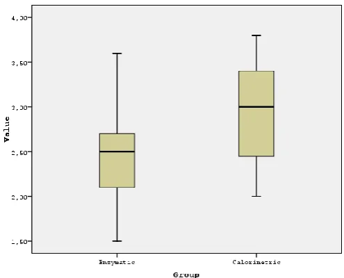
Figure 2:
A graph of citrate levels by two methods shows lower citrate concentration for both groups by enzymatic method.Colorimetric method is also better than enzymatic method according to recovery studies; R% was found 93% of colorimetric method, while it was 87% of enzymatic method.
Discussion
Hypocitraturia, a low amount of citrate in the urine, is an important risk factor for kidney stone formation (7). Citrate in the urine has long been recognized as an inhibitor of calcium salt crystallization (7). Citrate is the dissociated anion of citric acid, a weak acid that is ingested in the diet and produced endogenously in the Tricarboxylic acid cycle. Citrate plays several important roles in the mechanism of urinary stone formation. First, citrate complexes to calcium ions in the urine, reducing calcium ion activity, which results in lowering the urinary supersaturating of calcium phosphate and calcium oxalate. This complexion action is not completely understood, but it has been shown to involve the formation of a calcium-citrate-phosphate species (7-10). One of the main contributing factors on urinary system stone formation is urine citrate level. Because of that there are many studies in these studies they are tried to find a reliable and sensitive method for determination of urinary citrate. In this studied we made a comparative study between two methods for determination of urinary citrate these are colorimetric and enzymatic method.
The colorimetric method was first developed by Millan (12) with a subsequent modification of Lewis (13) and Mezzour (14) and Seker (15); in this study we used the last modification by Mezzour with changes in this method.
PH of the reaction mixture is very important for citrate- Fe +3
International Educational Applied Research Journal
Volume: 2 | Issue: 1 | Jan 2018
4
INTERNATIONAL EDUCATIONAL APPLIED RESEARCH JOURNAL
adjusting pH to 2.0 as done in this study. In order to avoid large pH variation after addition of ferric chloride solution the solution was prepared in deionized water instead of HCl. In this study hypocitrateuria happing was found as 25%and 28%by enzymatic and colorimetric methods, respectively, in previous studies it was found 34% (18) and 26% (15). In previous studies it was found that the urinary citrate concentrations in patients with kidney stone disease are significantly lower than that of healthy persons. In this study we found that the urinary citrate concentrations of patients were lower than that in healthy persons by both of methods (figure 2) . The difference was significantly in colorimetric method (p<0.05), where it was insignificantly in enzymatic method.
In this study the colorimeric method shows a good performance, lower limit of detection, linearity and high CV% at lower levels of citrate, because lower urinary concentrations are clinically more important.
Using this colorimetric method for measurements of urinary citrate is a suitable, easy and more sensitive .In comparison with other methods it more cheap.
Conclusion
In conclusion colorimetric method for determination of citrate concentration was found to be better than enzymatic method according to the analytical and clinical performances , colorimetric method inexpensive and sensitive and has a good performance especially it can be efficiently detects lower concentration of urinary citrate in urolithiasis patients.
Colorimetric method is so cheap in comparison with enzymatic. Both methods can be applied to routine clinical laboratory practice without the presence of more complex equipments.