JNS 2 (2012) 287-293
Electrospun Nitrocellulose and Composite Nanofibers
B. Naderizadeh
a*, A. Moghimi
b, M. Shahi
ca
Sama Technical and Vocatinal Training College, Islamic Azad University, Kermanshah Branch, Kermanshah, P.O. Box 671791-7855, Iran.
b
Department of Chemistry, Imam Hossein University, Tehran, P.O. Box 16575-416, Iran.
C
Department of Chemistry, Tehran North Branch, Islamic Azad University, Tehran, P.O .Box 191367-4711, Iran.
Abstract
Electrospinning was applied to nitrocellulose (NC) solutions in DMF, DMSO and Ethyl methyl keton to obtain NC nanofibers.
Electrospraying process was observed when DMF was used as
solvent. In comparison to NC/DMF, NC/DMSO solution resulted in
the formation of a coating film as indicate by SEM. Two different electrospun nanofibers, smooth nanofibers with 90 to 150 nm
diameters and porous nanofibers with 400 to 500 nm diameter, was noticed when EMK was used. Electrospun explosive composite
nanofibers were also successfully prepared from NC to modify the
properties of NC and their morphology were investigated by SEM.
Explosive materials such as nitrotriazolone (NTO), its sodium salt, picric acid and nanosilver were used as the second component of the
composites. A reduction in the diameters of the composite nanofibers involving Na- NTO was noticed comparing to the parent NC
nanofibers. The diameters of the other explosive composite nanofibers
were in the range of 50-300 nm indicating no significant diameter
change for NC nanofibers upon the addition of 0.1 wt% NTO and picric acid. Composite nanofibers containing nanosilver particles were
also prepared and their antibacterial activities were tasted against E. coli and S. aureus. These nanofibers successfully inhibited the growth
of both bacteria.
2012 JNS All rights reserved
Article history:
Received 5/8/2012 Accepted 10/12/2012 Published online 1/12/2012
Keywords:
Electrospinning Nanofiber Nnitrocellulose Explosive
Antimicrobial activity
*Corresponding author:
E-mail address:
288 B. Naderizadehet al./ JNS 2 (2012) 287-293
1.
Introduction
Electrospinning is an interesting process for
producing non-woven fibers with the average
diameters in the range of micro-to nanometers while traditional melts spun fibers range in a
diameter of about 5 to 200 µm [1, 2].
Electrospinning is a unique, simple, low-cost and effective approach not only for the preparation of
polymer nanofibers [3] but also for the ceramic
oxide materials [4] and the composite nanofibers involving metals and metal oxides [5-12]. In this
process, a continuous filament is drown from a
polymer solution or a melt through a spinneret by high electrostatic forces to deposit it on a
grounded-metal collective screen [13].
Electrospinning of a variety of synthetic as well as natural polymer fibers for different applications
have also been reported in the literature [14]. A search in the literature indicated that different
polymers such as chitosan, dextral,
polyacrylonitrile, polyvinyl alcohol, poly(methyl
methacrylate), polystyrene, polycaprolactone, poly(vinyl pyrrolidone), biodegradable
poly(L-lactide), nylon 6 and cellulose acetate [2-12] do
electrospinning as well as different conducting polymers.
Incorporation of nanoparticles in polymer
nanofibers has attracted a considerable attention due to possibilities of fabricating suitable materials
for applications such as catalysts, drug deliveries,
tissue engineering scaffolds, medical gauzes, fiber- based sensors and membranes [3-10,15]. Since the
first report on electrospinning method [16], many
scientists have used this process to incorporate different species. The appearance of the collected
fibers depends on several factors including;
viscosity of the solution, average molecular weight of the polymer, surface tension, concentration of
solution, gravitational force which is dependent on
solution density, electrostatic force which has been
found to depend on the applied electrostatic field
and the conductivity of polymer, and ambient
parameters such as temperature and humidity [17-20]. One attractive feature of electrospinning is the
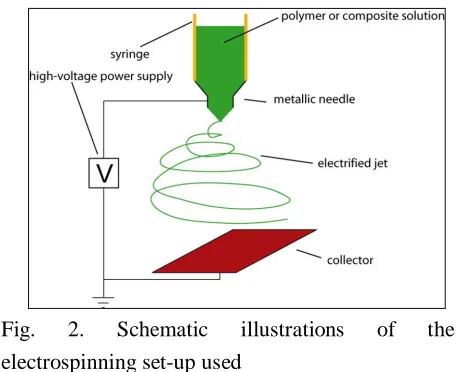
simplicity and inexpensive nature of the set up. The typical electrospinning set up consist of a
syringe pump, a high voltage source and a
collector.
A lack of research in the field of electrospun fibers in the literature promoted this research group
to prepare and characterize a new class of electrospun explosive nanofibers (EENF). Such
explosive fibers are expected to behave differently
from the corresponding common fibers and show
different properties from the explosive point of view. Also, of interest to us has been the
preparation of electrospun explosive composite nanofibers (EECNF) involving both NC and other
known explosive materials. In this paper three such
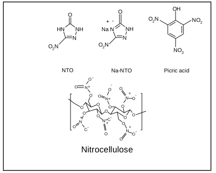
explosives ei. NTO, Na-NTO and Picric acid have been used and examined. The structure of these
components is observed in Fig. 1. The presence of
these components in NC electrospun nanofibers are expected to influence the properties of the resulting
composites.
2. Experimental procedure
2. 1. Materials and Instrumentation
Commercially available NC (12.6% N) was
obtained from Parcheen Chemical Industries, Tehran. NTO was prepared according to the
reported procedure [21], Dimethylsulfoxide
(DMSO) 99% was obtained from Fluka. Picric acid, N, N- dimethylformanide (DMF) 99.5% and
Ethyl Methyl Keton (EMK) 99% were purchased
from Merck.
To characterize the morphology of the
N NH Na N
O
O2N
Na-NTO NTO
NO2
OH
O2N NO2
Picric acid N NH N H O
O2N
+ -O O O O O O OO O O O N O O N O O N O O N O O N O O N O O -+ + + + + + Nitrocellulose
coated with gold and examined at different
accelerating voltages and Scanning Electron
Microscope (SEM) images were obtained by a
Philips XL-30. The thermal behavior of the composite nanofibers was studied by STA 1500
from 20 to 700 °C under argon flow with at a heating rate of 10°C/min.
Fig. 1. Chemical structures of NC, NTO, Na- NTO
and picric acid
2. 2. Synthesis of Na-NTO
The synthetic procedure reported in the
literature, with some minor modification, was followed to prepare Na-NTO [22]. NTO (1.3g) was
added to a basic solution containing NaOH (0.4g)
in water at 60 °C. The yellow color was turned
orange and orange precipitate was formed. The precipitate was filtered off and dried.
2. 3. Preparation of NC nanofibers and
composite nanofibers involving NC
Generally, viscose solutions were prepared by dissolving appropriate amount of dried NC, in an
oven at 65 °C for 2h, and additives, if needed, in
solvents and transferred into hypodermic syringes. Then, appropriate voltages were applied between
two electrodes and the webs of nanofibers were
accumulated on the surface of the aluminum foil,
as collector. As the jet accelerated towards the
cathode, the solvent evaporated and fibers were
deposited on the collector cathode.
2.3.1. Preparation of NC electrospun nanofibers NC electrospun nanofibers were obtained by
the preparation of NC solutions, using DMSO,
DMF and EMK as solvent. Typically, 1 ml of each
solvent was added to a container containing 0.1 g NC and stirred to obtain clear homogeneous
solution. The solution was then transferred into a syringe. The voltage applied between the tip of
syringe and collector was 18 kV and the distances
were 8 cm for DMSO and DMF and 11 cm for
EMK.
2. 3. 2 Preparation of NTO/NC and Na-NTO/NC electrospun composite nanofibers
For the preparation of NTO/NC electrospun
composite nanofibers, NTO compound (0.05g) was added to a solution containing NC (0.5g) in 6 ml
EMK and electrospinning was performed at high
voltage (20 kV) when the distances between two electrodes was set at 11 cm.
For the preparation of Na-NTO/NC electrospun
composite nanofibers, 0.1 g of NC was dissolved in 1 ml EMK and the resulting solution was added
to 0.1 g of Na-NTO and stirred until a clear viscose
solution was obtained. The resulting solution was then transferred to a syringe. Upon applying high
voltage (20 kV) and the 7 cm distance between
anode and cathode, a fluid jet was ejected from the capillary.
2. 3. 3. Preparation of Picric acid/NC electrospun
composite nanofibers
EMK (5 ml) was added to a mixture of NC (0.5
290 B. Naderizadehet al./ JNS 2 (2012) 287-293
min at room temperature. The homogeneous
solution was then transferred to a syringe and the
electrospinning was performed at 20 kV and 7.5
cm distance between two electrodes.
2. 3. 4. Preparation of Ag/NC electrospun composite nanofibers
The NC nanofibers prepared by the procedure
described in part a, was dipped and dried three
times in an aqueous solution containing 2000 ppm nanosilver. Also, NC nanofibers were immersed in
the same nanosilver solution while irradiating with UV irradiation.
2. 4. Antimicrobial Activity Study of Ag/CN
Composite Nanofibers
The antimicrobial activities of composite
nanofibers impregnated with Ag were examined against Gram- negative Escherichia coli (E. coli)
and Gram-positive Staphylococcus aureus (S.
aureus). Briefly, sterilized Luria- Bertani (LB) broth was measured (5 ml) into sterile tubes. The
impregnated fibers with nanosilver were
introduced into the LB broth solution containing E. coli. The mixtures were cultured at 37 ˚C in a
shaking incubator for 24 h. A tube without
nanofibers was also tested as blank control. This test was repeated for S. aureus too. 100 µL of each
microorganism was seeded onto LB agar using a
surface spread plate technique. The plates were incubated at 37 ˚C for 2h.
3. Results and Discussion:
3. 1. NC electrospun nanofibers
NC was dissolved in DMF and transferred into a
syringe. Then, both high voltage and the distance
between the syringe needle and aluminum foil collector were adjusted until a jet was ejected from
the surface of a charged NC solution indicating the
overcome of applied electric field strength to the
surface tension and consequently the electrostatic
repulsion on the surface of the fluid. The ejected
jet traveled rapidly to the aluminum foil collector target and was collected in the form of solid
polymer filament. During the flight time, a series of electrically driven bending instabilities that
gives rise to a series of looping and spiraling
motions was applied to the jet and therefore fine
fibers were obtained (Fig. 2).
Fig. 2. Schematic illustrations of the
electrospinning set-up used
The morphologies of collected NC pads on the
Al foil were investigated by SEM. The results, as shown in Fig.3, indicate that electrospinning of the
NC solution in DMF produce NC nanoparticles
with diameters of 50 nm to about 1µm. In
comparison to DMF, the morphology of collected NC was found to be different when DMSO was
used as solvent. As can be seen in Fig. 4, a coating film of NC on aluminum surface has been formed.
This indicates that DMF could be a good candidate
if one plans to prepare NC nanoparticles. The
ejected particles might be directed toward a container containing water to dissolve DMSO
carrier and prevent coagulation of NC particles.
solvent from the jet has been introduced in the
literature [23].
Fig. 3. SEM images of NC/DMF nanoparticles
Fig. 4. SEM images of NC/DMSO nanofibers
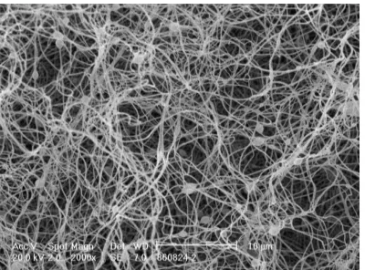
Fig. 5 demonstrates SEM image of nanofibers
electrospun from 10 wt% NC solution in EMK. As is clear two different morphologies could be
detected among the generated fibers. The surface of the nanofibers with 90-150 nm diameters
appears smooth while some pores are visible on the
surface of the fibers with greater diameters range
from 400 to 500 nm. So, among three solvents used, EMK was found as an appropriate solvent for
the preparation of NC nanofibers.
Fig. 5. SEM images of NC/EMK nanofibers
3. 2. NC\ NTO and NC/ Na-NTO Electrospun Composite Nanofiber
In order to modify the properties of NC from the explosive properties point of view, the preparation
of NC composites involving some known explosives was planned. As the first step NTO, Na-
NTO and Picric acid were chosen. So, based on the
results discussed earlier, EMK was used to
dissolve both NC and Na/NTO and electrospinning was applied to the solution. It has to be noticed that
a great deal of caution should be undertaken while working with explosives and a protective shield is
needed for safety purpose. The SEM image shown
in Fig. 6 presents nanofibers with diameters
ranging from 50 to 150 nm. It is seen that the average diameters of composite nanofibers have
been decreased in comparison to the Na-NTO free NC nanofibers in Fig. 5. The presence of Na-NTO
salt may have increased the solution polarity and
conductivity and these led to the formation of
nanofibers with reduced average diameters. Combination of NC and picric acid in EMK
solvent was also tried and smooth composite
nanofibers as shown in Fig. 7 were obtained successfully. However, the range of prepared
composite nanofibers (80- 300 nm) is greater than the range of NC nanofibers prepared under the
292 B. Naderizadehet al./ JNS 2 (2012) 287-293
Fig. 6. SEM image of NC/Na- NTO composite
nanofibers
Fig. 7. SEM images of NC/Picric acid composite
nanofibers
3. 3. Ag/NC Composite Nanofibers
As discussed in pervious section, the second
components in each of the composite nanofibers were introduced in the pre electrospinning setup.
For the preparation of Ag/NC composite there
were possible ways to introduce Ag particles; in situ, encapsulation and immersion of NC
nanofibers in to a solution containing nanosilver.
The last one, however, was chosen by dipping-drying NC nanofibers three times into an aqueous
solution containing 2000 ppm nanosilver.
The antimicrobial activities of the Ag/NC composite nanofibers were examined against E.
coli and S. aureus. The numbers of colonies of E.
coli and S. aureus were significantly reduced in the
case of composite after 24h incubation at 37 ˚C.
This not only clarified surface treatment by silver,
also indicated the successfully inhibition of growth of mentioned two bacteria.
3. 4. DSC Analysis of NC Electrospun
Nanofibers
In order to investigate the thermal behavior of
NC electrospun nanofibers, the heating temperature range of 25 to 550 °C with heating
rate of 10 °C/min and dinitrogen flow of 50 ml/min was chosen. Fig. 8 shows the resulting thermogram
with a strong exothermic peak at 212 °C. This
exothermic peak is consistent with the results
obtained for normal NC and indicating thermal stability of the resulting NC electrospun
nanofibers.
Fig. 8. Thermal analysis of NC nanofibers
4. Conclusions
It has been shown that NC can easily be used to
prepare electrospun NC nanofibers using EMK as
an appropriate solvent. The known explosive materials such as NTO, Na-NTO and picric acid
have been added to the dissolved NC in EMK and
electrospinning process have been applied to the resulting solution to obtain electrospun explosive
composite nanofibers successfully.
with electrospun nanofibers and researchers
working with explosive materials. The properties
of the obtained explosive nanofibers are currently
under investigation. The preparation of other EENP as well as EECNP is also in progress.
Acknowledgment
Financial support by Imam Hossein University is
gratefully acknowledged.
References
[1] S. Megelski, J.S. Stephens, D.B. Chasea and J.F.
Rabolt. 35 (2002) 8456-8466.
[2] D.H. Renker and I. Chun. 7 (1996) 216-223.
[3] a) M.M. Demir, I. Yilgor, E. Yilgor and B.
Erman. Polymer. 43 (2002) 3303-3309. b) J.M.
Deitzel, J.D. Kleinmeyer, J.K. Hirvonen and N.C.B. Tan, Polymer. 42 (2001) 8163-8170.
[4] J. Yuh, L. Perez, W.M. Sigmund and J.C. Nino, 37 (2007) 254-259.
[5] Q.B. Yang, D.M. Li, Y.L. Hong, Z.Y. Li, C.
Wang, S.L. Qiu and Y. Wei, Synth. Met. 137
(2003) 973-974.
[6] W.K. Son, J.H. Youk and W.H. Park,
Carbohydr. Polym. 65 (2006) 430-434.
[7] X. Xu, Q. Yang, Y. Wang, H. Yu, X. Chen and
X. Jing, Eur. Polym. J. 42 (2006) 2081-2087.
[8] S. Li, C. Shao, Y. Liu, S. Tang and R. Mu, J.
Phys .Chem .Solids. 67 (2006) 1869-1872. [9] C. Shao, H. Guan, Y. Liu, J. Gong, N. Yu and
X. Yang, J. Cryst.Grow. 267 (2004) 380-384. [10] K.H. Hong, J.L. Park, I.H. Sul, J.H. Youk and
T.J. Kang, J. Polym. Sci. B. 44 (2006)
2468-2474.
[11] Y. Hong, D. Li, J. Zheng and G. ZouIn. 22 (2006) 7331-7334.
[12] Y. Lu, M. Yu, M. Drechsler and M. Ballauff.
254 (2007) 97-102.
[13] T. Jarusuwannapoom, W. Hongrojjanawiwat,
S. Jitjaicham, L. Wannatong, M. Nithitanakul,
C. Pattamaprom, P. Koombhongse, R.
Rangkuoan and P. Supaphol, Eur. Polymer. J. 41 (2005) 409-421.
[14] J.M. Deitzel, J. Kleinmeyer, D. Harris and
N.C. Beck Tan, Polymer. 42 (2001) 261-272.
[15] X. Wang, C. Drew, S.H. Lee, K.J. Senecal, J.
Kumar and L.A. Samuelson, Nano Letters. 2
(2002) 1273-1275.
[16] A. Formhals, US Patents. 1,975,504, 1934.
[17] Q.P. Pham, U. Sharma and A.G. Mikos, Tissue
Engineering. 12 (2006) 1197.
[18] C.J. Buchko, L.C. Chen, Y. Shen and D.C.
Martin, Polymer. 40 (1999) 7397-7407.
[19] H. Fong, I. Chun and D.H. Renker, Polymer.
40 (1999) 4585-4592.
[20] B. Duan, C.H. Dong, X.Y. Yuan and K.D. Yao, J. Biomat. Sci. Polym. E. 15 (2004)
797-811.
[21] W. Smith Matthew and D. Cliff Matthew,
Report No. DSTO- TR- 0796, (1999).
[22] G. Singh and S.P. Felix, J. Hazard. Mater. A.
90 (2002) 1-17.
[23] G. Viswanathan, S. Murugesan, V. Pushparaj, O. Nalamasu, P.M. Ajayan and R.J. Linhardt,