Basic Study of the Human Foot
V.D.AMBETH KUMAR, S.VAISHALI
and B.SHWETA
Computer Science and Engineering, Panimalar Engineering College, Chennai, India.
DOI: http://dx.doi.org/10.13005/bpj/632
(Received: April 15, 2015; accepted: May 29, 2015)
ABSTRACT
The sole purpose of life in today's world, as seen by a common man, is to run in a meaningless race for attainment of a luxurious life and ample amount of money. As a result, he faces problems in maintaining and examining his health conditions on a regular basis leading to an outbreak of several diseases which slowly magnifies into a state which is beyond any chances of reversal. One such deadly disease which enormously endangers life if gone unnoticed at the right time is cancer. Cancer is caused due to abnormal cell growth which rapidly spreads to neighboring regions depending on the intensity. It is found to occur in several areas such as, bone, brain, skin, breast, lung, etc. Foot cancer too has been noted as a serious and painful tumour. Without timely diagnosis, it leads into a serious condition. In this paper, we describe the different regions of the foot, its evolution and types in the former half and the diseases that affect the foot with a small introduction to foot cancer in the latter half.
Key words: Foot; Types of Foot; Classification; Disease; Tumour.
INTRODUCTION
In the present scenario people mainly focus on building wealth at the cost of their health. The ultimate purpose of modern life has seen a major shift. But they have failed to realize that at the end of the day most of their wealth is being used to regain their lost health which can never be completely reacquired. The emergence of various kinds of new diseases has been at an increasing rate in the past few decades. These are the ultimate results of the negligence of people towards their way of living. People have easily adapted themselves to the fast food era but their body has not. A survey suggests that almost 62% of the world’s population is suffering from diabetes and similar is the condition in the case of blood pressure too. In recent times cancer has become one of the commonly occurring diseases. It has been triggered to this great extent mainly because of our changed lifestyles and negligence. Cancers commonly occurring are breast cancer, lung cancer, kidney cancer, melanoma etc. foot cancer and foot tumors is also one among such diseases found in people. Here we mainly analyze the various kinds of foot and their morphological structure and the
commonly occurring foot diseases and further make a study on foot tumors.
Evolution of the human foot
The human foot today, with bones, extrinsic and intrinsic muscles is found to be evolved through a series of changes over millions of years.
The first ever ‘foot’ dates from the Devonian period(four million years ago)1 when the
tetrapods developed four-limb locomotory structure derived from a special type of lobe-finned fish called Sarcopterygii from the class of Ichthyostega. The paired fins(pectoral and pelvic) were joined to the body by a single bone which was thereafter developed as the four limbs of the tetrapods.
Then came the semi-aquatic organisms, the amphibians which moved by means of limbs and by sinuous body movements. They had reduced limbs and mainly depended on body movements for rapid locomotion.
Further evolution led into the amniotes who laid eggs on land and slowly began to be occupants of the land. They had tarsal bones which were fused to form the astragalus and the calcineum which were later called the talus and calcaneus respectively. Their locomotory structures contained upright limbs attached better suited for walking.
The amniotes further developed into the great class of mammals with more defined locomotory structures of feet and tails. Some of these Mammals are said to have been evolved from certain reptile species called Pelycosaurus which contained sprawled limbs emerging from the sides of the body.
The next mammal-like ‘reptiles’ to evolve from the Pelycosaurus were the Therapsids which were endothermic and walked more upright. Other than the monotremes, all other 4000 species of mammals came from either of the two monophyeletic lineages: Marsupials and the Placentals.
Marsupial young ones were incompletely developed and hence nursed on nipples within an abdominal pouch. The placentals, on the other hand , were gestated for a much longer period and hence born with completely developed foot. Placentals contain many clades such as the Insectivora, Chiroptera, Rodentia, etc. One of these clades is the class of Primates.
Evolution in the primates led to the Palocene and the Eocene primates. Only in the Eocene Epoch period(55-34 million years ago), primitive primates showed a foot morphology that has shared derived features with extant primates. These primitive primates can be considered as the group that had ancestral form of the modern primate foot.
These protoprimates retained an aboreal lifestyle through evolution with their feet more suited for grasping purposes with pads and claws for climbing. One of the earliest evolution from these protoprimates is the monkey which was the dominant primate. Through evolution, monkeys derived new characteristics to be classified as apes and then further evolved into ‘hominids’3 who are
our reference of the direct ancestors of man.
The hominins then segregated into Humans(Homo) and Chimpanzees(Pan) around 5.4 to 6.3 million years ago. The diversification depended on the speciation changes that ranged over 4 million years when the Humans developed a complete bipedialism locomotory structure.
Even though they retained a number of primitive structures(opposable hallux or flexible mid foot), they also contained some derived (human like) structures like ankle morphology and the ability to dorsiflex the toes.
Thus, the foot was modified from a flexible structure with a function to grasp, to a rigid structure working as a lever. Such a highly specialized foot is now present in the Homo sapiens. Locomotion is said to be bipedialistic which is a terrestrial locomotive adaptation characterized by upright
Fig. 1: Initial stages of foot evolution in living organisms
walking by means of rear limbs or legs with the terminal feet mounted on the ground.
Morphological structure: biological background of foot
Foot is one of the most flexible structures of the body. It is mainly built up of nine types of bones and various muscles and joints. It basically consists of 33 bones which constitutes about 1/4th of the total bones present in the body. It also consists of about 33 joints and more than a 100 muscles , ligaments and tendons and also a network of blood
vessels ,nerves ,soft tissues and skin6. Tendons are
basically fibrous tissues that connect muscles to bones and ligaments are fibrous tissues that connects the bones with other bones
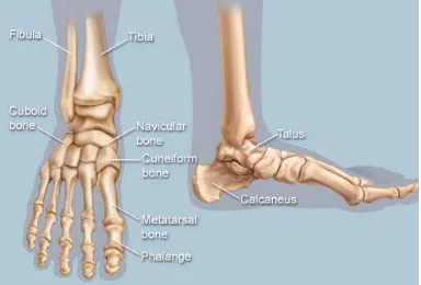
Bones
The various bones7 that are present in the foot are.
• tibia , fibula
• tarsus : talus, calcaneus, cuneiformes, cuboid, and navicular
• metatarsus : first, second, third, fourth, and fifth metatarsal bone
• phalanges
There can be many sesamoid bones near the metatarsophalangeal joints, although they are only regularly present in the distal portion of the first metatarsal bone.
Arches
The human foot has two longitudinal arches and a transverse arch maintained by the interlocking shapes of the foot bones, strong ligaments, and pulling muscles during activity3. The
slight mobility of these arches when weight is applied to and removed from the foot makes walking and running more economical in terms of energy4.
The medial longitudinal arch curves above the Fig. 3: Therapsid (mammal-like reptiles)
Fig. 4: Skeletal structure of an early ape Fig. 5: Skeletal structure indicating the evolution of foot
ground. This arch stretches from the heel bone over the ankle bone to the three medial metatarsals. In contrast, the lateral longitudinal arch is very low. With the cuboid serving as its keystone, it redistributes part of the weight to the calcaneus and the distal end of the fifth metatarsal8 The two
longitudinal arches serve as pillars for the transverse arch which run obliquely across the tarsometatarsal joints3. Excessive strain on the
tendons and ligaments of the feet can result in fallen arches or flat feet.
Muscles
The muscles present in the foot can be classified into two types. The extrinsic muscles which are originating on the anterior or posterior aspect of the lower leg and the intrinsic muscles which are originating on the dorsal (top) or plantar (base) aspects of the foot10.
Extrinsic
All muscles originating on the lower leg except the popliteus muscle11 are attached to the
bones of the foot. The tibia and fibula and the interosseous membrane separate these muscles into anterior and posterior groups, in their turn subdivided into subgroups and layers.
Anterior group
The anterior group of muscles is further subdivided into two groups namely the extensor group and the peroneal group.
Extensor group
The main muscles of the extensor11 group
are :
• tibialis anterior
• Extensor digitorum longus •Extensor hallucis longus
Fig. 8: The various muscles of human foot
Peroneal group: •peroneus longus •peroneus brevis • malleolus
Posterior group
The main muscles of the posterior group are:
• triceps surae •plantaris. •soleus • gastrocnemius • femur •Achilles tendon •tibialis posterior •flexor hallucis longus •flexor retinaculum • popliteus Intrinsic muscles •extensor digitorum brevis • extensor hallucis brevis • aponeurosis
Muscles of the big toe • abductor hallucis • flexor hallucis brevis •Adductor hallucis Muscles of the little toe •abductor digiti minimi •flexor digiti minimi •opponens digiti minimi Central muscle group • lumbricals
• Quadratus plantae •Flexor digitorum brevis • dorsal and plantar interossei The foot is sub-divided three parts •Fore foot
•Mid foot •Hind foot
Fore foot
The forefoot is composed12 of five toes
called the phalanges and their connecting long bones called metatarsals. Each toe is made up of several small bones. The big toe also known as hallux has two phalanx bones. They are the distal and the proximal. It has one joint calles the interphalangeal joint. The big toe articulates with the head of the first metatarsal and is called the
metatarsophalangeal joint. Underneath the first metatarsal head there are two tiny round bones called sesamoids. The other four toes each have three bones and two joints. The phalanges are connected to the metatarsals by five metatarsal phalangeal joints at the ball of the foot. The forefoot bears half the body’s weight and balances pressure on the ball of the foot.
Mid foot
The midfoot12 has five irregularly shaped
tarsal bones, forms the foot’s arch, and serves as a shock absorber. The bones of the midfoot are connected to the forefoot and the hindfoot by muscles and the plantar fascia which is also known as arch ligament.
Hind foot
The hindfoot12 is composed of three joints
and links the midfoot to the ankle which is also called the talus. The top of the talus is connected to the two long bones of the lower leg which are the tibia and fibula which forms a hinge that allows the foot to move up and down. The heel bone called the calcaneus is the largest bone in the foot. It joins the talus to form the subtalar joint. The bottom of the heel bone is cushioned by a layer of fat.
Thus these parts form the foot. Any structural flaw or malfunction in any one part can result in the development of problems elsewhere in the body such as the back pain. Abnormalities in other parts of the body can also lead to problems in the feet.
Foot types
The structure of the foot is relatively unique to every individual, and differs within a single individual from one side to the other.
Foot can be classified under two types: 1. Foot Arches
2. Toes
Classification based on foot arches
Evaluation of the type of foot which depends on the arches can be done statically or dynamically.
High-arch foot
This foot type has a curved point.But however, the mid-foot is generally rigid, lacking a good plie, also is not a good shock absorbant when running or while landing from jumps. It also tends to be associated with hammer toes,9 a condition
where the toes remain partly flexed. This causes bruising in the area of the ball of the foot.
Low-arch foot
This stresses the muscles supporting the arch which causes the person to tend to roll in, causing strain on the inside of foot and knee8.
Often flat feet are found to be hypermobile and get tired easily. However, they are better shock absorbers than high-arched feet.
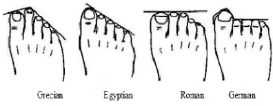
Classification based on toes Grecian foot
Also known as Morton’s Foot – The Greek13
Foot is one where the second toe is longer than all the other toes of the foot. Often a big space is found between the big toe and the second toe5 This foot
structure has a tendency to be unstable and people with this structure tend to have foot problems as they tend to shift their weight to the inside of the foot, and that repeated winged position lift can lead to injury.
Egyptian foot
The Egyptian Foot tends to be a narrow foot13. It has a longer big toe and the rest of the toes
taper down from longest to shortest.[5] It is the most functional of all the foot structures
Since the first metatarsal and big toe are long, there is a lot of pressure on that area. This limits motion or acts as a cause of arthritis of metatarsal joints.
Romanfoot
In this type, also called the squared foot13,
the toes are similar in length (either the first till the fourth, or the second toe, third toe, and fourth toe). There is a wide ball and narrow heel.
Germanic (pesant) foot
The Peasant Foot13 (also called a Giselle
Foot) has at least three toes of the same length and
tend to be stubby and short. This is a very stable and functional foot.
Diseases
Diseases and disorders is always a condition of painful or irritable experience. Foot being the sole organ of locomotion in the human body, it is subjected to a lot of friction and mechanical action as a result of which it slowly wears and withers causing damage.
Being the most neglected part of the body, many of the problems associated with it go unnoticed. Here are some of the diseases that are acute in the foot of the human body
Foot tumours
Foot tumor is one of the rarely occurring tumors14 It may be either malignant or benign. There
are a number of benign tumors specific to the foot that affect the skin, soft tissue and bone. Primary malignant tumors of the foot represent less than 1% of malignant tumors. The Kaposi’s sarcoma and malignant melanoma are the two most common malignant tumors found in the foot. These two malignant tumours are normally found in immune suppressed patients or in patients older than 85 years of age15. Though it is of rare occurrence, it
must be detected and diagnosed properly to prevent it from leading into a serious condition. Detection can be done by means of using the MRI technique as used in detecting brain tumour.
CONCLUSIONS
1. “The evolutionary history of the human foot” Author: Kristiaan D’Août, Peter Aerts 2. “Essentials of Orthopaedics and Applied
Physiotherapy”
3. http://www.ncbi.nlm.nih.gov/pmc/articles/ PMC1571304/
4. h t t p : / / o r t h o i n f o . a a o s . o r g / t o p i c . cfm?topic=a00168
5. http://www.slideshare.net/misterpeepers/ types-of-feet-slide-show
6. “Anatomy of the foot and ankle”. Podiatry Channel. Retrieved August 2009
7. “Foot Bone Anatomy ”Author : Vinod K Panchbhavi, MD, FRCS, FACS; Chief Editor: Thomas R Gest, PhD.
8. http://www.foot.com/site/arch-types.
REFERENCES
9. D’Août et alia Experimentally Generated Footprints in Sand: Analysis and Consequences for the Interpretation of Fossil and Forensic Footprints, American Journal of Physical Anthropology 141:515.
10. http://en.wikipedia.org/wiki/Foot
11. h t t p : / / w w w. i n n e r b o d y. c o m / a n a t o m y / muscular/leg-foot .
12. http://www.webmd.com/feet ——forefoot hind foot
13. http://curingchronicpain.com/your-feet-what-they-look-like-gives-valuable-information. 14.
http://www.myfootshop.com/article/tumors-of-the-foot