* To whom all correspondence should be adressed. [email protected]
Comparative Study Of Edge Detection Algorithms
on Medical Images
K.K. Thanammal1 and J.S. Jaya Sudha2
1Department of MCA, S.T.Hindu College, Nagercoil, India. 2Computer Science and Engineering,
Sree Chitra Tirunal College of Engineering, Tiruvananthapuram, India.
doi: http://dx.doi.org/10.13005/bbra/1569
(Received: 03 October 2014; accepted: 24 November 2014)
Detection of edge is a terminology in image processing and computer vision particularly in the areas of feature detection and extraction to refer to the algorithms which aims at identifying points in a digital image at which the image brightness changes sharply or more formally has discontinuities. Edge is a basic feature of image. The image edges include rich information that is very significant for obtaining the image characteristics by object recognition. Edge detection refers to the process of identifying and locating sharp discontinuities in an image. So, edge detection is a vital step in image analysis and it is the key of solving many complex problems. This paper, describes edge detection algorithms for image segmentation using various computing approaches which have got great fruits. Experimental results prove that Canny operator is better than Prewitt and Sobel for the selected image. Subjective and Objective methods are used to evaluate the different edge operators. The performance of Canny, Sobel and Prewitt Edge Detection are evaluated for detection of edges in digital images.
Key words: edge detection, mathematical morphology, feature detection
Edge detection is a fundamental tool, which is commonly used in many image processing applications to obtain information from images and frames1. The separation of the image into object
and background is a critical step in image interpretation2. An edge may be regarded as
boundary between two dissimilar regions in image; edge detection is a terminology in image processing and computer vision, particularly in areas of feature
detection and feature extraction. Mathematical morphology is a tool for extracting image
with small patterns, called ‘structuring element’, of varying sizes and shapes. This procedure results in non-linear image operators which are well suited to explore geometrical and topological structures. They do provide the strong visual clues that can help the recognition process.
Edge detection produces an edge map that contains important information about the image. Edge detection is difficult in noisy images, since both the noise and the edges have high frequency. Noisy images are typically larger in scope, so they pixels. This result is less accurate localization of the detected edges. Not all edges involve a step change in intensity. Effects such as refraction or poor focus can result in object boundaries defined by a gradual change in intensity3. The operator needs to be chosen to be
responsive to such a gradual change in those cases. So, there are problems due to noise etc. Therefore, the objective is to compare various edge detection techniques and analyze their performance in different conditions.
The rest of the paper is organized as follows. In section 2, the types of edges, edge detection operators Robert’s operator, sobel operator, zero crossing operator, Laplacian operator, Gausian edge detector, prewitt operator, Canny operator. In section 3, the results of edge detection on lung CT scan images. In section4,comparision graph of edge detection operators. In section5 concludes the paper.
Edge Detection For Image Segmentation
There are many techniques in the literature used for edge detection some of them are based on error minimization, maximizing an object function, fuzzy logic, wavelet approach, morphology, genetic algorithms, neural network and Bayesian approach.
Color image segmentation techniques can be roughly classified into four types such as histogram based approaches, neighborhood based approaches, clustering based approaches and hybrid based approaches. Histogram thresholding is widely accepted and easily computable technique in which the images are composed of regions with different gray level ranges. The main disadvantage of this technique is the lack of spatial relationship information of the pixels. The neighborhood based approach applies the
uniformity criteria to segment the image i.e., the neighboring pixels within the region should have similar values in intensity, color or texture. E.g. Region based techniques. Clustering based approach uses a fuzzy logic to define membership of the pixels. Hybrid based techniques improve the segmentation result by combining all above methods for segmentation4 CT scan is more
appropriate for showing the detailed information of the parts of human body and it is used for various applications such as detection, classification etc. The analysis of lungs in CT image is used to detect the airway and the vessel present in the lungs5.
Edge Detection Operators The Roberts Detection
In Robert cross algorithm the horizontal and vertical edges bring out individually and then they put together for the resulting edge detection.
Types of Edges
Table1. The Roberts Detection
+1 0
The two individual images Gx and Gy are combined using the approximation equation G = Gx + Gy or by using G=sqrt (Gx * Gx + Gy* Gy) to get the exact magnitude values. As the Roberts Cross Kernels are relatively small, they are highly susceptible to noise
Sobel Edge Detection
The Sobel edge detection technique is similar to that of the Robert Cross algorithm. Despite the design of Sobel and Robert are common, the main difference is the kernels that each uses to obtain the image is different. The Sobel Kernels are more suitable to detect edges along the horizontal and vertical axis whereas the Robert’s able to detect edges run along the vertical axis of 450 and 13502
and ISET (Shen-Castan). It is very time consuming and very complex for computation.
Prewitt Edge Detector
Prewitt operator edge detection masks are the one of the oldest and best understood methods of detecting edge in images. The strength of the edge at given location is then the square root of the sum of the squares of two derivatives.
Canny Edge Detector
The popular edge detection algorithm Canny was first presented in 1986. The problem with this type of traditional edge detection approach is that a low threshold produces false edges, but a high threshold misses important edges. First requires that the image be smoothened with a Gaussian mask, which cuts down significantly on the noise within the image. Then the image is run through the Sobel algorithm and as discussed before, this process is hardly affected by noise. Lastly, the pixel values are chosen based on the angle of the magnitude of that pixel and its neighboring pixels. Unlike Roberts Cross and much like Sobel, the canny operation is not very susceptible to noise. If the Canny detector worked properly it would be the superior to both Sobel and Roberts Cross, the only drawback is that it takes more time to compute.
The steps in the Canny edge detector are as follows:
Smooth the image with a two dimensional Gaussian. In most cases the computation of a two dimensional Gaussian is costly, so it is approximately by two one dimensional Gaussians, one in the X direction and the other in the Y direction.
Take the gradient of the image. This shows changes in intensity, which indicates the presence of edges. This actually gives two results, the gradient in the X direction and the gradient in the direction.
Non-maximal suppression. Edges will occur at points where the gradient is at a maximum. Therefore, all points not at a maximum should be suppressed. In order to do this, the magnitude and the direction of the gradient is computed at each pixel. Then for each pixel check if the magnitude of the gradient is greater at one pixel’s distance away in either the positive or the negative direction perpendicular to the gradient. If the pixel is not greater than both, suppress it.
0 +1
-1 0
Gy
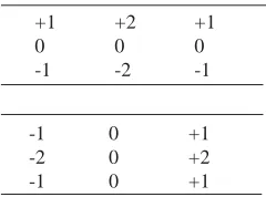
+1 +2 +1
0 0 0
-1 -2 -1
-1 0 +1
-2 0 +2
-1 0 +1
Table 2. Sobel Edge Detection
Zero Crossing
It uses second derivative and it includes Laplacian operator. It is having fixed characteristics in all directions and sensitive to noise. Haralick proposed the use of zero-crossing of the second directorial derivative of the image intensity function.
Laplacian of Gaussian (LOG)
It was invented by Marr and Hilderth (1980).The Gaussian filtering is combined with Laplacian to break down the image where the intensity varies to detect the edges effectively. It finds the correct place of edges and testing wider area around the pixel. The disadvantage of LOG operator is that it cannot finds orientation of edge because of laplacian filter5.
Gaussian Edge Detectors
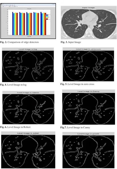
Fig. 2. Comparison of edge detectors Fig. 3. Input Image
Fig. 4. Level Image in log Fig. 5. Level Image in zero cross
Fig. 6. Level Image in Robert Fig.7. Level Image in Canny
Edge Thresholding. The method of thresholding used by the Canny Edge Detector is referred to as “hysteresis”. It makes use of both a high threshold and a low threshold. If a pixel has a value above the high threshold, it is set as an edge pixel. If a pixel has a value above the low threshold and is the neighbor of an edge pixel, it is set as an edge pixel as well. If a pixel has a value above the low threshold but is not the neighbor of an edge pixel, it is not set as an edge pixel. If a pixel has a value below the low threshold, it is never set as an edge pixel.
Mathematical Morphological Operators
Mathematical Morphology is one of the most productive areas in image processing6. The
content of mathematical morphology is based on the set theory. A structuring element is a special mask filter that enhances input images. It can be of different sizes and of different shapes (square, diamond, and circle). Following are the main mathematical morphological operators:
1. Dilation 2. Erosion 3. Opening 4. Closing 1. Dilation
Dilation is defined as maximum value in the window. Hence the image after dilation will be brighter or increased in intensity. It also expands the image and mainly used to fill the spaces. Dilation process expanding image objects by changing pixels with value of”0" to “1”.
Erosion
Erosion is just opposite to dilation. It is defined as the maximum value in the window. The image after dilation will be darker than the original image. It shrinks or thins the image. Erosion process shrinking objects or images by changing pixels with a value of”1" to “0”.
Opening and Closing
Both parameters are formed by using dilation and erosion. In opening, firstly image will be eroded and then it will be followed by dilation. And in case of closing, firstly image will be dilated and then followed by erosion.
CONCLUSION
From the simulation results, it is concluded that the edge detection using mathematical morphology is more efficient than the traditional methods. From the results and comparison of the different methods of edge detection, it is concluded that the mathematical morphological edge detection is better than the traditional method. In this work, edges of images using mathematical morphological and traditional method like Sobel, Canny, Prewitt and Laplician of Gaussian method has been studied and compared on subjective basis as well as objective manner.
REFERENCES
1. T.A.Mohmoud;S.Marshal,”Edge Detected Guided Morphological Filter For Image sharpening,” Hindawi Publishing corporation, EURASIP Journal on image and video processing, 2008.
2. S.Lakshmi,Dr.V.Sankaranarayanan,”A Study of Edge Detection Techniques for Segmentation Computing Approaches,” IJCA special issue on “Computer Aided Soft Computing Techniques for imaging and Biomedical Applications,” CASCT 20.
3. E.Argyle, “Techniques for edge detection,”
Proc.IEEE, 1971;59, pp.285-286.
4. First Arkansan Deshmukh, Member IAENG, Second B.Ganesh Shinde,”Adaptive Color Image Segmentation Using Fuzzy Min-Max Clustering,” Engineering Letters, Advance online Publication, Aug-2006.
5. Mohammed Roushdy,”Comparative Study of Edge detection Algorithms on Grayscale Noisy image using Morphological Filter,” GVIP Journal, 6(4); 2006.
6. Sluimer.I, Schilham.A, Prokop.M and Van Ginnekan.B, “Computer Analysis of Computed tomography scans of the lung: a Survey,” IEEE transactions on Medical Imaging, 2006; 25(4), 385-405.