Volume-5 Issue-1
International Journal of Intellectual Advancements and
Research in Engineering Computations
Detection of lung cancer from ct image using image processing
1.
S.Brindha, 2.P.Priya, 2.K.Sathya Bama, 2.P.Selvapriya, 2.S.Vijay Anand
1.
Assistant professor, 2.UG students, Department of Electronics and Communication Engineering,
Nandha Engineering college, Erode, TamilNadu, India.
ABSTRACT:
Lung cancer is the leading cancer
among both men and women. Detection of lung
cancer is important inorder to improve the
survival rate. Presence of lung cancer can be
diagnosed with the help of a CT image of the
lung. The proposed system is used to detect the
lung cancer in three steps. The steps include
preprocessing stage, feature extraction stage
and lung cancer cell identification stage. First
step is capturing the input image. Second step is
preprocessing. Preprocessing includes two
processes such as image enhancement and
image segmentation. The Weibull segmentation
is used to describe the texture contrast, shape,
scale and texture. The output from the image
segmentation goes to the feature extraction
stage. These extracted features are used to
identify the abnormalities in the lung.
Keywords: Lung cancer, CT scan image,
Log gabor filter, Weibull segmentation.
INTRODUCTION:
Lung Cancer is the wild development
of unusual cells, start off in one or both lungs,
generally in the line the air passages. The
unusual cells do not grow into healthy lung
tissue; they provide fast and form tumours.
According to American cancer society the cases
of lung cancer increase very quickly and about
14% newly diagnose cancers are a lung cancer
and also the major reason of cancer death
worldwide. The earlier study of analysis show
that the majority of the lung cancer patients
belong to the age of 60 years.
Lung Cancer is one of the most serious
human body problems in the world. The
fatality rate of lung cancer is the maximum of
all other type of cancer. The survival rate of
lung tumour is extremely least amongst all
type of tumour. So, there is a need to
propose a computational intelligence based
approach to identify the lung cancer because
the survival from lung cancer is openly
connected to its expansion at its detection
time. If we identify lung cancer at early
survive the patients. It is also showed from
previous study that cigarettes smoke are the
major cause of lung cancer. It is observed that
an estimated 85% of lung cancer cases in
males and 75% lung cancer cases in females
where cigarette smoking is the key cause.
The lung cancer discovery can be
done by taking a screening using Computed
Tomography (CT) Scan. The CT Scan
outcome then observed on morphological
guide of lung cancer as the diagnostic criteria
such as the tumor size, enhancement, irregular
spiculated margin , lobulated, water
bronchograms, ground glass opacity, and
heterogeneous density . The lung cancer
identification by using CT Scan image which
conducted by a radiologist may result in an
error subjective by the blur of anatomical
structures surrounding the lung area, the tiny
size of lesion, and also the diverse experiences
of the radiologist create a altered
interpretation . To avoid the errors and to
develop the accuracy and consistency, a
computer-based digital image processing is
essential as the second opinion to read the CT
Scan image result. In this study, the primary
stage of image processing is preprocessing
to crop Region of Interest (RoI),
segmentation, feature extraction and
classification.
METHODOLOGY:
Lung cancer detection system can be
developed by using this image processing
technique. Lung cancer recognition system has
three steps to discover the occurrence of cancer
nodule in lung. Pre-processing, feature
extraction and lung cancer cell identification
are the steps. Pre-processing step include image
enhancement and image segmentation.
Enhancement is removing the unnecessary
images and noise by the log-gabor filter.
Enhanced CT image of lung is then passed
through segmentation phase. The segmentation
is done by the Weibull segmentation process.
From the segmented output characteristics are
extracted to calculate the presence of defect of
lung. By means of these extracted features
sort the lung as normal lung or abnormal
lung.
BLOCK DIAGRAM:
IMAGE PRE-PROCESSING:
It is the second module in lung cancer
recognition system. The reason of
pre-processing stage is to develop the image data
done by enhancing the data which are essential
for additional processing. Image enhancement
and image segmentation are the procedure
performed in pre-processing stage.
Image enhancement: Image enhancement is
used to develop the interpretability of
information in the image to the human viewers.
Image enhancement is of two type, spatial
domain methods and frequency domain
methods. Spatial domain process deals with the
pixels in the image. Enhancement is performed
by altering the pixel values. That means,
changing the grey level value and alter the
contrast of the image. In frequency domain
enhancement is achieved by varying the
orthogonal transform of image. That means,
based on the frequency domain the processing
are performed.
Log Gabor filter: Log gabor filter was
proposed by Field in 1987. Log gabor filter is
the higher edition of gabor filter. Some changes
are made on the gabor function is the log gabor
function. Log gabor filter has two features, one
is it has no DC components and the next one is
the transfer function of log gabor function has
an extended tail at the high frequency end.
IMAGE SEGMENTATION:
Image segmentation algorithms are
based on discontinuity and similarity. These
two are the properties of intensity values. The
purpose of image segmentation is to partition
the image into meaningful region and to
identify the object or relevant information
from the digital image. Image segmentation
highlights the information which we are needed
for further processing from the image and thus
make it easy to analyze.
Weibull segmentation: Weibull segmentation
is one of the segmentation method used in
medical images. Distribution parameters of
weibull distribution describe the texture
contrast, scale, shape, and generate a
six-stimulus basis for texture perception. So it
can be treated as a good model. Variation in the
weibull parameters produces verity of
distribution, which includes exponential and
Gaussian and Raleigh. In this technique the
image is assumed to have a weibull
distribution. A complex image is represented
by Cm,n. Histogram of the resulted image is
plotted and the minimum grey level value and
maximum grey level value are selected from
the histogram. Then according to the number of
classes entered from the input the image is
segmented into regions.
FEATURE EXTRACTION AND CANCER
CELL IDENTIFICATION:
Feature extraction is the important stage in this
work. It uses different methods and algorithms
to extract the features from the segmented
image. Based on the extracted features
normality and abnormality of the lung are
decided. The features which we are extracted
are area, perimeter, and average intensity.
Segmented images have only two values 1 and
0. Nodule part will be represented with value
1. Then area of the nodule can be calculated by
finding number of pixel with value 1. Perimeter
of the nodule means the number of pixels in the
intensity is another feature which is used for the
purpose of cancer detection. Select two
threshold values for mean intensity values, and
then calculate the average intensity value for
the candidate region. If the average intensity
value is between the threshold values then this
part is assumed to be cancerous otherwise not.
Based on area of the nodules the cancer caused
nodules are identified. If the nodule size is
greater than 25mm then it is assumed as
abnormal image. If the nodule size is less than
25mm then it can be assumed as a normal
image.
RESULTS AND DISCUSSION:
The experiments are conducted on
the lung cancer detection system (LCDS)
with the inputs are CT images of lung. CT
image is successfully processed by each step in
lung cancer detection system and the resulted
was obtained. CT image of lung is given to
image enhancement technique such as
log-gabor filter and the output is obtained. The
resultant enhanced image is shown in Fig 1.
Output from image enhancement technique is
used as the input to the image segmentation
module. In this work output from log gabor
filter is used as input. For image segmentation
weibull segmentation technique is used.
Resultant output is generated and evaluated.
Obtained results are shown in Fig 2.
Outputs from the segmentation
techniques are processed under the feature
extraction and cancer cell identification
module. Cancer cell identification module
identifies the cancer caused part in lung and
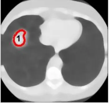
marked with red colour as shown in Fig 3.
Fig. 1 Log-gabor enhanced image
Fig. 2 Weibull segmentation
REFERENCES:
Wei-Hua Andrew Wang and Man-Ching Lin, “A Fast Method in
Reconstruction 3D CT Medical
Images” -
978-1-4244-6484-5110/$26.00 ©2010 IEEE
http://ieeexplore.ieee.org/document/63
91662/
A. Chaudhary and S. S. Singh, “Lung
cancer detection on ct images by using
image processing," in Computing
Sciences (ICCS), 2012 International
Conference on. IEEE, 2012, pp.
142-146.
N. Hadavi, M. Nordin, A. Shojaeipour et al., “Lung cancer diagnosis using
ct-scan images based on cellular learning
automata," in Computer and
Information Sciences (ICCOINS), 2014
International Conference on. IEEE,
2014, pp. 1-5.
N. Otsu, "A Threshold Selection
Method from Gray-level Histograms",
IEEE Trans. Syst. Man Cybernetics,
vol. 9, (1), 1979, pp. 62-66.
Lee,S.L.A.,Kouzani,A.Z.,Hu.E.J. “Empirical evaluation of
segmentation algorithm for lung
modeling”. IEEE International
Conference on Systems Man and
Cybernetics (2008) 719-724.
D. Lin And C. Yan, "Lung Nodules
Identification Rules Extraction With Neural
Fuzzy Network", Ieee, Neural Inf ormation
Processing, Vol. 4, (2 002)
T.M. Cover, and P.E. Hart, ―Nearest neighbor pattern classification‖, IEEE
Transactions on Information Theory, 13, pp.
21–27, 1967
S.K.V. Anand, “Segmentation coupled
Textural Feature Classification for Lung
Tumor Prediction,” Commun. Control
Comput. Technol.(ICCCCT), IEEE Int.