Early childhood lung function is a stronger
predictor of adolescent lung function in cystic
fibrosis than early Pseudomonas aeruginosa
infection
Jessica E. Pittman1*, Hannah Noah2, Hollin E. Calloway3, Stephanie D. Davis4, Margaret W. Leigh5,6, Mitchell Drumm7, Scott D. Sagel8, Frank J. Accurso8, Michael R. Knowles6,
Marci K. Sontag8
1 Washington University School of Medicine, Division of Pediatric Allergy, Immunology, and Pulmonary
Medicine, St. Louis, MO, United States of America, 2 University of North Carolina at Chapel Hill School of Medicine, Chapel Hill, NC, United States of America, 3 Stanford University School of Medicine, Department of Otolaryngology Head & Neck Surgery, Palo Alto, CA, United States of America, 4 Indiana University School of Medicine/Riley Hospital for Children, Section of Pediatric Pulmonology, Allergy, and Sleep Medicine, Indianapolis, IN, United States of America, 5 University of North Carolina at Chapel Hill,
Department of Pediatrics, Chapel Hill, NC, United States of America, 6 University of North Carolina at Chapel Hill, Marisco Lung Institute, Chapel Hill, NC, United States of America, 7 Departments of Pediatrics and Genetics and Genome Sciences, Case Western Reserve University, Cleveland, OH, United States of America, 8 Department of Pediatrics, Children’s Hospital Colorado and University of Colorado School of Medicine, Aurora, CO, United States of America
Abstract
Objective
Pseudomonas aeruginosa has been suggested as a major determinant of poor pulmonary outcomes in cystic fibrosis (CF), although other factors play a role. Our objective was to investigate the association of early childhood Pseudomonas infection on differences in lung function in adolescence with CF.
Methods
Two populations of subjects with CF were studied: from the Gene Modifier Study (GMS), 346 F508del homozygotes with severe vs. mild adolescent lung disease, and from the Colo-rado Newborn Screen Study (NBS) 172 subjects diagnosed with CF by newborn screening. Associations of Pseudomonas infection and lung function in early childhood with lung func-tion in adolescence were investigated using multivariate linear regression analyses.
Results
Among GMS subjects, those with severe adolescent lung disease had worse lung function in childhood (FEV125 percentage points lower) compared to subjects with mild adolescent
lung disease, regardless of early childhood Pseudomonas status. Among NBS subjects, those with lowest adolescent lung function had significantly lower early childhood lung func-tion and faster rate of decline in FEV1than subjects with highest adolescent lung function; a1111111111 a1111111111 a1111111111 a1111111111 a1111111111 OPEN ACCESS
Citation: Pittman JE, Noah H, Calloway HE, Davis
SD, Leigh MW, Drumm M, et al. (2017) Early childhood lung function is a stronger predictor of adolescent lung function in cystic fibrosis than early Pseudomonas aeruginosa infection. PLoS ONE 12(5): e0177215.https://doi.org/10.1371/ journal.pone.0177215
Editor: Dominik Hartl, University of Tu¨bingen,
GERMANY
Received: October 19, 2016
Accepted: April 24, 2017
Published: May 15, 2017
Copyright:©2017 Pittman et al. This is an open access article distributed under the terms of the
Creative Commons Attribution License, which permits unrestricted use, distribution, and reproduction in any medium, provided the original author and source are credited.
Data Availability Statement: All relevant data are
within the paper and its Supporting Information files.
Funding: CTSA UL1 TR001082 (MKS, SDS, FJA): http://www.ncats.nih.gov/ctsa; CFF PITTMA10A0 (JEP): https://www.cff.org/Our-Research/For-Researchers/Research-Awards/; CFF
early Pseudomonas infection was not associated with rate of FEV1decline. The strongest
predictor of adolescent lung function was early childhood lung function. Subjects with a higher percentage of cultures positive for Pseudomonas before age 6 or a lower BMI at 2–4 years old also had lower adolescent lung function, though these associations were not as strong as with early childhood lung function.
Conclusions
In separate analyses of two distinct populations of subjects with CF, we found a strong cor-relation between lower lung function in early childhood and adolescence, regardless of early childhood Pseudomonas status. Factors in addition to early Pseudomonas infection have a strong impact on lung function in early childhood in CF. Further exploration may identify novel underlying genetic or environmental factors that predispose children with CF to early loss of lung function.
Introduction
Cystic Fibrosis (CF) is a chronic, life-limiting genetic illness in which dysfunction of the CF transmembrane conductance regulator (CFTR) causes impaired mucociliary clearance, lead-ing to chronic pulmonary disease, among other symptoms[1–4]. Lung disease in CF begins in infancy and is characterized by a cycle of chronic inflammation, infection, and airway damage causing progressive obstructive airways disease and loss of lung function[1–11]. Despite the early onset of pathology, patients with CF show marked variability in phenotype, disease sever-ity, and survival[12–16].
Previous studies have tried to explain this heterogeneity by investigating factors associated with lower lung function in CF, including environmental (lower socioeconomic status, tobacco exposure)[17–20], nutritional[15,21,22], infectious (bacterial, viral, and fungal)[15,
21–32], and genetic (CFTRgenotype and effects of various modifier genes)[12,33–35].
Pseudo-monas aeruginosainfection, in particular, has been associated with more rapid decline in lung function and more severe lung disease; earlier acquisition ofPahas been associated with poorer lung function in adulthood and higher risk of death in childhood. This has led to wide-spread adoption ofPaeradication protocols in children with CF[36,37].
We have previously shown in a large, retrospective case-control study that earlier age ofPa
infection (before 5 years of age) was strongly associated with severe (vs. mild) CF lung disease in adolescence and adulthood[29]. However, we hypothesized that multiple other factors are involved in determining lung function in early childhood in CF, and that earlyPainfection alone would not account for differences in lung function in early childhood that persist into adolescence, even in a cohort of children diagnosed by newborn screening with subsequent aggressive early management of CF lung disease. Our primary objective was to investigate the association between early childhood lung function, earlyPainfection, and adolescent lung function in subjects with CF. We also aimed to investigate other factors associated with early lung function in children with CF. To accomplish this, we tested our hypothesis in two sepa-rate datasets: the Gene Modifier Study (GMS), a retrospective, case-control study with annual-ized data, used in our original publication[29], and the Colorado Newborn Screen (NBS) Study, a prospectively collected, encounter-based study.
funding/index.htm; CFF SONTAG07A0 (MKS):
https://www.cff.org/Our-Research/For-Researchers/Research-Awards/.
Competing interests: Dr. Stephanie Davis has
Materials and methods
Populations & variables
Gene Modifier Study (GMS). A multicenter, retrospective case-control study of subjects
with CF, all F508del homozygotes, classified as having either severe or mild adolescent lung function (defined as FEV1in the highest or lowest quartiles for FEV1percent predicted by
birth cohort, respectively–an extremes of phenotype design), using annualized data from the CF Foundation Patient Registry (CFFPR)[29,35]. Written informed consent was obtained for all subjects. This study was approved by the Institutional Review Board at the University of North Carolina, Chapel Hill. Analysis was restricted to subjects in our previous publication (n = 629)[29] with a) spirometry (PFTs) at 6–8 years of age, b) respiratory culture data before 6 years of age, and c) classification of adolescent lung disease severity at age 12 or older (to avoid overlap with early childhood FEV1data).
Definitions of key variables:
• Pastatus: ever/never recorded as having a culture positive forPabefore 6 years of age.
• Early childhood lung function: defined as the mean FEV1percent predicted (FEV1) between
6–8 years of age (Wang)[38]. Best FEV1between 6 and 8 years of age was also calculated.
Calculations utilized all available spirometric data from the CFFPR.
• Mild vs. severe adolescent lung disease: defined based on GMS enrollment criteria[35].
Colorado Newborn Screen database (NBS). The NBS database consists of prospectively
collected, encounter-based clinical data (supplemented by CFFPR data) for subjects diagnosed with CF from 1982–2010 at the Children’s Hospital Colorado [39,40]. Informed consent was obtained for all subjects. This study was approved by the Colorado Multiple Institutional Review Board, University of Colorado, Denver. For this study, analysis was restricted to sub-jects who a) were pancreatic insufficient, b) had BMI data available at 2–4 years of age, c) had respiratory culture data available before 6 years of age, d) had PFT data between 6 and 8 years of age, and e) had PFT data available at age 10 or older.
Definitions of key variables:
• Early childhood lung function: defined as the mean FEV1percent predicted (FEV1) at 6–8
years of age (Wang)[38]. Best FEV1at 6 to 8 years of age was also calculated. Calculations
uti-lized all available spirometric data from the CFFPR.
• Adolescent lung function: defined as the mean FEV1percent predicted (Hankinson)[41] for
the last 3 years of available PFT data beginning at 10 years of age or older. Subjects were divided into even quartiles by adolescent lung function for bivariate analysis. This variable was treated as continuous for regression analysis.
• Number of spirometric measures: counts defined separately for both early childhood and adolescent lung function variables.
• Mean age at PFTs: defined separately for early childhood and adolescent variables.
• Early childhood infection status:
Pastatus: ever/never culture-positive forPabefore 6 years of age,
Percentage of cultures positive forPaprior to 6 years of age,
Percentage of cultures positive forS.aureusprior to 6 years of age,
Number of respiratory cultures before 6 years of age.
• Early childhood nutritional status:
Mean BMI Z-score from 2 to 4 years of age,
Mean height Z-score from 2 to 4 years of age,
Mean weight Z-score from 2 to 4 years of age[42].
• Absolute change in FEV1percent predicted from early childhood to adolescence: defined as
mean early childhood FEV1percent predicted minus the mean adolescent FEV1percent
predicted;
• Rate of change in FEV1(percent predicted/year): defined as the absolute change in FEV1
per-cent predicted / (mean age at adolesper-cent PFTs–mean age at early childhood PFTs)
Other variables included for both analyses were gender, race (Caucasian vs. non-Cauca-sian), ethnicity (Hispanic vs. non-Hispanic), year of birth, age at diagnosis, means of diagnosis (newborn screen, meconium ileus at presentation, and symptoms [respiratory, gastrointesti-nal, or failure to thrive]),CFTRgenotype, and age of first recorded respiratory culture.
Analyses (NBS, GMS)
Bivariate analyses used Student’s t-test andχ2testing to compare means or proportions, respectively. Multiple comparisons were accounted for in the NBS cohort using ANOVA test-ing with Bonferroni correction. Comparisons consisted of:
1. Comparison of population characteristics by adolescent lung function group (GMS: severe vs. mild adolescent lung function; NBS: adolescent FEV1quartile).
2. GMS: Comparison of early FEV1in subjects with severe vs. mild adolescent FEV1, by early
childhoodPastatus.
3. GMS: Comparison of early FEV1in subjects who werePapositive vs. negative in early
childhood, by severe vs. mild adolescent lung function group.
4. NBS: Comparison of early FEV1by adolescent FEV1quartile and by early childhoodPa
status.
5. NBS: Comparison of early FEV1in subjectsPapositive vs. negative in early childhood, by
adolescent FEV1quartile.
6. NBS: Comparison of rate of change and absolute change in FEV1percent predicted from
early childhood to adolescence by adolescent FEV1quartile and early childhoodPastatus.
7. NBS: Comparison of rate of change and absolute change in FEV1percent predicted from
early childhood to adolescence in subjectsPapositive vs. negative in early childhood by adolescent FEV1quartile.
no), age at CF diagnosis, genotype (F508del homozygous vs. other), number of discrete PFTs included in early childhood FEV1variable, number of discrete PFTs included in adolescent
FEV1variable, number of discrete cultures prior to 6 years of age, percentage of cultures
posi-tive forPaprior to 6 years of age, percentage of cultures positive forS.aureusprior to 6 years of age, mean BMI percentile at 2 to 4 years of age, and number of discrete BMI values between 2 and 4 years of age. Variables were removed from the model based on evaluation of p values (p<0.05) and partial F testing.
Results
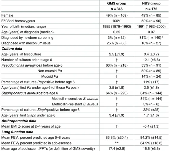
Table 1presents population characteristics for the GMS and NBS datasets (datasets available as
supporting information files 1 & 2). NBS subjects were, on average, born later, diagnosed at a younger age (median 0.07 years vs. 0.35 years), and had respiratory culture data earlier than GMS subjects. Approximately 50% of NBS subjects were F508del homozygous, vs. 100% of GMS subjects. Approximately 17% (27/172) of the NBS cohort were diagnosed by meconium ileus and 4/172 were missed on the newborn screen and diagnosed later. The mean age at
Table 1. Population characteristics of GMS and NBS subjects.
GMS group NBS group
n = 346 n = 172
Female 49% (n = 169) 49% (n = 85)
F508del homozygous 100% 52% (n = 90)
Year of birth (median, range) 1985 (1979–1993) 1991 (1982–2000)
Age (years) at diagnosis (median) 0.35 0.07
Diagnosed by newborn screening 3% (n = 12) 81% (n = 140)*
Diagnosed with meconium ileus 25% (n = 88) 16% (n = 27)
Culture data
Age (years) at first culture 2.5 (±1.9) 0.4 (±0.7)
Number of cultures prior to age 6 † 12.1 (±6.6)
Pseudomonas aeruginosa before age 6 63% (n = 218) 53% (n = 91)
Non-mucoid Pa † 52% (n = 89)
Mucoid Pa † 14% (n = 24)
Percentage of cultures Pa positive before age 6 † 11% (±17) Age (years) first Pa under age 6 (of those Pa pos.) 3.5 (±1.8) 2.5 (±1.8)
Staphylococcus aureus before age 6 64% (n = 223) 84% (n = 144) Methicillin-sensitive S. aureus † 84% (n = 144) Methicillin-resistant S. aureus † 3% (n = 6) Percentage of cultures Staph positive before age 6 † 32% (±25) Age (years) first Staph under age 6 3.4 (±1.9) 1.7 (±1.6) Anthropometric data
Mean BMI Z-score at 2–4 years of age † -0.4 (±1.3)
Lung function data
Mean FEV1percent predicted age 6–8 years 86.8% (±20.4) 94.2% (±14.5) Mean FEV1percent predicted in adolescence ** 84.9% (±18.8) Mean age of adolescent PFTs (or definition of GMS severity) 17.4 (±2.9) 15.5 (±3.6)
*Additional 5 subjects w/ false-negative newborn screening, remainder diagnosed with meconium ileus † Limited data available (annualized CF Registry data)
definition of adolescent lung function was 15.5 and 17.4 years for NBS and GMS subjects, respectively.
GMS population
Of the 629 F508del homozygous subjects initially enrolled and described elsewhere[29], 346 met inclusion criteria (Fig 1). Over 60% of subjects had at least one culture positive forPa
prior to 6 years of age.
GMS subjects were stratified into four subgroups byPainfection status at age 6 (ever vs. neverPapositive prior to 6 years of age) and adolescent lung disease group (severe vs. mild). Subjects in the four sub-groups did not differ significantly by gender distribution, age at diag-nosis, age of definition of adolescent lung function, or presence of other respiratory pathogens in early childhood (data not shown). Age of first culture was younger in subjects who werePa
positive in early childhood whether they had mild or severe lung function in adolescence (Mean age (years) at first culture in the mild adolescent lung disease group was 2.22 vs. 3.51 forPapositive and negative subjects, respectively, p<0.05. Mean age at first culture in the severe adolescent lung disease groups were—2.18 vs. 3.51 forPapositive and negative subjects, respectively, p<0.05). Similarly,Papositive subjects had a higher mean number of respiratory cultures before age 6 regardless of lung function in adolescence (number of cultures among subjects with mild adolescent lung disease—4.56 vs. 3.11 forPapositive and negative subjects, respectively, p<0.05. Number of cultures among subjects with severe adolescent lung disease —4.86 vs. 2.57 forPapositive and negative subjects, p<0.05).
Fig 2illustrates differences in early lung function among the four GMS subgroups. Among
subjects who werePapositive in early childhood, mean FEV1at 6–8 years of age was lower in
those with severe vs. mild adolescent lung disease (72.4%±15.8 and 99.4%±13.1, respectively) (p<0.0001). AmongPa-negative subjects, there was a similar difference in early childhood FEV1between those with severe vs. mild adolescent lung disease (79.1%±16.7 and 103.2%±
14, respectively) (p<0.0001). In comparing subjects with severe lung disease later in life, mean early childhood FEV1was ~8 percentage points lower in those subjects who werePa-positive
before age 6 (p = 0.01); however, there was no significant difference in early FEV1betweenPa
positive and negative subjects with mild adolescent lung disease.
NBS population
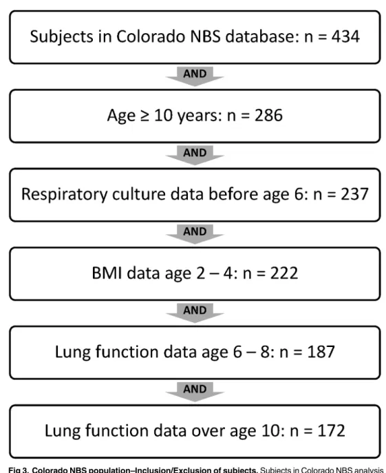
Of 434 subjects included in the Colorado NBS database, 172 were included in final analysis; the majority excluded were not old enough to have “adolescent” lung function data available at 10 years of age or older (Fig 3).
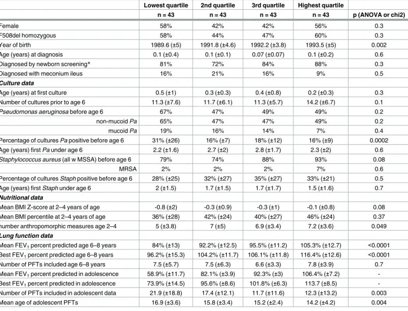
Comparison by adolescent lung function quartile. NBS subjects were first divided into
even quartiles by adolescent lung function (n = 43 in each quartile), then further divided byPa
status in early childhood. When comparing NBS subjects by lung function quartile in adoles-cence, subjects with lower adolescent lung function were born earlier and had a higher per-centage of cultures positive forPaprior to age 6 than adolescents with higher lung function (Table 2). There were no significant differences in nutritional status, age of first culture, num-ber of cultures before age 6, percentage of subjectsPa-positive before age 6, or age at firstPa -positive culture between the adolescent lung function quartile groups. Subjects with lower ado-lescent lung function had significantly lower FEV1at 6 to 8 years of age than those in the
higher adolescent lung function quartiles (Table 2).
adolescent lung function who werePapositive before age 6 (mean early childhood FEV178%
(±13), 89% (±7), 94% (±11), and 107% (±12) for lowest to highest adolescent lung function groups, p<0.0001). Though a similar pattern was seen in thePanegative group, differences did not reach statistical significance (mean early childhood FEV193% (±7), 95% (±16), 97%
(±11), and 103% (±13) for lowest to highest adolescent lung function group, p = 0.065). When comparingPapositive and negative subjects by adolescent lung function quartile,Papositive subjects in the lowest adolescent lung function quartile had significantly lower early childhood lung function thanPanegative (78% (±13) vs. 93% (±6), p<0.001); differences in early child-hood FEV1betweenPapositive and negative subjects in the other quartiles were not
statisti-cally significant (p = 0.09, 0.18, 0.86 for 2ndlowest, 3rd, and highest quartiles, respectively). There was a significant decline in FEV1percent predicted from early childhood to
adoles-cence in the two lowest adolescent lung function quartiles, with a decrease of 25 percentage
Fig 1. GMS population: Inclusion/Exclusion of subjects. Subjects from the parent GMS study included in
current analysis were limited to those who a) had lung function data between age 6 and 8, b) were enrolled in the GMS study at 12 years of age or greater, and c) had respiratory culture data available prior to age 6. Definition of adolescent lung function occurred at time of GMS enrollment; age cutoff was created to separate time of definition of adolescent lung function from early childhood lung function.
https://doi.org/10.1371/journal.pone.0177215.g001
Fig 2. GMS population: Early childhood lung function by Pa status before age 6 and severe vs. mild lung disease in adolescence.
Mean FEV1percent predicted at 6 to 8 years of age is plotted by classification of lung disease in adolescence (severe vs. mild) and Pa infectious status up to age 6 (ever vs. never culture positive for Pa).
points and 10 percentage points in lowest and 2ndlowest quartiles, respectively (p<0.0001 for both). There was no significant change in mean FEV1from early childhood to adolescence in
the higher two adolescent lung function quartiles (Table 3). Similarly, rate of decline in FEV1
from childhood to adolescence (percent predicted per year) was significantly higher in subjects in the two lowest adolescent lung function quartiles (Table 3).
Table 4shows annual rate of decline (percent predicted per year) in FEV1from early
child-hood to adolescence by adolescent lung function quartile and early childchild-hoodPastatus. Sub-jects with the lowest adolescent lung function had significantly faster annual rates decline in FEV1than all other subjects; mean FEV1percent predicted in the two highest quartiles
appeared nearly stable from childhood to adolescence (by both percent predicted and rate of
Fig 3. Colorado NBS population–Inclusion/Exclusion of subjects. Subjects in Colorado NBS analysis
were limited to those 10 years of age or older at the time of analysis in order to have “adolescent” lung function data available. Subjects included in analysis were required to have data on respiratory cultures before age 6, BMI at age 2 to 4, lung function at age 6 to 8, and lung function over age 10.
decline). Importantly, rate of decline did not differ significantly within adolescent lung func-tion quartiles when comparing subjects by early childhoodPastatus (Table 4).
For all comparisons discussed above, similar patterns were seen when comparing best early childhood FEV1as opposed to mean (data not shown).
Linear regression. Variables included in the final linear regression model (after
back-wards elimination of covariates) were: adolescent FEV1, early childhood FEV1, BMI Z-score
at 2–4 years of age, percentage of cultures positive forPabefore age 6, number of cultures obtained before age 6, number of PFTs included in the early childhood lung function variable, number of PFTs included in the adolescent lung function variable, gender, year of birth, and age at diagnosis. We found a strong association between lower early childhood FEV1and
lower adolescent FEV1(p<0.001,Table 5). Higher percentage of cultures positive forPaprior
to age 6 was also significantly associated with lower adolescent FEV1(p = 0.02), though the
effect appeared markedly less than that of early childhood FEV1. Contribution to the explained
Table 2. Colorado NBS population characteristics by adolescent lung function quartile.
Lowest quartile 2nd quartile 3rd quartile Highest quartile
n = 43 n = 43 n = 43 n = 43 p (ANOVA or chi2)
Female 58% 42% 42% 56% 0.3
F508del homozygous 58% 44% 47% 60% 0.3
Year of birth 1989.6 (±5) 1991.8 (±4.6) 1992.2 (±3.8) 1993.5 (±5) 0.002
Age (years) at diagnosis 0.1 (±0.4) 0.1 (±0.1) 0.07 (±0.07) 0.1 (±0.2) 0.6
Diagnosed by newborn screening* 81% 72% 84% 88% 0.3
Diagnosed with meconium ileus 16% 21% 16% 9% 0.5
Culture data
Age (years) at first culture 0.5 (±1) 0.3 (±0.3) 0.4 (±0.8) 0.2 (±0.3) 0.3
Number of cultures prior to age 6 11.3 (±7.6) 11.7 (±6.1) 11.3 (±5.7) 14.2 (±6.7) 0.1
Pseudomonas aeruginosa before age 6 67% 47% 49% 49% 0.2
non-mucoid Pa 65% 47% 47% 49% 0.2
mucoid Pa 19% 16% 14% 7% 0.4
Percentage of cultures Pa positive before age 6 31% (±26) 16% (±7) 18% (±12) 16% (±9) 0.0002 Age (years) first Pa under age 6 2.2 (±1.6) 2.7 (±2) 2.8 (±1.7) 2.3 (±2) 0.6
Staphylococcus aureus (all w MSSA) before age 6 79% 74% 88% 93% 0.08
MRSA 2% 2% 2% 7% 0.6
Percentage of cultures Staph positive before age 6 28% (±25) 32% (±27) 35% (±27) 33% (±21) 0.5 Age (years) first Staph under age 6 2 (±1.5) 1.7 (±1.5) 1.7 (±1.7) 1.5 (±1.6) 0.7 Nutritional data
Mean BMI Z-score at 2–4 years of age -0.8 (±2) -0.3 (±0.9) -0.3 (±1) -0.1 (±0.8) 0.08 Mean BMI percentile at 2–4 years of age 36% (±28) 42% (±24) 40% (±27) 46% (±24) 0.37 number anthropomorphic measures age 2–4 5 (±3.8) 7 (±5) 6.9 (±3.4) 7.2 (±3.6) 0.049 Lung function data
Mean FEV1percent predicted age 6–8 years 84% (±13) 92.2% (±12.5) 95.5% (±11.2) 105.3% (±12.7) <0.0001 Best FEV1percent predicted age 6–8 years 96.2% (±15.3) 104.2% (±11.7) 106.1% (±11.8) 116.4% (±12.6) <0.0001 Number of PFTs included age 6–8 years 7.5 (±5.7) 7.5 (±6.3) 6.6 (±3.3) 7.8 (±3.9) 0.7 Mean FEV1percent predicted in adolescence 58.9% (±11.7) 82.1% (±3.9) 92.3% (±3) 106.4% (±7.2) -Best FEV1percent predicted in adolescence 73.9% (±14.5) 95.6% (±8.6) 101.8% (±6.3) 113.7 (±8.5) -Number of PFTs included in adolescent data 21.9 (±18.8) 17.4 (±12.1) 11.7 (±11.6) 12.3 (±13.2) 0.003 Mean age of adolescent PFTs 16.9 (±3.6) 15.8 (±3.4) 15.2 (±2.4) 14.2 (±4.2) 0.004
variance of the full model was 0.1957 for mean early childhood FEV1vs. 0.0157 for percentage
of cultures positive forPabefore age 6. In this model, predicted mean adolescent FEV1
in-creased by 19.7 percent predicted when early childhood FEV1was increased from the 25th
per-centile (85.5% predicted) to the 95thpercentile (117.4%), while it decreased by only 5.8% when percentage of cultures positive forPawas increased from the 25thpercentile (0%) to the 95th percentile (36.8% positive) (seeFig 5). Lower BMI in early childhood also showed a significant association with lower adolescent lung function (p = 0.005). Neither the simple presence ofPa
prior to age 6 (yes/no dichotomous variable for at least one positive culture) nor the presence or percentage of cultures positive forS.aureusprior to age 6 were significantly associated with adolescent FEV1.
Discussion
We have shown in two separate, distinct cohorts of CF subjects that lower FEV1in early
child-hood (6 to 8 years of age) is strongly associated with lower FEV1in adolescence, and that this
association is only partially explained by infection withPabefore age 6; thus, other genetic and/or environmental factors, likely in early childhood, must be playing a major role. Impor-tantly, we have also shown a striking divergence in loss of lung function among CF patients, as
Fig 4. Colorado NBS Population: Early childhood lung function by adolescent lung function quartile and early childhood Pa status. Early childhood lung function (mean FEV1at 6–8 years of age) by adolescent lung function quartile and early childhood Pa status (ever/never Pa positive before age 6). Subjects were first divided into even quartiles by adolescent lung function (n = 43 in each quartile), then further divided by Pa status in early childhood. Among Pa positive subjects, those in the lowest adolescent lung function quartile had significantly lower early childhood lung function compared to all other quartiles; differences in early childhood lung function among Pa negative subjects did not reach statistical significance. When comparing subjects within individual adolescent lung function quartiles, Pa positive subjects in the lowest adolescent lung function quartile had significantly lower early childhood lung function than Pa negative (p<0.001); otherwise there were no significant differences in early childhood lung function by Pa status within adolescent lung function quartiles.
subjects with worse lung function in early childhood had a significantly faster rate of FEV1
decline from childhood to adolescence compared to those with better early childhood lung function. This finding holds even for those diagnosed in infancy, was the majority of NBS sub-jects presented for management and treatment prior to the development of respiratory symp-toms, and those with worse lung function early still had a faster rate of decline. Our findings suggest that predisposing genetic and/or environmental factors and events in infancy and pre-school in children with CF may have a strong impact on lung function from early childhood through adolescence and adulthood, even in children diagnosed by newborn screening.
Our results are consistent with Burns’ finding that 97% of infants with CF in their clinic population had serologic or microbiologic evidence ofPaexposure before age three[43], which would suggest that growth ofPaon respiratory culture cannot be the driving force in
Table 3. Colorado NBS Population: Absolute change and annual rate of change (percent predicted per year) in FEV1percent predicted from early childhood to adolescence by adolescent lung function quartile. Absolute change in FEV1was defined as mean FEV1percent predictedage 6–8minus mean FEV1percent predictedadolescent. Annual rate of change was defined as absolute change in FEV/ (mean age adolescent PFTs–mean age early child-hood PFTs).
Mean FEV1 Best FEV1
mean p mean p
LOWEST QUARTILE
change in % predicted (child—adolescent) -25.1 <0.0001 -22.3 <0.0001
annual rate of change -2.8 (±2.1) *† -1.1 (±1.6) *†
2nd QUARTILE
change in % predicted (child—adolescent) -10.1 <0.0001 -8.6 <0.0001
annual rate of change -2.4 (±3.3) ‡ -1 (±1.5)
3rd QUARTILE
change in % predicted (child—adolescent) -3.2 0.06 -4.4 0.03
annual rate of change -1.7 (±2.1) -0.5 (±2.1)
HIGHEST QUARTILE
change in % predicted (child—adolescent) 1.1 0.5 -2.7 0.09
annual rate of change -1.1 (±2.7) -0.5 (±1.8)
*p value for difference among any of the 4 quartiles (ANOVA)<0.001 † p value comparing lowest quartile to all other quartiles<0.001 ‡ p value comparing 2nd quartile to highest quartile<0.003 https://doi.org/10.1371/journal.pone.0177215.t003
Table 4. Colorado NBS Population: Annual rate of change in FEV1percent predicted from childhood to adolescence by adolescent lung function quartile and Pa status before 6 years of age.
Adolescent Lung Function Quartile Lowest quartile
n = 43
2nd quartile n = 43
3rd quartile n = 43
Highest quartile n = 43
Mean (SD) Pa negative
n = 14
Pa positive
n = 29
Pa negative
n = 23
Pa positive
n = 20
Pa negative
n = 22
Pa positive
n = 21
Pa negative
n = 22
Pa positive
n = 21
FEV1age 6–8 92.9 (±6.8)* 79.7
(±13.2)
94.6 (±15.6) 89.5 (±7.1)†
97.1 (±11.5) 93.9 (±11)†**
103.2 (±13) 107.4 (±12.4)†**
Annual rate of change in FEV1% predicted
-2.6 (±0.9) -2.8 (±2.5) -1.2 (±1.9) -0.9 (±1.1) -0.4 (±1.6)† -0.1 (±1.4)† 0.6 (±1.5)† -0.1 (±1.7)†
*p<0.05 comparing Pa positive and negative groups within adolescent lung function quartile † p<0.05 compared to lowest adolescent lung function quartile group with same Pa status
Table 5. Colorado NBS population: Linear regression, association with mean adolescent FEV1percent predicted. Results of linear regression
inves-tigating association of early childhood events/exposures with mean adolescent FEV1percent predicted. All variables with p<0.05 are shown; the model was also adjusted for gender, age at diagnosis, number of respiratory cultures prior to age 6, and number of PFTs used to calculate early childhood FEV1. Vari-ables eliminated from the model included genotype (F508del homozygous yes/no), diagnosis by newborn screening, percentage of cultures positive for S.
aureus prior to age 6, and number of measures included in BMI value.
95% CI
VARIABLE ß coeff SE Lower 95% Upper 95% p value
Mean FEV1%predicted age 6 to 8 0.616 0.077 0.465 0.768 <0.001
Percent of cultures Pa positive before age 6 -0.159 0.070 -0.297 -0.021 0.024
Year of birth 1.03 0.319 0.397 1.66 0.002
BMI z-score age 2 to 4 2.37 0.826 0.743 4.01 0.005
Number of PFTs used to calculate mean adolescent FEV1 -0.324 0.078 -0.478 -0.17 <0.001 Also adjusted for gender, age at diagnosis, number of cultures age 0–6, number of PFTs age 6–8 (these variables were also included in regression analysis, but without significant p value).
https://doi.org/10.1371/journal.pone.0177215.t005
Fig 5. NBS linear regression: Predicted adolescent lung function is more strongly associated with early childhood FEV1than with percentage of cultures positive for Pa in early childhood. Subject values for mean early childhood and adolescent FEV1are shown in scatterplot, with subjects Pa positive in early childhood represented by gray triangles, Pa negative by black circles. Superimposed lines represent NBS linear regression results showing predicted adolescent FEV1(with 95% confidence intervals—dashed lines) by early childhood FEV1. The black line shows predicted adolescent FEV1by early childhood FEV1when percentage of cultures positive for Pa prior to age 6 is held at 0% (25
th
percentile); the grey line represents the predicted adolescent FEV1when percentage of cultures positive for Pa prior to age 6 is held at 30% (approximately the 90thpercentile for our cohort). These values were all adjusted for gender, year of birth, age at diagnosis, number of cultures before age 6, BMI Z score at 2 to 4 years of age, and number of PFT tests included in early childhood and adolescent values (all held at mean). Predicted mean adolescent FEV1increased by 19.7 percent predicted when early childhood FEV1was increased from the 25
th
percentile (85.5% predicted) to the 95thpercentile (117.4%), while it decreased by only 4.8% when percentage of cultures positive for Pa was increased from the 25thpercentile (0%) to the 92ndpercentile (30% positive), or 5.8% when increased to 36.8% positive (95th percentile). Contribution to the explained variance of the full model was 0.1957 for mean early childhood FEV1vs. 0.0157 for percentage of cultures positive for Pa before age 6.
differences in early lung function. In the GMS dataset, subjects with severe adolescent lung dis-ease had mean early childhood FEV1approximately 25 percentage points lower than
adoles-cent subjects with mild disease, regardless of early childhoodPastatus. In the NBS dataset, early lung function was lower in subjects with lower adolescent lung function. We acknowl-edge that there is an association between earlyPainfection, early childhood lung function, and adolescent lung function, as subjects with lower adolescent lung functionandearlyPa infec-tion appeared to have worse early childhood lung funcinfec-tion in both study populainfec-tions. This confirms that infection withPais one of many factors associated with lower lung function in adolescence, but our findings emphasize that, even when adjusting for chronicity or prevalence of positive cultures,Pais not themajorcause of lower lung function in children that persists into adolescence.
Previous studies have shown that lung function in healthy subjects remains relatively stable when comparing percent-predicted values from childhood to adolescence[44,45]. Studies in children with CF have noted an association with higher baseline FEV1and more rapid rate of
decline over the next 3–6 years[27,46]. However, we found that subjects with CF with lower adolescent lung function showed significantly greater rate of decline in FEV1percent predicted
andannual rate of decline in FEV1percent predicted from early childhood to adolescence,
while those subjects with higher adolescent lung function showed no significant decline from early childhood to adolescence. This suggests that children with CF and lower FEV1are not
simply tracking along a given lung function trajectory, but have active progression and wors-ening of their lung disease in comparison to their healthier counterparts with CF. Our findings may differ from previous studies in follow-up time from early childhood to adolescence was relatively longer–perhaps uniformly bridging adolescence in our population resulted in differ-ent patterns of lung function decline. The NBS population was also limited to a single state population, and thus our study population was smaller and possibly more homogenous. Finally, our analysis approaches may have differed.
The strongest predictor of low lung function in adolescence was low lung function in early childhood. Importantly, there were no significant differences in rate of decline when compar-ingPapositive and negative subjects within adolescent lung function quartiles. Instead, sub-jects with lower adolescent lung function (in bothPapositive and negative groups) both started with lower early childhood lung functionandshowed a faster rate of decline in lung function than their counterparts with higher adolescent lung function. Those subjects with a higher percentage of respiratory cultures positive forPaprior to age 6 also had lower lung function in adolescence, though that association was not as strong as that with early childhood lung function, and the dichotomous presence/absence ofPaprior to age 6 was not associated with adolescent lung function. This suggests that infectious burden, and possibly inflammatory response, plays a role in outcomes, though compliance with prescribed preventative treatments such as chest physiotherapy could also explain persistent decline in some children. Our find-ings are consistent with previous studies implicating chronicPainfection in poorer pulmonary outcomes in patients with CF[15,27,29,47], and implicating inflammation as a strong predic-tor of lung function decline[48].
The importance ofS.aureusin CF lung disease has been a source of recent debate[28,30,
31,49,50]; we found no significant association betweenS.aureusinfection in early childhood
and adolescent lung function, though we were unable to investigate the association with methi-cillin-resistantS.aureusdue to low prevalence in our NBS population and lack of consistent reporting in our GMS population.
subjects would have been older when adolescent lung function was recorded, but could also reflect advances in management and treatment. Our findings suggest early childhood lung function, early nutritional status, and density or persistence of earlyPainfection were most strongly associated with adolescent lung function in subjects with CF.
Our two distinct datasets show consistent results, even though they represent different birth cohorts (straddling initiation of widespread use of inhaled tobramycin for treatment of
Painfection), had different genetic makeups and geographic origins, and likely reflected dif-ferent treatment regimens (based on clinical advances from GMS to NBS birth cohorts). Both datasets had distinct advantages and complement the other well. The GMS dataset was large, representing CF centers across the U.S., and genetically homogenous (all F508del homozygotes), but was limited by use of annualized data, relatively scant data in infancy and preschool, and the extremes of phenotype design. The NBS dataset was smaller regional, but contained the full spectrum of CF lung disease (not extremes of phenotype), a representative distribution of CF genotypes, and used encounter-based, non-annualized data. Initiation of CF-directed care such as chest physiotherapy at diagnosis, and prior to development of respiratory symptoms, is another advantage in the NBS dataset and eliminates a potential confounder.
Our study had several limitations. Both GMS and NBS datasets are comprised of clinically collected data; there was not uniformity in data collection frequency or variables collected. PFT data at 6–8 years of age was the earliest age at which we could determine “early” childhood lung function. Because of the annualized data in the GMS study and the extremes of phenotype design, we were not able to explore associations between early childhood events, early child-hood lung function, and adolescent lung function in great detail in this population. The NBS study population had more extensive early childhood data; however, there was still substantial variability as to how much data was available on individual subjects. We were unable to in-clude mucoidPaor methicillin-resistantS.aureusin either analysis due to low prevalence in the NBS population, and limited data available from the CFFPR for the majority of GMS sub-jects. Similarly, we had limited data on other organisms such asHaemophilus influenza, Asper-gillus fumigatus, orStenotrophomonas maltophilia, and were not able to include these in our analyses, nor were we able to include differences in therapies and treatments. Finally, socio-economic status was not reliably collected and was therefore not included in our regression model, yet socioeconomic status has been shown to be associated with worse outcomes in CF [18]. Despite these limitations, we believe the consistency of our results in two distinct popula-tions of subjects lend support to our findings.
In summary, we have shown in two distinct populations of subjects with CF that lung func-tion in early childhood is more strongly associated with lung funcfunc-tion in adolescence in CF than infection withPseudomonas aeruginosabefore age 6. Most strikingly, we found that sub-jects with lower lung function in adolescence had lower lung function in early childhoodanda more rapid rate of decline of FEV1from childhood to adolescence than those subjects with
Supporting information
S1 Table. GMS dataset. Variables for all subjects included in the final analysis of Gene
Modi-fier Study population. Please contact Dr. Pittman with questions or for more details. (XLSX)
S2 Table. NBS dataset. Variables for all subjects included in the final analysis of the Newborn
Screen Study population. Please contact Dr. Pittman with questions or for more details. (XLSX)
Author Contributions
Conceptualization: JEP MRK MKS MD SDD.
Data curation: JEP HEC HN.
Formal analysis: JEP MKS HN HEC.
Funding acquisition: MRK MKS MD SDS FJA JEP.
Methodology: MKS JEP.
Resources: MRK MD MKS SDS FJA.
Supervision: MRK MKS SDD MWL.
Visualization: JEP.
Writing – original draft: JEP MRK MKS.
Writing – review & editing: JEP MRK MKS SDD MWL MD SDS FJA.
References
1. Bush A. Six of the best: cystic fibrosis. Paediatr Respir Rev. 2001; 2(4):287–93. Epub 2002/06/08. https://doi.org/10.1053/prrv.2001.0162PMID:12052299
2. Cutting GR, Zietlin P.L. Genetics and Pathophysiology of Cystic Fibrosis. In: Chernick V, Boat T.F., Wil-mott R.W., Bush A., editor. Kendig’s Disorders of the Respiratory Tract in Children: Saunders Elsevier; 2006.
3. Davies JC, Alton EW, Bush A. Cystic fibrosis. BMJ. 2007; 335(7632):1255–9. Epub 2007/12/15.https:// doi.org/10.1136/bmj.39391.713229.ADPMID:18079549
4. Davis PB. Pulmonary Disease in Cystic Fibrosis. In: Chernick V, Boat T.F., Wilmott R.W., Bush A., edi-tor. Kendig’s Disorders of the Respiratory Tract in Children. Philadelphia: Saunders Elsevier; 2006. p. 873–86.
5. Moss RB. Infection, inflammation, and the downward spiral of cystic fibrosis lung disease. J Pediatr. 2009; 154(2):162–3. Epub 2009/01/20.https://doi.org/10.1016/j.jpeds.2008.09.042PMID:19150671
6. Rosenfeld M, Gibson RL, McNamara S, Emerson J, Burns JL, Castile R, et al. Early pulmonary infec-tion, inflammainfec-tion, and clinical outcomes in infants with cystic fibrosis. Pediatr Pulmonol. 2001; 32 (5):356–66. Epub 2001/10/12. PMID:11596160
7. Armstrong DS, Hook SM, Jamsen KM, Nixon GM, Carzino R, Carlin JB, et al. Lower airway inflamma-tion in infants with cystic fibrosis detected by newborn screening. Pediatr Pulmonol. 2005; 40(6):500– 10. Epub 2005/10/07.https://doi.org/10.1002/ppul.20294PMID:16208679
8. Davis SD, Fordham LA, Brody AS, Noah TL, Retsch-Bogart GZ, Qaqish BF, et al. Computed tomogra-phy reflects lower airway inflammation and tracks changes in early cystic fibrosis. Am J Respir Crit Care Med. 2007; 175(9):943–50. Epub 2007/02/17.https://doi.org/10.1164/rccm.200603-343OCPMID: 17303797
10. Linnane BM, Hall GL, Nolan G, Brennan S, Stick SM, Sly PD, et al. Lung Function in Infants with Cystic Fibrosis Diagnosed by Newborn Screening. Am J Respir Crit Care Med. 2008. Epub 2008/09/13.
11. Lum S, Gustafsson P, Ljungberg H, Hulskamp G, Bush A, Carr SB, et al. Early detection of cystic fibro-sis lung disease: multiple-breath washout versus raised volume tests. Thorax. 2007; 62(4):341–7. Epub 2006/11/24.https://doi.org/10.1136/thx.2006.068262PMID:17121870
12. Accurso FJ, Sontag MK. Gene modifiers in cystic fibrosis. J Clin Invest. 2008; 118(3):839–41. Epub 2008/02/23.https://doi.org/10.1172/JCI35138PMID:18292812
13. Corey M, Edwards L, Levison H, Knowles M. Longitudinal analysis of pulmonary function decline in patients with cystic fibrosis. J Pediatr. 1997; 131(6):809–14. Epub 1998/01/15. PMID:9427882
14. Davis PB. The decline and fall of pulmonary function in cystic fibrosis: new models, new lessons. J Pediatr. 1997; 131(6):789–90. Epub 1998/01/15. PMID:9427876
15. Emerson J, Rosenfeld M, McNamara S, Ramsey B, Gibson RL. Pseudomonas aeruginosa and other predictors of mortality and morbidity in young children with cystic fibrosis. Pediatr Pulmonol. 2002; 34 (2):91–100. Epub 2002/07/12.https://doi.org/10.1002/ppul.10127PMID:12112774
16. Gan KH, Heijerman HG, Bakker W. Correlation between genotype and phenotype in patients with cystic fibrosis. N Engl J Med. 1994; 330(12):865–6; author reply 6–7. Epub 1994/03/24.
17. Schechter MS, Shelton BJ, Margolis PA, Fitzsimmons SC. The association of socioeconomic status with outcomes in cystic fibrosis patients in the United States. Am J Respir Crit Care Med. 2001; 163 (6):1331–7. Epub 2001/05/24.https://doi.org/10.1164/ajrccm.163.6.9912100PMID:11371397
18. Schechter MS. Non-genetic influences on cystic fibrosis lung disease: the role of sociodemographic characteristics, environmental exposures, and healthcare interventions. Semin Respir Crit Care Med. 2003; 24(6):639–52. Epub 2005/08/10.https://doi.org/10.1055/s-2004-815660PMID:16088580
19. Collaco JM, Vanscoy L, Bremer L, McDougal K, Blackman SM, Bowers A, et al. Interactions between secondhand smoke and genes that affect cystic fibrosis lung disease. JAMA. 2008; 299(4):417–24. Epub 2008/01/31.https://doi.org/10.1001/jama.299.4.417PMID:18230779
20. Wolfenden LL, Schechter MS. Genetic and non-genetic determinants of outcomes in cystic fibrosis. Paediatr Respir Rev. 2009; 10(1):32–6. Epub 2009/02/11.https://doi.org/10.1016/j.prrv.2008.04.002 PMID:19203742
21. McPhail GL, Acton JD, Fenchel MC, Amin RS, Seid M. Improvements in lung function outcomes in chil-dren with cystic fibrosis are associated with better nutrition, fewer chronic pseudomonas aeruginosa infections, and dornase alfa use. J Pediatr. 2008; 153(6):752–7. Epub 2008/09/02.https://doi.org/10. 1016/j.jpeds.2008.07.011PMID:18760423
22. Zemel BS, Jawad AF, FitzSimmons S, Stallings VA. Longitudinal relationship among growth, nutritional status, and pulmonary function in children with cystic fibrosis: analysis of the Cystic Fibrosis Foundation National CF Patient Registry. J Pediatr. 2000; 137(3):374–80. Epub 2000/09/02.https://doi.org/10. 1067/mpd.2000.107891PMID:10969263
23. Schaedel C, de Monestrol I, Hjelte L, Johannesson M, Kornfalt R, Lindblad A, et al. Predictors of deterio-ration of lung function in cystic fibrosis. Pediatr Pulmonol. 2002; 33(6):483–91. Epub 2002/05/10. https://doi.org/10.1002/ppul.10100PMID:12001283
24. Rosenfeld M, Ramsey BW, Gibson RL. Pseudomonas acquisition in young patients with cystic fibrosis: pathophysiology, diagnosis, and management. Curr Opin Pulm Med. 2003; 9(6):492–7. Epub 2003/10/ 10. PMID:14534401
25. Goss CH, Mayer-Hamblett N, Aitken ML, Rubenfeld GD, Ramsey BW. Association between Stenotro-phomonas maltophilia and lung function in cystic fibrosis. Thorax. 2004; 59(11):955–9. Epub 2004/11/ 02.https://doi.org/10.1136/thx.2003.017707PMID:15516471
26. Li Z, Kosorok MR, Farrell PM, Laxova A, West SE, Green CG, et al. Longitudinal development of mucoid Pseudomonas aeruginosa infection and lung disease progression in children with cystic fibrosis. JAMA. 2005; 293(5):581–8. Epub 2005/02/03.https://doi.org/10.1001/jama.293.5.581PMID:
15687313
27. Konstan MW, Morgan WJ, Butler SM, Pasta DJ, Craib ML, Silva SJ, et al. Risk factors for rate of decline in forced expiratory volume in one second in children and adolescents with cystic fibrosis. J Pediatr. 2007; 151(2):134–9, 9 e1. Epub 2007/07/24.https://doi.org/10.1016/j.jpeds.2007.03.006PMID:17643762
28. Sagel SD, Gibson RL, Emerson J, McNamara S, Burns JL, Wagener JS, et al. Impact of Pseudomonas and Staphylococcus infection on inflammation and clinical status in young children with cystic fibrosis. J Pediatr. 2009; 154(2):183–8. Epub 2008/09/30.https://doi.org/10.1016/j.jpeds.2008.08.001PMID: 18822427
30. Dasenbrook EC, Merlo CA, Diener-West M, Lechtzin N, Boyle MP. Persistent methicillin-resistant Staphylococcus aureus and rate of FEV1 decline in cystic fibrosis. Am J Respir Crit Care Med. 2008; 178(8):814–21. Epub 2008/08/02.https://doi.org/10.1164/rccm.200802-327OCPMID:18669817
31. Levy H, Kalish LA, Cannon CL, Garcia KC, Gerard C, Goldmann D, et al. Predictors of mucoid Pseudo-monas colonization in cystic fibrosis patients. Pediatr Pulmonol. 2008; 43(5):463–7https://doi.org/10. 1002/ppul.20794PMID:18361452
32. Waters V, Atenafu EG, Salazar JG, Lu A, Yau Y, Matukas L, et al. Chronic Stenotrophomonas maltophi-lia infection and exacerbation outcomes in cystic fibrosis. Journal of cystic fibrosis: official journal of the European Cystic Fibrosis Society. 2012; 11(1):8–13. Epub 2011/08/19.
33. McKone EF, Emerson SS, Edwards KL, Aitken ML. Effect of genotype on phenotype and mortality in cystic fibrosis: a retrospective cohort study. Lancet. 2003; 361(9370):1671–6. Epub 2003/05/28.https:// doi.org/10.1016/S0140-6736(03)13368-5PMID:12767731
34. Braun AT, Farrell PM, Ferec C, Audrezet MP, Laxova A, Li Z, et al. Cystic fibrosis mutations and geno-type-pulmonary phenotype analysis. J Cyst Fibros. 2006; 5(1):33–41. Epub 2005/11/09.https://doi.org/ 10.1016/j.jcf.2005.09.008PMID:16275171
35. Drumm ML, Konstan MW, Schluchter MD, Handler A, Pace R, Zou F, et al. Genetic modifiers of lung disease in cystic fibrosis. N Engl J Med. 2005; 353(14):1443–53. Epub 2005/10/07.https://doi.org/10. 1056/NEJMoa051469PMID:16207846
36. Ramsey BW, Pepe MS, Quan JM, Otto KL, Montgomery AB, Williams-Warren J, et al. Intermittent administration of inhaled tobramycin in patients with cystic fibrosis. Cystic Fibrosis Inhaled Tobramycin Study Group. N Engl J Med. 1999; 340(1):23–30. Epub 1999/01/08.https://doi.org/10.1056/
NEJM199901073400104PMID:9878641
37. Gibson RL, Emerson J, McNamara S, Burns JL, Rosenfeld M, Yunker A, et al. Significant micro-biological effect of inhaled tobramycin in young children with cystic fibrosis. Am J Respir Crit Care Med. 2003; 167(6):841–9. Epub 2002/12/14.https://doi.org/10.1164/rccm.200208-855OCPMID: 12480612
38. Wang X, Dockery DW, Wypij D, Fay ME, Ferris BG Jr. Pulmonary function between 6 and 18 years of age. Pediatr Pulmonol. 1993; 15(2):75–88. Epub 1993/02/01. PMID:8474788
39. Abman SH, Ogle JW, Harbeck RJ, Butler-Simon N, Hammond KB, Accurso FJ. Early bacteriologic, immunologic, and clinical courses of young infants with cystic fibrosis identified by neonatal screening. The Journal of pediatrics. 1991; 119(2):211–7. Epub 1991/08/01. PMID:1907318
40. Hammond KB, Abman SH, Sokol RJ, Accurso FJ. Efficacy of statewide neonatal screening for cystic fibrosis by assay of trypsinogen concentrations. The New England journal of medicine. 1991; 325 (11):769–74. Epub 1991/09/12.https://doi.org/10.1056/NEJM199109123251104PMID:1870650
41. Hankinson JL, Odencrantz JR, Fedan KB. Spirometric reference values from a sample of the general U. S. population. Am J Respir Crit Care Med. 1999; 159(1):179–87. Epub 1999/01/05.https://doi.org/10. 1164/ajrccm.159.1.9712108PMID:9872837
42. Kuczmarski RJ OC, Guo SS, et al. CDC growth charts for the United States: Methods and development. In: Statistics. NCfH, editor. Washington, DC: Vital Health Stat; 2000.
43. Burns JL, Gibson RL, McNamara S, Yim D, Emerson J, Rosenfeld M, et al. Longitudinal assessment of Pseudomonas aeruginosa in young children with cystic fibrosis. J Infect Dis. 2001; 183(3):444–52. Epub 2001/01/03.https://doi.org/10.1086/318075PMID:11133376
44. Stern DA, Morgan WJ, Wright AL, Guerra S, Martinez FD. Poor airway function in early infancy and lung function by age 22 years: a non-selective longitudinal cohort study. Lancet. 2007; 370(9589):758–64. Epub 2007/09/04. PubMed Central PMCID: PMC2831283.https://doi.org/10.1016/S0140-6736(07) 61379-8PMID:17765525
45. Hibbert ME, Hudson IL, Lanigan A, Landau LI, Phelan PD. Tracking of lung function in healthy children and adolescents. Pediatric Pulmonology. 1990; 8(3):172–7. Epub 1990/01/01. PMID:2349010
46. Cogen J, Emerson J, Sanders DB, Ren C, Schechter MS, Gibson RL, et al. Risk factors for lung function decline in a large cohort of young cystic fibrosis patients. Pediatr Pulmonol. 2015; 50(8):763–70.https:// doi.org/10.1002/ppul.23217PMID:26061914
47. Kosorok MR, Zeng L, West SE, Rock MJ, Splaingard ML, Laxova A, et al. Acceleration of lung disease in children with cystic fibrosis after Pseudomonas aeruginosa acquisition. Pediatr Pulmonol. 2001; 32 (4):277–87. Epub 2001/09/25. PMID:11568988
49. Rosenfeld M, Ramsey B. Evolution of airway microbiology in the infant with cystic fibrosis: role of non-pseudomonal and non-pseudomonal pathogens. Semin Respir Infect. 1992; 7(3):158–67. Epub 1992/09/01. PMID:1475540