doi: 10.3389/fnhum.2017.00170
Edited by: Jose Luis Contreras-Vidal, University of Houston, USA
Reviewed by: Brian H. Dalton, University of British Columbia Okanagan, Canada Wei Peng Teo, Deakin University, Australia
*Correspondence: Chang S. Nam [email protected] Jason R. Franz [email protected]
Received:15 December 2016 Accepted:22 March 2017 Published:10 April 2017 Citation: Wittenberg E, Thompson J, Nam CS and Franz JR (2017) Neuroimaging of Human Balance Control: A Systematic Review. Front. Hum. Neurosci. 11:170. doi: 10.3389/fnhum.2017.00170
Neuroimaging of Human Balance
Control: A Systematic Review
Ellen Wittenberg1, Jessica Thompson2, Chang S. Nam1* and Jason R. Franz2*
1Edward P. Fitts Department of Industrial and Systems Engineering, North Carolina State University, Raleigh, NC, USA,2Joint Department of Biomedical Engineering, University of North Carolina at Chapel Hill and North Carolina State University, Chapel Hill, NC, USA
This review examined 83 articles using neuroimaging modalities to investigate the neural correlates underlying static and dynamic human balance control, with aims to support future mobile neuroimaging research in the balance control domain. Furthermore, this review analyzed the mobility of the neuroimaging hardware and research paradigms as well as the analytical methodology to identify and remove movement artifact in the acquired brain signal. We found that the majority of static balance control tasks utilized mechanical perturbations to invoke feet-in-place responses (27 out of 38 studies), while cognitive dual-task conditions were commonly used to challenge balance in dynamic balance control tasks (20 out of 32 studies). While frequency analysis and event related potential characteristics supported enhanced brain activation during static balance control, that in dynamic balance control studies was supported by spatial and frequency analysis. Twenty-three of the 50 studies utilizing EEG utilized independent component analysis to remove movement artifacts from the acquired brain signals. Lastly, only eight studies used truly mobile neuroimaging hardware systems. This review provides evidence to support an increase in brain activation in balance control tasks, regardless of mechanical, cognitive, or sensory challenges. Furthermore, the current body of literature demonstrates the use of advanced signal processing methodologies to analyze brain activity during movement. However, the static nature of neuroimaging hardware and conventional balance control paradigms prevent full mobility and limit our knowledge of neural mechanisms underlying balance control.
Keywords: static and dynamic balance control, temporal and spatial dynamics of brain activation, mechanical perturbation, sensory degradation, susceptibility to cognitive dual tasks, movement artifacts
INTRODUCTION
Static and dynamic human balance control, and changes thereof due to intrinsic and extrinsic factors, have garnered considerable scientific and clinical attention. Walking balance control is especially dynamic, involving coordinated adjustments in posture (i.e., head and trunk stabilization) and foot placement from step to step (Bauby and Kuo, 2000; Kay and Warren, 2001; Donelan et al., 2004; Rankin et al., 2014). Particularly in unpredictable and challenging environmental conditions, these adjustments depend on the integration of reliable sensory feedback and the planning and execution of appropriate motor responses (O’Connor and Kuo, 2009; O’Connor et al., 2012; Francis et al., 2015; Franz et al., 2015, 2016; Goodworth et al., 2015). Accordingly, sensory and mechanical perturbations are increasingly used to study corrective motor responses in standing and walking and the onset and progression of balance deficits. Sensory perturbations may include those of visual (e.g., optical flow) (O’Connor and Kuo, 2009; O’Connor et al., 2012; Francis et al., 2015; Franz et al., 2015, 2016), somatosensory (e.g., tendon vibration) (Gurfinkel et al., 1976; Hay et al., 1996; Bove et al., 2003; Mullie and Duclos, 2014), or vestibular feedback (e.g., galvanic stimulation) (Day et al., 1993; Fitzpatrick et al., 1994; Bent et al., 2002; Dakin et al., 2007; Dalton et al., 2014), whereas mechanical perturbations most frequently incorporate support surface translations (Sinitksi et al., 2012; Aprigliano et al., 2016). Cortical activity and high-order cognitive processes are highly involved in the planning and execution of these motor responses. Indeed, dual tasks during standing and walking elicit cognitive-motor interference that compromise metrics of balance control (Dubost et al., 2006; Priest et al., 2008; Plummer et al., 2015).
Human balance control investigations have primarily focused on quantifying motor responses based on kinematic measurements such as movement variability and dynamic stability and/or kinetic measurements such as center of pressure and angular momentum (O’Connor and Kuo, 2009; McAndrew et al., 2011; Francis et al., 2015; Sheehan et al., 2015). These studies are rapidly accelerating our scientific understanding of human balance control, with exciting translational implications for the diagnosis and rehabilitation of people at risk of falls. However, this promising translational potential is currently limited by our relatively incomplete understanding of central mechanisms involved in human balance control and ultimately changes thereof due to aging or disease. Advancements in the use of neuroimaging, and in particular the development of advanced mobile measurements, are now providing previously inaccessible insight into brain connectivity during functional movement and balance tasks.
REVIEW OBJECTIVES
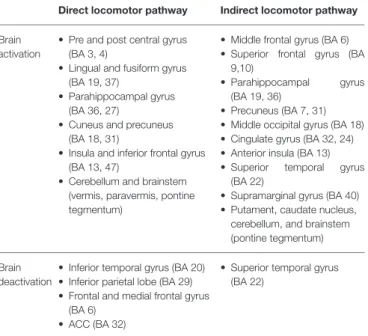
Evidence from neuroimaging studies reveals cortical involvement in standing and walking, with modalities having high spatial resolution, such as functional magnetic resonance imaging (fMRI), single-photon emission computed tomography (SPECT), and positron emission tomography (PET), leading to the identification of a supraspinal locomotor network (la Fougere et al., 2010; Zwergal et al., 2013). Within this supraspinal network,la Fougere et al. (2010)identified a direct locomotor pathway, used primarily during gait execution, and the indirect
TABLE 1 | Motor pathways inla Fougere et al. (2010).
Direct locomotor pathway Indirect locomotor pathway
Brain activation
• Pre and post central gyrus (BA 3, 4)
• Lingual and fusiform gyrus (BA 19, 37)
• Parahippocampal gyrus (BA 36, 27)
• Cuneus and precuneus (BA 18, 31)
• Insula and inferior frontal gyrus (BA 13, 47)
• Cerebellum and brainstem (vermis, paravermis, pontine tegmentum)
• Middle frontal gyrus (BA 6) • Superior frontal gyrus (BA
9,10)
• Parahippocampal gyrus (BA 19, 36)
• Precuneus (BA 7, 31) • Middle occipital gyrus (BA 18) • Cingulate gyrus (BA 32, 24) • Anterior insula (BA 13) • Superior temporal gyrus
(BA 22)
• Supramarginal gyrus (BA 40) • Putament, caudate nucleus, cerebellum, and brainstem (pontine tegmentum)
Brain deactivation
• Inferior temporal gyrus (BA 20) • Inferior parietal lobe (BA 29) • Frontal and medial frontal gyrus
(BA 6) • ACC (BA 32)
• Superior temporal gyrus (BA 22)
BA represents the Brodmann Area.
locomotor pathway, activated during gait planning (seeTable 1). The main difference between the pathways involves activating the pre and post central gyri in the direct pathway in contrast to activating the dorsolateral prefrontal cortex (DLPFC) and basal ganglia in the indirect pathway. Furthermore,Zwergal et al. (2013)and others have observed activation of brain regions of the indirect locomotor pathway gait execution in populations with neurological disorders.
Understanding the neural underpinnings of human balance control is an essential next step in addressing slip and fall risks in the general population. While spatial brain activation during steady-state walking has been reviewed byHamacher et al. (2015), to our knowledge, there is no current review analyzing changes in brain activity due to static and dynamic balance control challenges, including spatial, temporal, and frequency analyses. Additionally, the impact of mobile neuroimaging hardware paired with non-restrictive balance control tasks has not yet been evaluated. Therefore, this study reviewed the current neuroimaging literature investigating the neural correlates of human balance control to address the following three research questions:
• Research Question (RQ) 1:What are the spatial and temporal
dynamics of brain activity when mechanical perturbations, cognitive tasks, and modulation of sensory inputs challenge static balance control?
Evaluation of human balance control uses a variety of paradigms to challenge motor, cognitive, and sensory components of balance (de Oliveira et al., 2008). Animal models, lesion studies, and neuroimaging evidence support cortical involvement in maintaining upright stance due to external perturbations (Jacobs and Horak, 2007). However, the spatial and temporal characteristics of brain activity evoked in varying balance control paradigms has yet to be analyzed.
• Research Question (RQ) 2:What are the spatial and temporal dynamics of brain activity when mechanical challenges, cognitive tasks, and modulation of sensory inputs test dynamic balance control?
While brain activity during walking has been recently reviewed (Hamacher et al., 2015), there is no current analysis on the impact of balance challenges on dynamic balance control.
• Research Question (RQ) 3:What methods have been used to identify and remove movement artifact from brain signals acquired during balance control tasks?
Neuroimaging modalities are sensitive to head and body movement, which introduces movement artifact into the acquired brain signal. The characteristics of movement artifact are related to both the neuroimaging modality and the research paradigm, with minimal artifact when subjects are completely still. However, it is reasonable to expect sudden movements to maintain static balance control or rhythmic walking during dynamic balance control to introduce variable movement artifact. In order to identify neural mechanisms involved in balance control, movement artifact must be correctly identified and removed. Therefore, it is important to identify the artifact removal methods used in static and dynamic balance control studies.
REVIEW METHOD
Search Strategy
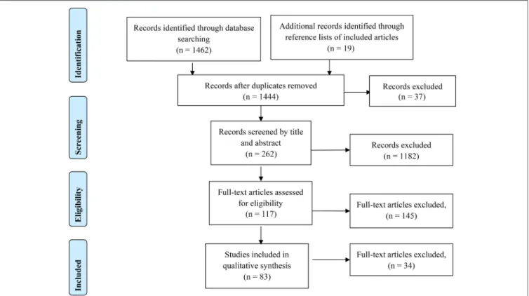
This systematic review utilized the Preferred Reporting Items for Systematic Reviews and Meta-Analyses (PRISMA; seeFigure 1)
approach (Liberati et al., 2009). The searches included detailed terms related to neuroimaging, static and dynamic balance control, and brain activity. The three search fields were connected with “AND” to ensure at least one term of each field could be found in the results. The terms in each of search field were linked with “OR.”
Search Terms
Neuroimaging search terms included variations of the following: fMRI, EEG, fNIRS, MEG, PET, and SPECT. Balance control search terms included variations of the following: Standing, balance, posture, gait, stepping, walking, plantarflexion, dorsiflexion, locomotion, and postural control. Brain activity search terms included variations of the following: Cortical activity, subcortical activity, neural activity, executive function.
Search Process
The primary information sources included in this review are: (1) IEEExplore and (2) Compendex, both to provide an engineering perspective, (3) ACM Library, to provide a computing and signal processing perspective, (4) PubMed, to provide a clinical perspective, and (5) Web of Science, to provide a cross disciplinary perspective. The database search included search terms found in the article title, abstracts, and keywords. The results from each database were added to Mendeley and checked for any duplicate results.
Inclusion and Exclusion Criteria
Search inclusion and exclusion were based on the characteristics and goals of the balance control tasks and the use and application of neuroimaging modalities. Literature included in this review aimed to investigate human balance control. Studies were included if they incorporated balance challenges (e.g., perturbations, eyes closed, dual-task, etc.). Only articles in English were considered. We excluded literature from this review when either the balance control paradigm or the neuroimaging paradigm did not align with the goals of this study. Studies that evaluate the effects of a therapy or intervention, including drug or hormone trials, rehabilitation, training, robotic-assisted walking, or development (in children and adolescents) were excluded. Studies that did not include active subject movement, such as those using motor imagery, brain volume correlation, or passive walking paradigms were excluded. Passive walking paradigms are those where the experimenter or specially designed equipment assisted the subject in moving their legs (Dobkin et al., 2004). Additionally, studies that use isolated joint movement and coordinated body movement (arms and legs) were excluded. Lastly, studies that aimed to evaluate technological advancements with no concurrent evaluation of human subjects were excluded.
Data Extraction
FIGURE 1 | PRISMA flow diagram of this review.
tasks. Studies in both groups that included methodologies for movement artifact identification and removal were pooled to address research question three. The subsequent data were then extracted from each article: Authors, year of publication, static or dynamic balance task, type of balance challenge (further categorized as sensory, mechanical or cognitive), treadmill or overground walking (for dynamic studies), subject characteristics, modality of brain imaging, and brain activity (including spatial, temporal, and/or frequency response). As well as extraction of pre-processing, spatial filtering, and artifact removal methods.
RESULTS AND DISCUSSIONS
Study Selection and Characteristics
Literature searches included relevant studies published on or before September 1, 2016, and resulted in 1,462 results, with 1,444 studies remaining after duplicate removal. After initial screening of the abstract, 117 studies were evaluated for inclusion in the static balance control or dynamic balance control category. The full-text articles for each of the studies was reviewed, a total of 38 studies were eligible for inclusion in the static balance control group and 47 for the dynamic group, with two studies including both static and dynamic balance control tasks. All 83 studies included in the static and dynamic groups were evaluated for inclusion in addressing research question three regarding identification and removal of movement artifact.Figure 1shows the PRISMA approach used in the present study.
Studies Selected for RQ1
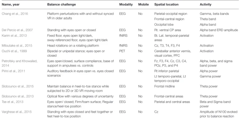
This review covered 38 studies investigating neural correlates of static balance control. The balance challenge paradigms include mechanical perturbations, cognitive, dual-task paradigms and sensory degradation, or impairment, which can be found in Tables 2–4, respectively.
Mechanical Challenges
Twenty-seven studies used mechanical perturbations to elicit a balance control response, 15 of which focused on the evoked cortical potentials (SeeTable 2). The negative potential occurring around 100 ms following an event, such as mechanical perturbations, is termed the N100 potential. The N100 response over the fronto-central area has been observed in a wide range of balance tasks, and N100 amplitude increases in challenging balance control tasks, including unpredictable or surprise perturbations, and in balance challenges with low sensory inputs (Adkin et al., 2006, 2008; Mochizuki et al., 2008; Huang et al., 2014; Varghese et al., 2014, 2015). However, Mochizuki et al. (2009a)observed no difference in N100 latency and amplitude in sitting and standing instability conditions, suggesting that there may be more general processes that underlie stability, regardless of sensory, motor, or postural aspects of response.
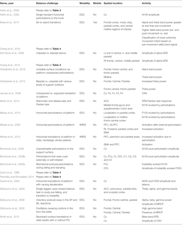
TABLE 2 | Brain activity due to mechanical challenges to static balance control.
Name, year Balance challenge Modality Mobile Spatial location Activity
Adkin et al., 2008 Please refer toTable 3
Adkin et al., 2006 Single transient horizontal perturbations to the trunk
EEG No Cz N100 amplitude
Bulea et al., 2014 Sit-to-stand transitions EEG Yes Frontal cortex, motor strip, parietal cortex, and central midline regions of interest
Alpha and theta band power greater at rest than pre-movement
Higher delta band power pre- and post-movement vs. rest
Classification of lower extremity movement intent based on pre-movement delta band signal
Chang et al., 2016 Please refer toTable 4
Del Percio et al., 2009 Unipedal vs. bipedal stance EEG No Lt and rt central, rt. and middle parietal
Amplitude of alpha ERD
Rt frontal, central, middle parietal Amplitude of alpha ERD
Huang et al., 2014 Please refer toTable 3
Hülsdünker et al., 2016 Unstable surface conditions via platform unexpected perturbations
EEG No Frontal, fronto-central, and fronto-parietal
Alpha band power
Midline Theta band power
Hülsdünker et al., 2015 Bipedal vs. unipedal with various levels of support surfaces
EEG No Frontal, Central, Parietal Increased theta power
Fronto-central, fronto-parietal Theta power
Jacobs et al., 2008 Unexpected vs. expected translation of platform
EEG No Cz, Pz, Fz, F3, F4 CNV
Marlin et al., 2014 Stand lean and release task and Flanker task
EEG No ACC ERN Flanker task response
Medial frontal gyrus and supplementary motor area
N100 evoked by perturbations
Mierau et al., 2015 Horizontal perturbations of platform EEG No Localization in parietal cortex P100 evoked by perturbations
Localization in midline fronto-central cortex
N100 evoked by perturbations
Mihara et al., 2008 Horizontal perturbations of platform fNIRS No PFC, DLPFC Activation after external perturbation
Rt. Posterior parietal cortex and SMA
Increased activation
Mihara et al., 2012 Horizontal translations of platform in older, hemiplegic stroke patients
fNIRS No PFC, premotor and parietal areas Increased activation due to perturbation
SMA and PFC Activation
Mochizuki et al., 2008 Unpredictable perturbations to the support surface
EEG No Cz N100 post-perturbation amplitude
Mochizuki et al., 2009b Perturbations that were cued externally or self-initiated
EEG No Cz, FCz, Fz, CPz, C1, C2, C3, and C4
N100 post-perturbation amplitude
Mochizuki et al., 2009a Mechanical postural perturbations during sitting and standing
EEG No FCz Instability evoked N100
CPz Amplitude of instability evoked P200
Ouchi et al., 1999 Please refer toTable 4
Petrofsky and Khowailed, 2014 Please refer toTable 4
Quant et al., 2005 Horizontal translations of platform with varying deceleration
EEG No Cz N200 and P200 amplitude and
latency
Slobounov et al., 2009 Single-legged, eyes-closed balance task to study pre-falling, and transition to instability
EEG No ACC, precuneus, parietal lobe, and occipital cortex
Theta, alpha, and gamma bands
Slobounov et al., 2008 Voluntary postural sway in the AP and ML directions
EEG No Frontal, Fronto-central, parietal Alpha, beta, gamma power
Amplitude of MRCP
Slobounov et al., 2005 Oscillatory swaying motions in the from the ankle
EEG No Frontal, Central High gamma band
Frontal, Central, Parietal Presence of MRCP
Smith et al., 2012 Backward surface translations in older adults with or without PD
EEG No Cz Beta band ERD
Cz Amplitude of CNV
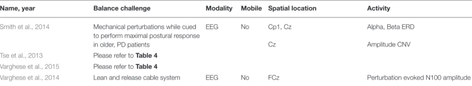
TABLE 2 | Continued
Name, year Balance challenge Modality Mobile Spatial location Activity
Smith et al., 2014 Mechanical perturbations while cued to perform maximal postural response in older, PD patients
EEG No Cp1, Cz Alpha, Beta ERD
Cz Amplitude CNV
Tse et al., 2013 Please refer toTable 4
Varghese et al., 2015 Please refer toTable 4
Varghese et al., 2014 Lean and release cable system EEG No FCz Perturbation evoked N100 amplitude
PD, Parkinson’s Disease; lt., left; rt., right; bi. lat., bilateral; PFC, prefrontal cortex; DLPFC, dorsolateral prefrontal cortex; SMA, supplemental motor area; ACC, anterior cingulate cortex; ERD, event related desynchronization; CNV, contingent negative variation; ERN, error related negativity potential; MRCP, movement related cortical potential.
to invoke a feet-in-place balance response [similar to those in: Adkin et al. (2006)andMochizuki et al. (2008)] and used dipole analysis to locate the N100 response. The ERN was localized to the ACC, as expected. However, the perturbation related N100 was localized to the medial frontal gyrus and supplementary motor area. These results indicate that perturbation evoked N100 is related to motor processes rather than a general error event.
Although there has been research in the early cortical response following perturbations, the role of later potentials is still unclear. In the auditory domain, for example, the event related potential (ERP) component around 200 ms post-stimulus (P200), represents a shift in attention toward the initial audio cue.Quant et al. (2005) used horizontal platform translations with quick or delayed deceleration to determine if P200 in the balance domain was related to a shift in attention or indicated sensory or motor processes. There was no difference in N200 and P200 amplitude or latency in immediate vs. delayed decelerations, suggesting that the motor reactions needed to maintain stability and later cortical responses are likely independent (Quant et al., 2005). Another event related potential is the contingent negative variation (CNV), a slow potential related to anticipatory attention, preparation, and motivation (Nagai et al., 2004). The CNV has been observed in response to unexpected surface perturbations in the midline and frontal areas (Jacobs et al., 2008) and to perturbations with an unexpected magnitude at Cz (Smith et al., 2012). Subjects in these studies are preparing for an unexpected surface perturbation, which explains the CNV response. However,Smith et al. (2014)provided amplitude cuing of the upcoming response in Parkinson’s Disease patients and did not observe a CNV response. Given that subjects successfully demonstrated balance control, these results indicate that another type of cortical response is involved in cued responses.
Thirteen EEG studies performed frequency analysis. As task difficulty increased (for example, decreased surface support due to standing on foam), alpha power in healthy controls decreased, indicating increased cortical activation (Petrofsky and Khowailed, 2014; Hülsdünker et al., 2016). Slobounov et al. (2008) observed a larger decrease in alpha power in central electrodes prior to sway in the medio-lateral direction compared to an anterior-posterior sway. While performance studies have found anterior-posterior sway magnitude and torque is larger than medio-lateral sway, these results suggest that cortical activity may be sway-direction dependent, with medio-lateral instability requiring more cortical control in self-initiated
postural movements. Lastly,Slobounov et al. (2009)saw a drop in alpha power in occipital region prior to a fall. However, when subjected to the challenging balance conditions, trans-tibial amputees exhibited an increase in alpha power (Petrofsky and Khowailed, 2014).Chang et al. (2016)challenged balance control of older adults in high and low fall risk groups by subjecting them to anterior-posterior motion via a Steward Platform. Two conditions were tested with and without a virtual-reality based scenery synchronized with the motion platform. Beta band power increased in the virtual-reality condition in the frontal and occipital regions for both groups.Petrofsky and Khowailed (2014) also observed an increase in beta power as balance challenges increased in central and parietal areas of both controls and amputee patients.Tse et al. (2013)also found increase in beta power with increase in balance challenge task (base of support or surface compliance) in the parietal and central areas.Smith et al. (2012)observed an increased in beta band event related desynchronization (ERD) at Cz for predictability predictable, small magnitude perturbations. However, in a similar paradigm with and without amplitude cuing,Smith et al. (2014)did not observe a significant difference in beta power between the two conditions. In comparing the demands of balance during medio-lateral and anterior-posterior sway,Slobounov et al. (2008)found that beta power in the central region dropped significantly more prior a self-initiated ML sway.
transitioned from sitting to standing posture, they were able to classify movement intent (i.e., if the subject was going to stand-up, sit-down, or remain at rest).
While neuroimaging analysis following EEG signal acquisition has high temporal resolution, it lacks spatial resolution. Therefore, PET and fNIRS have been used to investigate the spatial characteristics of a balance response. Ouchi et al. (1999) observed the hemodynamic response following bipedal or unipedal stance with eyes open or closed using PET, finding an increased activation of the cerebellar anterior vermis and posterior lobe lateral cortex during unipedal stance and increased activation of the cerebellar anterior lobe and right visual cortex during bipedal stance. The visual cortex and vermis were activated during standing with feet together with eyes on a target as well (Ouchi et al., 1999). These findings provide spatial insights into neural correlates of balance control, mainly that the cerebellar vermis and visual cortex may be involved in maintaining and regulating standing posture. Studies using fNIRS also reveal cortical involvement in balance control. Mihara et al. (2008) observed an activation of the prefrontal cortex (PFC) and dorsolateral prefrontal cortex (DLPFC) after anterior-posterior and medio-lateral horizontal perturbations accompanied by increased activation of the right posterior parietal cortex and supplementary motor area in conditions with auditory warning signals. Similarly, Mihara et al. (2012) observed activation of prefrontal cortex, premotor, and parietal areas following anterior-posterior and medio-lateral horizontal perturbations in older, hemiplegic stroke patients. These findings point to involvement of the prefrontal, premotor, supplementary motor, and parietal cortex in standing balance control.
Cognitive Challenges
Seven studies required subjects to maintain balance while performing a cognitive dual-task paradigm (SeeTable 3). Dual-task paradigms necessitate allocation of attentional resources to perform both the cognitive and balance tasks, and performance outcomes have correlated with the integrity of balance control.
A decrease in N100 amplitude was observed in visual working memory task and visuo-motor track task conditions (Quant et al., 2004; Little and Woollacott, 2015). Similarly,Huang et al. (2014) provided subjects with visual feedback to facilitate maintaining balance on a tilt platform, resulting in decreased N100 amplitude over the motor cortex and sensorimotor areas. These studies found a decrease in N100 in the dual-task conditions, indicating their efficacy to split attentional resources between balance and cognitive tasks. In contrast, Adkin et al. (2008) evoked an emotional response (fear; anxiety) by placing subjects at a prescribed height off the ground. An N100 response was observed prior to an unpredictable perturbation, and N100 amplitude increased with increasing height. Mirelman et al. (2014) had subjects stand still and perform serial seven subtractions, a common cognitive loading task, resulting in increased activation in the left and right frontal lobe compared to single-task standing. Fujita et al. (2016) also observed increased brain activity in a cognitive DT paradigm using fNIRS. High and low memory span groups performed one or two leg standing with a Stroop task. Increased right dorsolateral prefrontal cortex activation was
observed in the high span group in both one and two leg standing dual-task conditions, compared to the low span group. Also, performance in both groups decreased during one leg, dual-task standing. Lastly,Lau et al. (2014)compared effective connectivity during standing or walking with or without a visual oddball discrimination response task. They found that connectivity was weaker during standing when performing a cognitive task in the prefrontal cortex, posterior parietal cortex, and ACC. However, effective connectivity was stronger in the standing conditions compared to the walking condition regardless of cognitive task. These findings suggest that more cognitive resources may be required to maintain standing posture, as compared to walking (Lau et al., 2014).
Sensory Challenges
Experimentally manipulating sensory inputs (vestibular, visual, or proprioceptive) is another method to challenge balance control. Eleven studies used a form of sensory removal or augmentation, including eyes closed conditions and the use of virtual reality (SeeTable 4). For example, subjects stood with eyes closed and feet together or in tandem (i.e., heel-to-toe), which is more challenging, and N100 was evoked prior to the need for a balance reaction. The N100 amplitude increased with increasing postural challenge (Varghese et al., 2015).
Chang et al. (2016)utilized frequency analysis and observed a modulation of alpha, beta, gamma, and theta band power in response to balance challenge in a virtual reality environment, particularly an increase in alpha power in occipital lobe in the virtual reality condition in both groups. Del Percio et al. (2007)also observed and larger amplitude of alpha band ERD in athletes in a closed-eye one and two leg balance task, compared to non-athletes. In contrast, Pirini et al. (2011) observed a decrease in alpha power during eyes-open task in the right inferior parietal area, which is in line with the increased attention required to perform a balance under degraded conditions. Petrofsky and Khowailed (2014) also observed an increase in activation with increased balance challenge, finding an overall signal power increased with a decrease in the amount of qality of sensory feedback. Likewise, Tse et al. (2013)observed an increase in beta and sigma bands in parietal and central areas with a more difficult balance challenge.
Using fNIRS,Karim et al. (2013)found increased activation of the bilateral temporal-parietal areas when both vision and perceptual information were degraded (eyes closed, swaying floors).Mitsutake et al. (2015) aimed to induce instability by requiring subjects to rotate their heads while on a rotating platform. Using fNIRS, they found that activation at the central, frontal and temporal cites were not significantly different during high or low speed rotations despite these differences in stability. This may indicate different types of instability may differentially affect cortical or spinal control mechanisms.
Studies Selected for RQ2
TABLE 3 | Brain activity due to cognitive challenges to static balance control.
Name, year Balance challenge Modality Mobile Spatial location Activity
Adkin et al., 2008 Perturbation under postural threat EEG No Cz Perturbation evoked N100
amplitude
Fujita et al., 2016 Stroop task during bipedal vs. unipedal standing
fNIRS No Rt. DLPFC Increased activation
Huang et al., 2014 Tilt platform using visual feedback EEG No Bi. lat. fronto-central and contralateral sensorimotor areas
Latency and amplitude of N100 for postural control
Bi. lat. fronto-central and ipsilateral temporal areas
Latency and amplitude of P200 for postural control
Lt. frontal-central area MRP for postural control
Lau et al., 2014 Visual oddball response task while standing or walking
EEG No Sensorimotor cortex Effective connectivity
PFC, posterior parietal cortex, ACC Effective connectivity
Little and Woollacott, 2015
Visual WM capacity during surface perturbations and walking
EEG No Lt. pre-motor and rt. sensory areas Amplitude of N100 ERP
Mirelman et al., 2014 Walking while counting forward, walking with serial 7’s and serial 7’s in standing
fNIRS No Fp1 and Fp2 Increased activation with
increased task difficulty
Quant et al., 2004 Horizontal translations platform with or without a visuomotor track task
EEG No Cz Amplitude of perturbation
evoked N100
lt., left; rt., right; bi. lat., bilateral; PFC, prefrontal cortex; ACC, anterior cingulate cortex; MRP, movement related potential.
TABLE 4 | Brain activity due to sensory challenges to static balance control.
Name, year Balance challenge Modality Mobile Spatial location Activity
Chang et al., 2016 Platform perturbations with and without synced VR in older adults
EEG No Parietal-occipital region Gamma, beta bands
Frontal-central region Theta band
Occipital lobe Alpha band
Del Percio et al., 2007 Standing with eyes open or closed EEG No Rt. ventral CP area Alpha band ERD amplitude
Karim et al., 2013 Fixed floor, eyes open light/dark, sway-referenced floor, eyes open light/dark
fNIRS No Bi. Lat. temporal-parietal areas
Activation
Mitsutake et al., 2015 Head rotations on a rotating platform fNIRS No Cz, T3, T4, F3, F4 Activation
Ouchi et al., 1999 Bipedal or unipedal stance; eyes open or closed
PET No Cerebellar anterior vermis, visual cortex, PFC
Activation
Petrofsky and Khowailed, 2014
Eyes open/closed, surface compliance, base of support in amputees vs. controls
EEG No Fz, F3, F4, Cz, C3, C4, POz, P3, and P4
Alpha, beta, and sigma band power
Pirini et al., 2011 Auditory feedback in eyes open vs. eyes closed scenarios
EEG No Rt inferior parietal Alpha power
Lt temporo-parietal, Lt temporo-occipital
Gamma power
Slobounov et al., 2015 Maintain balance in heel-to-toe stance while subjected to 2D or 3D VR moving room
EEG No Frontal midline Theta power
Slobounov et al., 2013 Optical flow with various degrees of uncertainty EEG No Frontal-central areas Theta power
Tse et al., 2013 Eyes open/ closed; Firm/foam surface; Regular stance/heel-toe position
EEG No Parietal and central areas Beta and Sigma band power
Varghese et al., 2015 Standing with eyes closed and feet together or feet heel-to-toe position
EEG No Cz Amplitude of N100 evoked
prior to balance reaction
TM, treadmill; OG, overground; VR, virtual reality; lt., left; rt., right; bi. lat., bilateral; ERD, event related desynchronization.
been thoroughly reviewed byHamacher et al. (2015), this review only includes the remaining 32 studies investigating brain activity under challenges to dynamic balance control. The balance challenge paradigms include mechanical perturbations, the use of cognitive dual-tasks, and the experimental manipulation of sensory inputs. Results can be found in Tables 5–7, respectively.
Mechanical Challenges
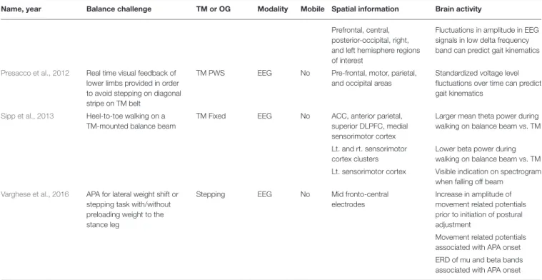
TABLE 5 | Brain activity due to mechanical challenges to dynamic balance control.
Name, year Balance challenge TM or OG Modality Mobile Spatial information Brain activity
Beurskens et al., 2016 ST vs. DT: Motor or cognitive interference
TM PWS EEG Yes FCz Alpha band activity decreased
during motor DT vs. ST
FPz, Fz Beta increased during motor vs. cognitive DT
Bradford et al., 2015 TM walking at specified levels of incline
TM Fixed EEG No Sensorimotor, posterior parietal, ACC clusters
Higher theta power fluctuations across gait cycle in inclined walking conditions
Lt. sensorimotor, ACC clusters
Greater gamma power during level walking
Lt. and rt. sensorimotor cluster
Distinct alpha and beta fluctuations dependent on gait cycle for both walking conditions
Bruijn et al., 2015 Laterally stabilized while TM walking
TM Fixed EEG No Bilateral premotor cortices Higher beta power during stabilized walking in left premotor area specifically around push-off
Bulea et al., 2015 Steady state walking using an active or a passive TM
TM: Fixed vs. feedback driven
EEG Yes PFC and posterior parietal cortex
Low gamma band power increased during double support and early swing phases in active TM
Sensorimotor cortex Mu and beta band
desynchronization during walking cycle
Clark et al., 2014b Carrying tray, obstacles, and weighted vest tasks while walking in older adults
OG fNIRS No PFC Increased activation in walking
phase
Haefeli et al., 2011 Obstacle navigation in dim lighting with audio cue to signal upcoming obstacle
TM Fixed EEG No Oribital gyrus (BA 11) and medial frontal gyrus (BA 10)
Activation in preparation phase prior to stepping over obstacle
Superior frontal gyrus (BA 9) Activation in performance phase
Jaeger et al., 2016 External load applied during stepping movements
Stepping fMRI No SMA-proper (BA4a), superior occipital gyrus (BA 18)
Activation in 0 load condition
Vermis, S1/M1 (left BA 6), Thalamus
Activation in 20 load condition
Insula, vermis, middle occipital gyrus, precuneus S2, thalamus, sup occ. gyrus
Activation in 40 load condition
Kurz et al., 2012 Forward vs. backward walking on TM
TM Fixed fNIRS No SMA, pre-central gyrus, sup. parietal lobule
Increased activation in backward walking
Pre-central gyrus and SMA Maximal activation correlated with stride-time intervals in forward walking
Lin and Lin, 2016 Overground walking with wide, narrow, or obstacle path with and without n-back task
OG fNIRS No PFC Increased activation at beginning
of task
Lu et al., 2015 Please refer toTable 6
Maidan et al., 2015 Walking patterns known to cause FoG in PD patients with FoG and healthy controls
OG fNIRS No Frontal activation (BA 10) Decreased activation during turns without FoG episode in PD group
Increased activation during anticipated turns before and during FoG episode
No changes in activation in controls
Presacco et al., 2011 Real time visual feedback of lower limbs provided in order to avoid stepping on diagonal stripe on TM belt
TM PWS EEG No Full scalp analysis Higher delta, theta, and low beta spectral power during walking vs. rest
TABLE 5 | Continued
Name, year Balance challenge TM or OG Modality Mobile Spatial information Brain activity
Prefrontal, central, posterior-occipital, right, and left hemisphere regions of interest
Fluctuations in amplitude in EEG signals in low delta frequency band can predict gait kinematics
Presacco et al., 2012 Real time visual feedback of lower limbs provided in order to avoid stepping on diagonal stripe on TM belt
TM PWS EEG No Pre-frontal, motor, parietal, and occipital areas
Standardized voltage level fluctuations over time can predict gait kinematics
Sipp et al., 2013 Heel-to-toe walking on a TM-mounted balance beam
TM Fixed EEG No ACC, anterior parietal, superior DLPFC, medial sensorimotor cortex
Larger mean theta power during walking on balance beam vs. TM
Lt. and rt. sensorimotor cortex clusters
Lower beta power during walking on balance beam vs. TM
Lt. sensorimotor cortex Visible indication on spectrogram when falling off beam
Varghese et al., 2016 APA for lateral weight shift or stepping task with/without preloading weight to the stance leg
Stepping EEG No Mid fronto-central electrodes
Increase in amplitude of movement related potentials prior to initiation of postural adjustment
Movement related potentials associated with APA onset
ERD of mu and beta bands associated with APA onset
TM, treadmill; OG, overground; PW, preferred walking speed; ST, single task; DT, dual task; APA, anticipatory postural adjustment; PD, Parkinson’s Disease; FoG, freezing of gait; BA, Brodmann Area; lt., left; rt., right; bi. lat., bilateral; PFC, prefrontal cortex; DLPFC, dorsolateral prefrontal cortex; ACC, anterior cingulate cortex; SMA, supplementary motor area; ERD, event related desynchronization.
walking with a weighted vest. Prefrontal cortex (PFC) activation increased prior to walking for all three challenges compared to steady state walking. Increased prefrontal cortex activation during the weighted vest and obstacle conditions was also observed. Lu et al. (2015) also had subjects carry an object while walking and observed an increase in prefrontal cortex activation during task initiation, an increase in supplementary motor area (SMA) and premotor cortex (PMC) activation during the task. In addition, an increase in premotor cortex and supplementary motor area activation correlated with decreased gait performance.
Using a truly mobile fNIRS hardware system contained in a backpack, subjects walked over narrow, wide, or obstacle path conditions (Lin and Lin, 2016). At the beginning of each trial, regardless of path condition, average prefrontal cortex activation was higher and had larger variability compared to the end of the trial. Lastly, in healthy controls and Parkinson’s Disease patients with freezing of gait (FoG),Maidan et al. (2015) monitored frontal activation during walking patterns known to cause FoG episodes. In the Parkinson’s patients, frontal activation was decreased during turns without FoG episodes but increased during anticipated turns before and during FoG episodes. In the healthy controls, there was no change in frontal activation during turns.
Eight studies utilized EEG paradigms to investigate mechanical challenges to dynamic balance control. Bradford et al. (2015)required subjects to walk on a treadmill at specified inclines and observed theta power fluctuations that increased at
TABLE 6 | Brain activity due to cognitive challenges to dynamic balance control.
Name, year Balance challenge TM or
OG
Modality Mobile Spatial information Brain activity
Al-Yahya et al., 2016 ST vs. DT (counting) walking in adults with chronic stroke
TM PWS fNIRS No PFC Increased activation in DT for both
groups
Beurskens et al., 2014 Walking with visual or verbal memory task in young and elderly adults
TM PWS fNIRS No PFC Decreased activation in DT (visual)
in elderly group
PFC Little change in PFC activation in DT in young group
Beurskens et al., 2016 ST vs. DT: motor or cognitive interference
TM PWS EEG Yes Cz Decreased alpha activity during
cognitive DT
FCz, Cz Decreased beta activity decreased during cognitive DT
Clark et al., 2014b Verbal task while walking in older adults
OG fNIRS No PFC Increased activation during walking
phase
De Sanctis et al., 2014 Evaluate walking load on response inhibition with Go/No-Go Task
TM Fixed EEG No O1/Oz/O2 Increase in P200 amplitude
between sitting and walking
FCz, Cz, CPz Reduction in N200 amplitude during walking vs. sitting
CPz P300 amplitude reduced for
walking
FCz, Cz P300 increased amplitude, reduced latency at higher walking speed
Doi et al., 2013 DT walking using verbal letter fluency task in older adults
OG fNIRS No PFC Increased activation during DT
walking
Holtzer et al., 2011 WWT DT in young and old adults OG fNIRS No PFC Increased activation in WWT
compared with ST walking
Greater activation in young vs. old group in DT condition
Holtzer et al., 2015 WWT DT in older adults OG fNIRS No PFC Increase in activation during WWT
condition
Holtzer et al., 2016 WWT DT in adults with and without neurological gait abnormalities
OG fNIRS No PFC Increased activation during WWT
Huppert et al., 2013 Lateral stepping based on congruent or incongruent information
Stepping fNIRS No BA 46, BA 6, BA 4 Increased activation in incongruent trials
Kline et al., 2014 Brooks spatial WM task at multiple speeds
TM Fixed EEG No Somatosensory
association cortex
Alpha power increased prior to stimulus presentation
Alpha power decreased during memory encoding
Rt. superior parietal lobule and posterior cingulate cortex
Theta power decreased around memory encoding
Lau et al., 2014 Respond to target while sitting or walking on TM, with or without cognitive DT
TM Fixed EEG No Sensorimotor Cortex Effective connectivity weaker for walking than standing regardless of cognitive task
PFC, posterior parietal cortex, ACC
Connectivity stronger for walking than standing only in cognitive DT condition
Lin and Lin, 2016 Please refer toTable 5
Lu et al., 2015 Walking with motor task (carry water on tray) or cognitive task (subtraction)
OG fNIRS Yes Left PFC Increase in activation during
preparation of DT conditions, maintained activation during cognitive task.
SMA and PMC Increased activation during both DT conditions
TABLE 6 | Continued
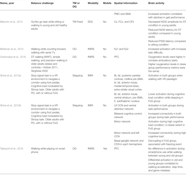
Name, year Balance challenge TM or
OG
Modality Mobile Spatial information Brain activity
PMC and SMA Increased activation correlated with declines in gait performance
Malcolm et al., 2015 Go/No-go task while sitting or walking in young and old healthy adults
TM Fixed EEG No Cz, FCz, and CPz Decreased N200 amplitude for DT condition in young adults
Reduced N200 latency for DT condition compared in young adults
Reduced P300 latency compared to sitting condition
Mirelman et al., 2014 Walking while counting forward, walking with serial 7’s
OG fNIRS No Fp1 and Fp2 Increased activation with increased task difficulty.
Osofundiya et al., 2016 DT walking (WWT), simple walking, and precision walking in older adults (obese and controls)—Holtzer 2011, Verghese 2002
OG fNIRS No PFC Oxygenation levels were higher in
complex ambulatory tasks Higher oxygenation levels in obese group (performance metrics were the same)
Shine et al., 2013a Stop-signal task in a VR environment to navigate a corridor using foot pedals. Cognitive load modulated by Stroop task. Older adults with PD, with or without FoG
Stepping fMRI No Bi. lat. posterior parietal cortices, midline pre-SMA, bi. lat. anterior insula, medial temporal lobes, extra-striate visual cortex
Activation in both groups when walking with VR paradigm
Bi. lat. anterior insula, ventral striatum, pre-SMA, lt. subthalamic nucleus
Lower activation during cognitive load condition while stepping in FoG group
Shine et al., 2013b Stop-signal task in a VR environment to navigate a corridor using foot pedals. Cognitive load modulated by Stroop task. Older adults with PD, with or without FoG
Stepping fMRI No Lft CCN and ventral attention network
Activation in both groups during task performance
Bilateral cognitive control network
Increased connectivity in both groups during task performance
Motor network Activation during high cognitive load condition, to lesser extent in FoG group
Motor network and left CCN
Increased connectivity during high cognitive load
Basal ganglia network and CCN in each hemisphere
Decoupling in FoG group, associated with freezing event
Takeuchi et al., 2016 Walking while playing on smart phone
OG fNIRS Yes PFC No difference in activation during
smartphone use while walking between young and old groups
Differential activation in old and young groups correlated to walking acceleration, step time, and game mistakes
TM, treadmill; OG, overground; PWS, preferred walking speed; ST, single task; DT, dual task; WWT, walking while talking; WM, working memory; VR, virtual reality; PD, Parkinson’s Disease; FoG, freezing of gait; BA, Brodmann Area; lt., left; rt., right; bi. lat., bilateral; PFC, prefrontal cortex; ACC, anterior cingulate cortex; SMA, supplementary motor area; CCN, cognitive control network.
signal amplitude was enhanced in the orbital gyrus and medial frontal gyrus, the area of the brain responsible for processing of environmental stimuli. Also, during the performance phase (stepping over the obstacle) enhanced EEG signal amplitude was observed in the superior frontal gyrus, the brain area for monitoring motor performance. Presacco et al. (2012) also observed differential brain activity during different phases of the walking cycle. Utilizing EEG signal captured while subjects walked on a treadmill using visual feedback to avoid stepping on a white line on the treadmill belt, the authors found that the
TABLE 7 | Brain activity due to sensory challenges to dynamic balance control.
Name, year Balance challenge TM or OG Modality Mobile Spatial
information
Brain activity
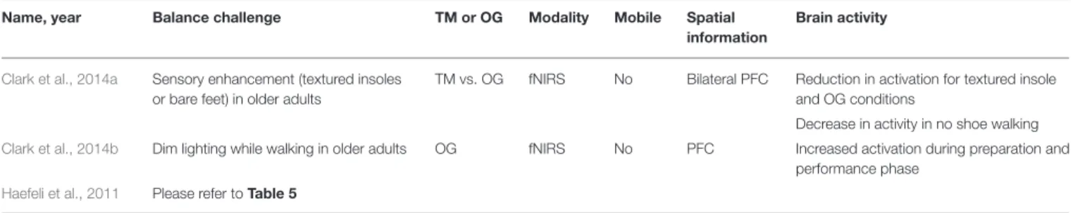
Clark et al., 2014a Sensory enhancement (textured insoles or bare feet) in older adults
TM vs. OG fNIRS No Bilateral PFC Reduction in activation for textured insole and OG conditions
Decrease in activity in no shoe walking
Clark et al., 2014b Dim lighting while walking in older adults OG fNIRS No PFC Increased activation during preparation and performance phase
Haefeli et al., 2011 Please refer toTable 5
TM, treadmill; OG, overground; PFC, prefrontal cortex.
during stepping in the fronto-central cortical areas. However, prior to the APA and during lateral weight shift, there was no difference in brain activity, suggesting cortical activity involved in predictive balance control is independent of context (weight shift vs. lateral step).
One study utilized an fMRI paradigm, applying various external loads during stepping movements in an fMRI to simulate ground reaction forces experienced during real walking (Jaeger et al., 2016). In the zero unloaded condition, activation was observed in the supplementary motor area, superior occipital gyrus. When 20% of the subject’s body weight was applied, primary somatosensory cortex (S1)/Primary motor cortex (M1), vermis, and thalamus activation was observed. Lastly, at a higher load condition (40% of body weight), activation was seen in the insula, vermis, middle occipital gyrus, precuneus secondary somatosensory cortex (S2), thalamus, superior occipital gyrus.
Cognitive Challenges
Twenty studies used cognitive dual-task paradigms to challenge dynamic balance control (See Table 6). Shine et al. (2013a,b) used fMRI-compatible steppers and a virtual reality hallway to evaluate walking in Parkinson’s disease patients with and without FoG. Shine et al. (2013a) found activation of the cognitive control network (bilateral posterior parietal cortices, midline pre-supplementary motor area, bilateral anterior insula, medial temporal lobes, extra-striate visual cortex) in Parkinson’s disease patients with and without FoG while walking. However, when cognitive load was modulated by a Stroop task, the FoG group had lower activation of the anterior insula, ventral striatum, pre-supplementary motor area, and subthalamic nucleus in FoG group compared to non-FoG group.
Shine et al. (2013b)found that Parkinson’s patients, both with and without FoG, used left cognitive control network (CCN) (left prefrontal cortex, dorsolateral prefrontal cortex, ventrolateral prefrontal cortex) and ventral attention network (anterior insula, dorsal cingulate) and exhibited increased connectivity between the bilateral cognitive control networks. There was increased connectivity of the motor network (pre and post-central gyrus, right supplementary motor area) and left CCN when walking with high cognitive load. Lastly, there was decoupling of the basal ganglia network (caudate, rostral cingulate) and CCN in each hemisphere in the FoG group which were associated with the occurrence of motor arrests, indicating impaired communication between these networks in FoG.Shine et al. (2013a,b)observed
activation of brain regions in the locomotor pathways. However, they also observed activation and connectivity with attention and cognitive control networks required to perform cognitive tasks. Additionally, impaired connectivity seen in FoG provides evidence toward the use of compensation mechanisms and/or additional brain regions in walking in those with neurological disorders.
in prefrontal cortex activation from single task to dual-task conditions.
More complicated paradigms have also been used to investigate brain activity in walking or stepping.Huppert et al. (2013) required subjects to step left or right according to incongruent information (conflicting location and direction of arrow on screen) or congruent information (location and arrow matched), and found increased activation in dorsolateral prefrontal cortex, premotor cortex, supplemental motor area, and precentral gyrus in incongruent trials, which require more attentional control.Lin and Lin (2016)had subjects walk over ground on wide, narrow, or obstacle pathways and with or without a cognitive load (n-back task), and found a decrease in prefrontal cortex activation at higher cognitive loads, similar to Beurskens et al. (2014)andShimada et al. (2013). Lastly,Takeuchi et al. (2016)observed no difference in prefrontal cortex activation between young and old groups while they played a game on their smartphone while walking. However, differential activation patterns in the right, left, and middle prefrontal cortex were observed to be correlated with gait speed, game mistakes, and step timing in the old and young groups.
Although these fNIRS studies investigate a range of cognitive dual-task conditions using both over ground and treadmill balance paradigms, there is no agreement on the activation of prefrontal cortex, supplementary motor area, and premotor cortex during dual-task conditions. Some studies found an increase in activation in dual-task conditions (Holtzer et al., 2011; Doi et al., 2013; Clark et al., 2014b; Al-Yahya et al., 2016), while others found a decrease (Shimada et al., 2013; Beurskens et al., 2014; Lin and Lin, 2016). In addition, although aging negatively affects balance control, Takeuchi et al. (2016) did not find a difference in prefrontal cortex activation between young and older adults while playing a game and walking.
Seven studies utilized EEG paradigms to investigate the impact of cognitive challenges on dynamic balance control. In a dual-task condition with a cognitive interference task, Beurskens et al. (2016)found a decrease in alpha band power over the Cz electrode and beta activity over the FCz and Cz electrodes compared to normal walking. However, beta band power increased during the cognitive dual-task over the FPz and Fz electrodes.Kline et al. (2014)had young healthy subjects perform a Brooks spatial working memory (WM) task at different treadmill speeds and found that there was an increase in alpha power prior to stimulus presentation followed by a decreased in alpha power during memory coding in the somatosensory association cortex. A decrease in theta power around memory encoding in the right superior parietal lobule and posterior cingulate cortex was also observed. Additionally, BA 7, BA 31, BA 5, and 6 exhibited power fluctuations time-locked to memory encoding during cognitive tasks. However, no distinct changes in brain activity or working memory task performance were observed due to different walking speeds.De Sanctis et al. (2014) evaluated the impact of walking on a go/no-go task that requires response inhibition in young adults, finding an increase in P200 amplitude in walking conditions (occipital lobe). A reduced N200 amplitude (at FCz, Cz, and CPz) and P300 amplitude (at CPz) was observed on trials requiring inhibition
while walking as compared to sitting. Increased P300 amplitude and decreased latency was observed in faster walking conditions (at FCz). These differences in ERPs were not accompanied by performance decrements in dual-task conditions, suggesting the use of compensation mechanisms to appropriately allocate attentional resources to perform both tasks (De Sanctis et al., 2014).Malcolm et al. (2015)also used a go/no-go task in young and older adults while walking or sitting, finding a decreased N200 amplitude for dual-task condition and earlier P300 latency over the Cz, FCz, and CPz electrodes in young adults, similar toDe Sanctis et al. (2014). Young adults exhibited a decreased N200 latency, while older adults showed an increase in P300 amplitude, indicating that older adults may have less flexibility of resource allocation in multi-task conditions (De Sanctis et al., 2014). Lau et al. (2014) investigated effective connectivity of the sensorimotor cortex and non-sensorimotor areas of the brain while subjects stood or walked on a treadmill with or without a cognitive challenge. They found that the effective connectivity of sensorimotor cortex was weaker for walking than standing, regardless of dual-task condition, which may point to a more automatic nature of walking execution. However, effective connectivity of non-sensorimotor areas (prefrontal cortex, posterior parietal cortex, and ACC) was stronger in the cognitive dual-task condition for walking than standing.
Sensory Challenges
The three studies investigating sensory challenges to balance control all observed modulation in prefrontal cortex activity (Haefeli et al., 2011; Clark et al., 2014a,b) (See Table 7). In a dimly lit walking environment, there was an increase in prefrontal cortex activity during preparation and performance of the walking task compared to a regularly lit condition (Clark et al., 2014b). While it has been shown that steady-state walking in older populations is less automatic and increases the need for attentional control and cognitive processes compared to younger populations,Clark et al. (2014a)had older adults with mild mobility deficits walk and mild somatosensory deficits walk on a treadmill and over ground while wearing normal shoes, and under enhanced somatosensory conditions wearing textured insoles, and with no shoes. There was a reduction in prefrontal cortex activity when subjects wore textured insoles compared to normal shoes for both treamill and overground walking. Also, walking with no shoes reduced prefrontal cortex activity for treadmill walking only compared to walking with normal shoes. Enhancing somatosensory information led to a reduction in prefrontal activation compared to control conditions, suggesting a more automatic process is being used perform the walking task. In contrast, under degraded visual conditions,Clark et al. (2014b) and Haefeli et al. (2011) observed increased prefrontal cortex activity.
Studies Selected for RQ3
Movement Artifact Identification and Removal
TABLE 8 | Neuroimaging studies investigating feasibility EEG signal acquisition during dynamic balance control tasks.
Name, year Balance challenge TM or OG Modality Mobile Spatial information Analysis
Gramann et al., 2010 Visual oddball response task while
standing/walking
TM Fixed EEG No (tether) Fz, Cz, Pz Identification of P300 and N100 amplitudes due to visual oddball stimulus
Gwin et al., 2010 Visual oddball task while walking or running
TM Fixed EEG No Mediofrontal clusters Identification of gait-related artifact in ERP during running
Oliveira et al., 2016 Auditory oddball task seated and walking
TM Fixed EEG Yes Fpz, F3, Fz, F4, C3, Cz, C4, P3, Pz, P4, O1, O2
Epoch rejection rate, pre-stimulus noise, signal-to-noise ratio, P300 amplitude
Lau et al., 2012 Response to target stimulus while walking
TM Fixed EEG No Global Identification of P300 response
in walking condition
Castermans et al., 2014 Barefoot walking TM Fixed EEG No Cz, Oz, T8 Harmonics in accelerometer and
EEG signals (delta, theta, alpha bands)
Snyder et al., 2015 Walking at set speeds with silicone cap
TM Fixed EEG No Global Movement artifact remains in
EEG signal following ICA and dipole fitting
Kline et al., 2015 Walking at set speeds with silicone cap
TM Fixed EEG Yes E12, A19, G11, C19, A1
Movement artifact varies with speed, subject and electrode location
Nathan and Contreras-Vidal, 2016
Walking at set speed TM Fixed EEG Yes Cz, Oz, T8 No large amplitude spikes in spectral signals corresponding stepping frequency
(accelerometer signal)
Strong wavelet coherence between delta band and accelerometer for higher walking speeds.
TM, treadmill; ERP, event related potential; ICA, Independent Component Analysis.
an ERP. In the walking condition, the ERP was nearly identical before and after movement artifact removal. However, in the running condition, the ERP was only identifiable after using a template regression artifact removal process. Gramann et al. (2010)also investigated the feasibility of using more mobile EEG hardware (a single tether connected electrodes to equipment) while subjects were walking or standing. The authors were able to identify the P300 and N100 ERP components evoked by a visual oddball paradigm over the Cz, Fz, and Pz electrodes. Oliveira et al. (2016) aimed to establish a protocol to validate the efficacy of EEG hardware and software systems in mobile neuroimaging using an auditory oddball paradigm in sitting and walking conditions. The authors recommended using epoch rejection rate, pre-stimulus noise, signal-to-noise ratio, and amplitude variance across the P300 event window to evaluate EEG hardware systems and artifact identification and removal efficacy in walking studies. In a different approach, Lau et al. (2012)used a weighted phase lag index across all channels and recovered a P300 response to a target stimulus while subjects stood or walked on a treadmill.
Four studies have focused on characterizing the walking related movement artifact. Castermans et al. (2014) used barefoot walking on a treadmill while collecting both EEG and accelerometer signals. The accelerometer was mounted on the subject’s head and showed increased activity corresponding
to completely remove movement artifact during walking. In contrast to the focus ofKline et al. (2015)andSnyder et al. (2015) on movement artifact only,Nathan and Contreras-Vidal (2016) analyzed the EEG signal and its relation to an accelerometer signal which was collected while subjects walked at various speeds. In this study, there were no large spectral amplitude spikes corresponding to the stepping frequency, which was obtained from the accelerometer signal. This finding refutes the analysis inCastermans et al. (2014). Wavelet coherence analysis revealed strong coherence between the EEG signal within the delta band and the accelerometer for higher walking speeds, which suggests the emergence of movement artifact within the EEG signal. However, after artifact subspace reconstruction, an automated artifact rejection process that uses baseline data and principal component analysis to remove transient and high amplitude artifact (i.e., muscle movements), this delta band coherence was not observed in the average subject data. This study suggests that movement artifact may not contaminate EEG data collected at slower walking speeds when using mobile neuroimaging equipment (Nathan and Contreras-Vidal, 2016).
Signal Processing of fNIRS Data
Table 9 summarizes the pre-processing and movement artifact removal methods of 20 studies using fNIRS for neuroimaging. Two studies used wavelet-minimum description length de-trending algorithms to remove global trends including artifacts due to respiration, heartbeat, and vasoconstriction. Five studies used principal (PCA) or independent (ICA) component analysis, or their combination, to remove environmental and equipment noise and signal drift. However, the majority of these studies did not use computational methods to remove artifacts from the acquired signal.
Signal Processing of EEG Data
Table 10 summarizes the pre-processing, spatial filtering, and movement artifact identification and removal methods used by the 54 EEG studies included in this review. For data pre-processing, all studies except three reported using high-pass, low-pass, or bandpass filters to exclude certain frequency ranges. Gwin et al. (2010)described criteria for removing noisy channels prior to data analysis: Standard deviation (SD) >1,000 µV,
kurtosis>5SDfrom mean, or poor correlation (r<0.4) with
neighboring channels for>1% of time samples. Ten additional
studies used these criteria for removing noisy channels prior to further analysis. Twenty-five studies used spatial filtering techniques, including Laplacian estimation (3), referencing to a common average (21), and bi-lateral referencing (1).
The literature reports a multitude of techniques to identify and remove ocular, muscular, and/or movement artifact during dynamic balance tasks, ranging from visual inspection (10) to sophisticated computational techniques. Petrofsky and Khowailed (2014) and Tse et al. (2013) used logistic discrimination function analyses tuned using an EEG database from sleep-deprived adults to identify eye blink regions, which were subsequently replaced by mean wavelet coefficients from nearby non-contaminated regions (Berka et al., 2007). Three studies defined and rejected time periods having substantial
artifact using a criterion (>0.8) on the z-transformed power
across all channels (Gwin et al., 2011; Sipp et al., 2013; Lau et al., 2014). Gwin et al. (2011) presented a moving average artifact removal method for EEG data collected during walking. Here, time-warped strides were averaged before and after a specific foot strike event. Then this time-warped average stride data was subtracted from the new data for a current stride. This was also used inKline et al. (2015)explored the combination of this moving average approach with a wavelet technique that removed signal content at frequencies below 8 Hz and applied the wavelets to the whole stride using Daubechies four wavelets.
However, approximately half the studies (i.e., a total of 23) used conventional ICA or adaptive mixture ICA (AMICA) to identify and remove artifacts from EEG data collected during walking.Hülsdünker et al. (2015)andHülsdünker et al. (2016) identified and removed ocular or muscular artifacts based on the cortical mapping, frequency spectrum, and time course components using ICA. Bruijn et al. (2015) described the following criteria for categorizing and removing components not associated with brain activity: Muscle artifact (50–100 Hz mean power larger than that in the beta and/or alpha bands), eye-blink artifacts (median frequency<3 Hz and the topo map
corresponded to eye components), and movement artifacts (frequency spectrum at the harmonics of stride frequency). After performing ICA or AMICA, several studies also used a source localization algorithm, DIPFIT, to refine artifact identification and removal.Gramann et al. (2010)evaluated each independent component for location within the head model and evaluated the residual variance between the scalp projection through a head model and scalp map.Gwin et al. (2011)used a similar approach but excluded components if the current dipole model to scalp accounted for less than 80% of the scalp variance - criteria used by six other studies (Sipp et al., 2013; Kline et al., 2014; Lau et al., 2014; Bradford et al., 2015; Bulea et al., 2015; Snyder et al., 2015). Lastly, 6 studies used PCA to identify and remove movement artifact (Bulea et al., 2014, 2015; Huang et al., 2014; Marlin et al., 2014; Luu et al., 2016; Nathan and Contreras-Vidal, 2016).
GENERAL DISCUSSION AND
CONCLUSION
This study reviewed 83 articles using neuroimaging modalities to investigate the neural correlates underlying static and dynamic human balance control, with aims to support future mobile neuroimaging research in the balance control domain. Images demonstrating the use of mobile neuroimaging modalities integrated with a dynamic balance control tasks can be found in Kline et al. (2015). Likewise, examples of study paradigms using non-mobile neuroimaging modalities within this domain can be found inChang et al. (2016)andGramann et al. (2010).
TABLE 9 | Signal processing and artifact removal methods for fNIRS studies.
Name, year Pre-processing Removal of movement artifact
Al-Yahya et al., 2016 Low-pass filter at 0.67 Hz cutoff frequency
Beurskens et al., 2014 Gaussian/Hemodynamic response function lowpass filter Wavelet-minimum description length algorithm
Caliandro et al., 2015 Low pass filter (0.1 Hz)
Doi et al., 2013 Low pass filter (0.5 Hz)
Fujita et al., 2016 Low pass filter (0.5 Hz)
High pass filter (0.01 Hz)
Holtzer et al., 2011 Low pass filter (FIR, 0.14 Hz) ICA and PCA
Holtzer et al., 2015, 2016 Low pass filter (FIR, 0.14 Hz) Inspection to remove signal artifact
Huppert et al., 2013 Low pass filter (0.8 Hz)
Series of discrete cosine transform terms
Karim et al., 2013 Series of discrete cosine transform terms
Kim et al., 2016 Gaussian smoothing. Wavelet minimum description length algorithm.
Koenraadt et al., 2014 Low pass filter (Butterworth, 1.25 Hz) Short separation channels and scaling factor used to normalize data per individual
High pass filter (Butterworth, 0.01 Hz)
Low pass filter (Butterworth, 1 Hz)
Kurz et al., 2012 High pass filter (0.01 Hz) PCA, removing components<0.25 correlation with reference waveform
Lin and Lin, 2016 Low pass filter (FIR, 0.2 Hz)
Lu et al., 2015 Removal of noisy channels using coefficient of variation Bandpass filter (0.01–0.2 Hz)
PCA and Spike Rejection
Maidan et al., 2015 Low pass filter (FIR, 0.14 Hz)
Mihara et al., 2008 High pass filter (0.05 Hz) Gaussian function
Mihara et al., 2012 High pass filter (0.03 Hz)
Mirelman et al., 2014 Low pass filter (FIR, 0.14 Hz)
Takeuchi et al., 2016 Bandpass filter (0.01–0.5 Hz) Rapid changes in oxyHb concentration were removed
FIR, finite impulse response; ICA, independent component analysis; PCA, principle component analysis; SD, standard deviation.
were performed on healthy, young adults. Analysis of brain activity during standing balance control included perturbation evoked ERPs and frequency analysis, with findings of increased activation during balance challenges, regardless of challenge type (sensory, cognitive, or mechanical). Activation of the prefrontal cortex (PFC), supplementary motor area (SMA), and Premotor cortex (PMC) frequently occurred in response to the static balance challenges. Although more common in dynamic balance control studies, seven of the static balance control studies invoking mechanical perturbations used advanced signal processing methodologies such as ICA to reduce movement artifacts (Mochizuki et al., 2009a; Smith et al., 2012, 2014; Marlin et al., 2014; Varghese et al., 2014, 2015; Hülsdünker et al., 2016).
In dynamic balance control paradigms, there were many cognitive dual-task conditions and very few sensory and mechanical balance challenges. The mechanical balance challenges included obstacles or challenge walking scenarios, such as using a balance beam. However, none of these studies used surface perturbations as seen in the static paradigms. Lastly, almost a third of the dynamic balance tasks included over ground walking instead of treadmill walking. Analysis of brain activity included activation of the prefrontal cortex, supplementary motor area, and SMC and frequency band analysis at different time points in gait cycle and task execution process. These areas of brain activation overlap with the direct and indirect locomotor pathways proposed byla Fougere et al. (2010). Given
the dual-task nature of dynamic balance control challenges, it is not surprising that there is involvement of the prefrontal cortex, which is associated with the indirect locomotor pathway (la Fougere et al., 2010; Zwergal et al., 2013; Hamacher et al., 2015). Lastly, the majority of the EEG studies in this review used advanced signal processing methods to identify and remove movement artifact in the acquired brain signal, specifically by using ICA. Several studies have explicitly investigated and attempted to characterize the movement artifact due to steady-state walking and the feasibility of using these advanced signal processing methods for research in this domain.