Scheme for
Streptococcus gallolyticus
subsp.
gallolyticus
J. Dumke, D. Hinse, T. Vollmer, C. Knabbe, J. Dreier
Institut für Laboratoriums- und Transfusionsmedizin, Herz- und Diabeteszentrum Nordrhein-Westfalen, Universitätsklinik der Ruhr-Universität Bochum, Bad Oeynhausen, Germany
Streptococcus gallolyticussubsp.gallolyticus(formerly known asS. bovisbiotype I) is a commensal of the gastrointestinal tract in animals and in up to 15% of healthy humans. Furthermore, it is a facultative pathogen that can cause infectious endocarditis, mastitis, and septicemia. The number of infections is increasing, but the transmission routes and zoonotic potential remain un-known. To assess the zoonotic potential and characterize the epidemiological structure ofS.gallolyticussubsp.gallolyticus, we established a multilocus sequence typing (MLST) scheme. We amplified and sequenced internal fragments of seven housekeep-ing genes. The resulthousekeep-ing sequences were analyzed with BioNumerics software 6.6 by ushousekeep-ing the unweighted-pair group method using average linkages algorithm. A total of 101S.gallolyticussubsp.gallolyticusstrains isolated from animals, humans, and en-vironmental samples were analyzed and divided into 50 sequence types. Our first results highlight the importance of this MLST scheme for investigating the epidemiology, transmission patterns, and infection chains ofS.gallolyticussubsp.gallolyticus.
S
treptococcus gallolyticussubsp.gallolyticusis a Gram-positive bacterium belonging to the Lancefield group D streptococci. Traditionally, it was classified as a member of theStreptococcus bovisbiotype I group. Depending on the bacterium’s ability to ferment mannitol, three biotypes ofS. boviswere distinguished, I, II/1, and II/2. The taxonomy ofS. bovisunderwent several amend-ments before mannitol-fermentingS. bovisbiotype I was reclassi-fied asS.gallolyticussubsp.gallolyticusin 2003 (1).S.gallolyticussubsp.gallolyticus, a commensal of the gastroin-testinal tract, is found in 2.5 to 15% of healthy humans (2). The organism can also act as a pathogen. This opportunistic bacterium may cause septicemia and meningitis in animals, as well as in humans (3,4). In 24% of cases of streptococcal endocarditis,S. gallolyticussubsp.gallolyticuswas identified as the causative agent (5–7). Furthermore, studies have shown a correlation between streptococcal endocarditis and colon cancer (8). Nevertheless,S. gallolyticussubsp.gallolyticushas been found in various animals, especially in pigeons, chickens, and cattle, where it can cause var-ious diseases (4,9,10). Particularly in dairy cows, it is often the causative agent of mastitis (11). Further studies have identified this facultative pathogen in milk and raw milk products (12–14). Indirect transmission by contact with a contaminated environ-ment or directly by smear or droplet infection from human to human or from animal to human can be assumed. However, the transmission pathways, as well as the pathogenic mechanisms, remain unexplained. Because of its presence in, e.g., poultry, ru-minants, and humans, it is suspected but has not been confirmed to have zoonotic potential (4,8–10,15).
Several multilocus sequence typing (MLST) schemes have been successfully developed for various bacterial species. MLST is a portable method based on the sequencing of housekeeping genes that provides accurate and comparable results for analyzing evo-lutionary structures and infection chains, which help explain the virulence of pathogenic bacteria (16).
Recently, an MLST scheme for the speciesStreptococcus gallo-lyticuswas established for investigation of its epidemiology and determination of its subspecies. There was no indication of patho-genic groups within the clusters ofS.gallolyticussubsp.gallolyticus
(17). To explore the zoonotic potential and characterize the epi-demiology ofS.gallolyticussubsp. gallolyticus, we established a subspecies-specific MLST scheme. This typing method permits the classification of different clusters ofS.gallolyticussubsp. gal-lolyticusisolates. Furthermore, this technique can provide evi-dence of the zoonotic potential of this facultative pathogen and may provide information about transmission routes.
MATERIALS AND METHODS
Bacterial strains and cultivation.One hundred oneS.gallolyticussubsp.
gallolyticusstrains (51 from animals, 33 from humans, 1 from an environ-mental sample, and 17 from unknown sources) were analyzed. Bacterial strains were obtained from the American Type Culture Collection (ATCC, LGC Standards GmbH, Wesel, Germany), the Deutsche Samm-lung von Mikroorganismen und Zellkulturen GmbH (Braunschweig, Germany), or the Belgian Coordinated Collections of Microorganisms (Ghent, Belgium), or were previously isolated from blood cultures or feces from patients at the Herz- und Diabeteszentrum NRW (Bad Oeynhausen, Germany) or from fecal samples from animals (e.g., poultry) (see Table S1 in the supplemental material). Eight strains were kindly given by the Na-tional Reference Center for Streptococci, Institute of Medical Microbiol-ogy, University Hospital, Aachen, Germany, and two strains were from LADR GmbH MVZ Dr. Kramer & Colleagues, Geesthacht, Germany (see Table S1 in the supplemental material). All isolates were characterized by matrix-assisted laser desorption ionization–time of flight mass spectrom-etry (MALDI-TOF MS) and partial sequencing of the
manganese-depen-dent superoxide dismutase (sodA) gene (18,19). Bacteria were grown on
Received27 November 2013Returned for modification20 January 2014
Accepted20 April 2014
Published ahead of print30 April 2014
Editor:P. Bourbeau
Address correspondence to J. Dreier, [email protected]. J. Dumke and D. Hinse contributed equally to this work.
Supplemental material for this article may be found athttp://dx.doi.org/10.1128 /JCM.03329-13.
Copyright © 2014, American Society for Microbiology. All Rights Reserved.
doi:10.1128/JCM.03329-13
on May 16, 2020 by guest
http://jcm.asm.org/
brain heart infusion agar (Oxoid Ltd., Cambridge, United Kingdom) at 37°C.
DNA extraction.The total DNA ofS.gallolyticussubsp.gallolyticus
strains was isolated with the QIAamp Blood minikit (Qiagen, Hilden, Germany). For extraction, approximately 10 single colonies were
inocu-lated into 180l of lysis buffer (containing 20 mg/ml lysozyme). The
suspension was incubated at 37°C for 30 min. DNA extraction was per-formed in accordance with the manufacturer’s instructions. DNA was
eluted in 50l of elution buffer. DNA integrity and concentrations were
measured with a NanoDrop 2000 (Thermo Scientific, Wilmington, DE).
Nucleotide sequencing of gene fragments.To establish an MLST
scheme, the published whole-genome sequences ofS.gallolyticussubsp.
gallolyticusstrains UCN 34 (GenBank accession no.FN597254), ATCC
43143 (accession no.AP012053.1), and ATCC BAA-2069 (accession no.
FR824043) were compared by using the EDGAR software to identify genes
they have in common (20–23). From the resulting gene pool, a set of 22
housekeeping genes were selected on the basis of the variability and length
(⬎500 bp) of DNA sequences or usage in other MLST schemes. For these
candidate genes, 22 primer pair systems were designed. Nine strains
dif-fering in their genetic characteristics (sodAsequence, DNA fingerprinting
profile) were selected to test these primer systems. All of the primers were adjusted to the same annealing temperature. Primers that provided no or too much DNA sequence variation were excluded.
Gene fragments from the chromosomal DNA of 101S.gallolyticus
subsp.gallolyticusstrains were amplified with primers for the
housekeep-ing genesaroE(shikimate-5-dehydrogenase),glgB(glycogen branching
enzyme),nifS(cysteine desulfurase),p20(acyl coenzyme AN
-acyltrans-ferase), tkt (transketolase), trpD (anthranilate
phosphoribosyltrans-ferase), anduvrA(excinuclease ABC subunit A). For the primers used, see
Table S2 in the supplemental material.
PCRs were carried out in a 50-l reaction volume that comprised
HotMasterTaqDNA polymerase (5Prime, Hamburg, Germany). For
se-quencing analyses, 5l of the PCR products was purified enzymatically
with 1l of exonuclease I solution and 1l of shrimp alkaline
phospha-tase (USB, Cleveland, OH).
Each cycle sequencing reaction mixture was prepared with the BigDye Terminator v1.1 Cycle Sequencing kit (Applied Biosystems, Darmstadt, Germany). Excess dye terminators and primers were removed by centrif-ugation with a spin column prepared with Sephadex G-50 (Amersham, Braunschweig, Germany). Finally, denaturation at 95°C for 120 s was performed. The sequences of both strands were determined with a 3500 Genetic Analyzer DNA sequencer (Applied Biosystems, Darmstadt, Ger-many). Detailed protocols concerning the PCR, purification of PCR
prod-ucts, and sequencing reactions are available atwww.pubmlst.org. All
se-quences were aligned and analyzed by BioNumerics software 6.6 (Applied Maths, Sint-Martens-Latem, Belgium), START version 2, and
eBURST version 3 (www.mlst.net) (24,25). To investigate the relatedness
of the strains, a dendrogram was constructed by the unweighted-pair group method using average linkages (UPGMA; BioNumerics). eBURST
(baseduponrelatedsequencetypes [STs]) was used to identify clonal
lineages. Clonal complexes were defined as groups when six out of seven
alleles were the same (most stringent definition) (24). The program
START version 2 (www.mlst.net) was used to determine the numbers of
nucleotide alterations causing amino acid changes (nonsynonymous, dN)
and silent mutations (synonymous, dS) (dN/dSratio), the polymorphic
sites, and the index of association (IA) (25). IAwas calculated to determine
the linkage disequilibrium among the alleles of seven housekeeping genes.
It was defined as the observed variance (vo) in the distribution of allelic
mismatches in all pairwise comparisons of the allelic profiles divided by
the expected variance (ve) in a freely recombining population minus 1
(26). The significance of IAwas estimated by comparing the voof the
actual data with the maximum variance (vmax) calculated by using 1,000
randomizations of data sets. The linkage disequilibrium was considered
significant if the vowas greater than the vmaxobtained in 1,000 trials;
otherwise, there was no evidence of a departure from the linkage
equilib-rium (26).
Simpson’s index of diversity (SID) was calculated on the basis of the mo-lecular pattern of the seven loci by using the Comparing Partitions website (http://darwin.phyloviz.net/ComparingPartitions). A SID and a 95% confi-dence interval (CIs) was calculated for a set of 101 strains. A value close to 1 reflects high diversity, and a value close to 0 indicates little diversity.
RESULTS
To establish an MLST scheme, the whole sequences of the three independentS. gallolyticussubsp.gallolyticusstrains were com-pared by using the EDGAR software (23). The seven housekeeping genes (loci) of 101S.gallolyticussubsp.gallolyticusstrains were successfully amplified and sequenced. Each locus was cut with trimming sequences to compare the internal fragments. An allelic profile was assigned to 101S.gallolyticussubsp.gallolyticusisolates (see Table S1 in the supplemental material). This study made use of the PubMLST website (http://pubmlst.org/) developed by K. Jolley (27). Data for the MLST scheme are available at www
.pubmlst.organd in Table S1 in the supplemental material (27). In
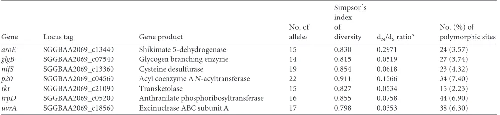
summary, 14 (glgB) to 22 (p20) different alleles were present at each locus, resulting in an average of 16.9 alleles in each internal fragment. The proportions of polymorphic sites ranged from 2.2% (tkt) to 7.4% (p20) and provided 1.3⫻1010genotypes. A dN/dSratio of⬍1 and an IAvalue of 2.4 were calculated, resulting in the detection of significant linkage disequilibrium (Table 1) (26).
[image:2.585.39.548.77.195.2]For each strain, the combination of the allelic numbers deter-mines the STs. The isolates were resolved into 50 different STs, and STs 45 and 50 were the most common. Within these two STs, multiple isolates from pigeons (pigeon lofts 1 and 2) and turkeys (turkey coop 1) were found. Thirty-five STs are represented by a single strain (Fig. 1; see Table S1 in the supplemental material). To TABLE 1Characteristics of MLST loci used forS.gallolyticussubsp.gallolyticus
Gene Locus tag Gene product
No. of alleles
Simpson’s index of
diversity dN/dSratioa
No. (%) of polymorphic sites
aroE SGGBAA2069_c13440 Shikimate 5-dehydrogenase 15 0.830 0.2971 24 (3.57)
glgB SGGBAA2069_c07540 Glycogen branching enzyme 14 0.815 0.0519 27 (3.74)
nifS SGGBAA2069_c13360 Cysteine desulfurase 19 0.854 0.0618 23 (4.32)
p20 SGGBAA2069_c04560 Acyl coenzyme AN-acyltransferase 22 0.911 0.1566 34 (7.40)
tkt SGGBAA2069_c21090 Transketolase 15 0.827 0.0534 15 (2.23)
trpD SGGBAA2069_c05200 Anthranilate phosphoribosyltransferase 16 0.855 0.0758 44 (6.90)
uvrA SGGBAA2069_c18560 Excinuclease ABC subunit A 17 0.798 0.0353 38 (6.30)
aRatio of nonsynonymous to synonymous substitutions.
MLST ofS.gallolyticussubsp.gallolyticus
on May 16, 2020 by guest
http://jcm.asm.org/
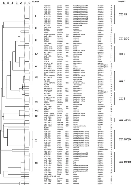
FIG 1UPGMA dendrogram of 101S.gallolyticussubsp.gallolyticusstrains. The phylogenetic tree shown was calculated with the allelic profile by using the UPGMA algorithm. The dashed line symbolizes the border defining the clusters. A linkage distance of 1.12 contributes to 11 clusters.
on May 16, 2020 by guest
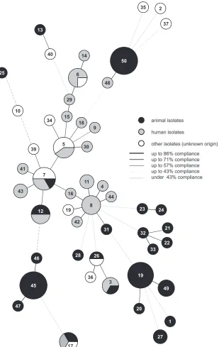
[image:3.585.71.529.43.668.2]illustrate the potential relatedness between the strains, a UPGMA dendrogram was constructed from the allelic distances of each strain. It showed a highly divergent population (Fig. 1). An aver-age SID of 0.84 (95% CI, 0.735 to 0.931) was defined as demon-strating the discriminatory power of theS.gallolyticussubsp. gal-lolyticus-specific MLST scheme (Table 1) (28,29). Therefore, 11 clusters (containing at least three isolates) are formed in the den-drogram based on a linkage distance of 1.12 (conforms SID⫽ 0.84) (Fig. 1;Table 1). STs 45, 46, 47, and 50 can be associated with cluster I or X and contain isolates from only one animal species. Clusters I and XI are the biggest groups and comprise 11 isolates from poultry, followed by cluster X with 10 isolates from pigeons (with one exception). In clusters I and XI, fecal isolates from dif-ferent pigeon lofts were found (Fig. 1). Furthermore, strains of fecal samples from a turkey coop are in cluster X. Clusters VI and VII comprise many isolates from humans. Several clusters (e.g., IV, V, and IX) comprise isolates from cattle and humans (Fig. 1). For a better characterization of the relatedness between strains and for more conclusive information about infection chains, ge-ography, or host specificity, a minimum spanning tree (MST) based on the MLST data set of 101 strains was generated (Fig. 2). Besides the UPGMA dendrogram and the MST, clonal complexes were defined. The 50 STs can be divided into 10 clonal complexes, and a total of 29 STs are included. Furthermore, 21 singletons were identified. The snapshot of the eBURST (zero out of seven alleles are in common) proposes ST 8 as the predicted primary founder of theS.gallolyticussubsp.gallolyticusstrain collection (data not shown). ST 8 belongs to clonal complex 8. This complex includes STs 4, 11, 16, 38, 42, and 48 (Fig. 1and2). With one exception, this lineage comprises only isolates from humans from different time points and countries.
All of the analysis methods used, MLST, MST, and eBURST, demonstrated the high diversity of our random collection ofS. gallolyticussubsp.gallolyticusstrains. Hence, no evidence of host or geographic specificity was found (Fig. 1and2; eBURST data not shown).
DISCUSSION
The host specificities, pathogenic mechanisms, and biochemical characteristics ofS.gallolyticussubsp.macedonicus,S.gallolyticus subsp.pasteurianus,S.gallolyticussubsp.gallolyticusdiffer widely. S.gallolyticussubsp.gallolyticusandS.gallolyticussubsp. pasteur-ianusare commensals of the gastrointestinal system and can cause endocarditis and meningitis (3,5–7,30–32). However,S. gallolyti-cussubsp.pasteurianuscan cause septicemia and is often associ-ated with, e.g., chronic liver disease or cirrhosis, especially in im-munocompromised patients (33,34). Besides human infections, to date there have been only two reported cases of septicemia due toS.gallolyticussubsp.pasteurianusin animals (ducklings, gos-lings) (35,36).S.gallolyticussubsp.macedonicusis often isolated from dairy products, e.g., cheese, and sour mash and is nonpatho-genic (12). In summary,S.gallolyticussubsp.gallolyticusshows a broader host range and differs in the spectrum of diseases. On the basis of the different characteristics of these three subspecies, we established a subspecies-specific MLST scheme forS.gallolyticus subsp.gallolyticus. This scheme offers the opportunity to charac-terize the phylogenetic structure, the zoonotic potential, and the transmission routes of this subspecies, as well as to assess its risks. We used bacteria from several strain collections and isolates from patients (e.g., blood cultures, feces) and from animal fecal
sam-ples. Housekeeping genes were chosen with a low dN/dSratio of ⬍1 and a high SID of 0.84 (95% CI, 0.735 to 0.931) to characterize 101S.gallolyticussubsp.gallolyticusisolates. A dN/dSratio of⬍1 was also calculated by Shibata et al. (17). The number of alleles in the subspecies-specific MLST scheme varies from 14 (glgB) to 22 (p20), which distinguishes more than 3.6⫻108STs. This range is comparable to those reported in other publications. For group B streptococci, a 1.2 to 2.5% range of allelic variation was identified (37). A comparable range of allelic variation (1.4 to 6.1%) was observed in the MLST scheme developed for group A streptococci (38). A comparison with the recently published MLST scheme presents from 15 (parC) to 24 (rpoD) allelic variations by using other genes (dpr, gmk, rpoD,parC, pta,pyrC, andrecN) for S. gallolyticus. However, in contrast to our work, the publication of Shibata et al. supports no SID calculations. Therefore, the descrip-tion of the divergent structure is based only on the calculated number of STs (17).
The distribution of the STs can be illustrated by calculating clusters or can be presented in the MST. The MST shows accumu-lations ofS.gallolyticussubsp.gallolyticus strains isolated from various animals. Otherwise, there are groups consisting of bacte-rial isolates from animals and humans but there is no evidence of host specificity and no suggestion of geographic-region-related occurrence. Remarkably, clonal complex 8 is dominated by man isolates. The occurrence of the ST 8 lineage especially in hu-man isolates may indicate that the STs involved are associated with human hosts. To gain better insights into the epidemiologic structure ofS.gallolyticussubsp.gallolyticus, cluster borders were based on the calculated average SID. One must acknowledge that the clusters of Shibata et al. are different. TheS.gallolyticusMLST scheme shows clusters based on the roots of the UPGMA dendro-gram. The clusters presented also containS.gallolyticus subsp. gallolyticusisolates from animals or humans, as well as from both animals and humans (17).
Moreover, on the basis of the IA, no epidemiological popula-tion structure can be observed referring to the detecpopula-tion of signif-icant linkage disequilibrium, which can be interpreted as a bacte-rial population with low rates of recombination. Recombination events were tested by using Sawyer’s run test in the MLST scheme for all three subspecies, whereby two genes with evidence of re-combination (rpoC,parC) were identified (17). However, this test is less sensitive for detecting recombination (39). Therefore, we calculated the IAfor our strain collection. For the comparison of population structures for theS.gallolyticusMLST scheme, an IA was not calculated (17). To answer questions concerning epide-miology, host specificity, and virulence, Shibata et al. defined clonal complexes. For this purpose, a more relaxed group defini-tion (five out of seven alleles are in common) was used and the 57 STs of 63 strains were divided into four lineages and 31 singletons (17).
Nevertheless, the established MLST schemes differ in focus. In addition to the epidemiologic application, Shibata et al. included all threeS.gallolyticussubspecies and strived to simplify subspe-cies classification (17). The 57 STs identified form five clusters (A to E), and three of these (A, B, and D) contain 41 STs ofS. gallo-lyticussubsp.gallolyticusstrains. Consequently, it is presumed that two of the threeS.gallolyticussubsp.gallolyticusclusters belong to a novel subspecies (17). In 2011, Hinse et al. published a reliable method for identifying isolates to the subspecies level by MALDI-MLST ofS.gallolyticussubsp.gallolyticus
on May 16, 2020 by guest
http://jcm.asm.org/
TOF MS andsodADNA sequencing, which was used in our re-search to identifyS.gallolyticussubsp.gallolyticusisolates (19).
Notwithstanding the fact that the typing schemes have differ-ent aims, both suggest zoonotic potdiffer-ential. In theS. gallolyticus MLST scheme, it is suggested byS.gallolyticussubsp.gallolyticus cluster A, which includes animal and human isolates (17). Fur-thermore, only virulent cluster C ofS.gallolyticussubsp. pasteur-ianus, which includes exclusively human patient isolates of ST 14, could be identified (17).
A comparison of these two different schemes shows that the
MLST method described here is focused explicitly on the zoonotic and epidemiological investigation of the distinct subspecies ofS. gallolyticusand does not aim at subspecies identification or deter-mination of virulence. The subspecies-specific focus on the epide-miological structure and risk assessment is confirmed by the spe-cific primer binding sites for the housekeeping genes for S. gallolyticussubsp.gallolyticus. On the basis of the observation of identical allelic profiles ofS.gallolyticussubsp.gallolyticusisolates and different species, we propose that there is zoonotic potential. Additionally, the transferability to other subspecies is quite lim-FIG 2Relatedness of 50 STs of 101S.gallolyticussubsp.gallolyticusstrains in an MST. The results were calculated by BioNumerics software on the basis of MLST data. Each ST is shown as a circle whose size is proportional to the number of strains included. Shading shows the origins of the isolates, and the lines represent the compliance levels of the strains.
on May 16, 2020 by guest
http://jcm.asm.org/
[image:5.585.141.450.66.555.2]ited because of the specificity of our MLST scheme forS. gallolyti-cussubsp.gallolyticus.
To prove the zoonotic potential ofS.gallolyticussubsp. gallo-lyticus, further studies are being performed. We are examining isolates from livestock in cooperation with the respective livestock owners to examine the transmission pattern of this bacterium and to try to assess the risk associated with this facultative pathogen. Our first results suggest thatS.gallolyticussubsp.gallolyticusmight act as a zoonotic agent. In summary, theS. gallolyticus subsp. gallolyticus-specific MLST scheme developed can be used for mo-lecular genetic characterization aiming for insight into its zoo-notic potential, epidemiology, and potential infection chains.
ACKNOWLEDGMENTS
We thank Jochen Schulz from the University of Veterinary Medicine,
Hannover, Germany, for severalS.gallolyticussubsp.gallolyticusisolates
from poultry. We also thank M. D. Fred Splittgerber for his linguistic advice.
This work was supported by the Ruhr-Universität Bochum Medizinis-che Fakultät (FoRUM).
REFERENCES
1.Schlegel L, Grimont F, Ageron E, Grimont PA, Bouvet A.2003.
Reap-praisal of the taxonomy of theStreptococcus bovis/Streptococcus equinus
complex and related species: description ofStreptococcus gallolyticus
subsp.gallolyticussubsp. nov.,S.gallolyticussubsp.macedonicussubsp.
nov. andS.gallolyticussubsp.pasteurianussubsp. nov. Int. J. Syst. Evol.
Microbiol.53:631– 645.http://dx.doi.org/10.1099/ijs.0.02361-0.
2.Sillanpää J, Nallapareddy SR, Qin X, Singh KV, Muzny DM, Kovar CL, Nazareth LV, Gibbs RA, Ferraro MJ, Steckelberg JM.2009. A collagen-binding adhesin, Acb, and ten other putative MSCRAMM and pilus family
proteins ofStreptococcus gallolyticussubsp.gallolyticus(Streptococcus bovis
group, biotype I). J. Bacteriol.191:6643– 6653.http://dx.doi.org/10.1128
/JB.00909-09.
3.Headings DL, Herrera A, Mazzi E, Bergman MA. 1978. Fulminant
neonatal septicemia caused byStreptococcus bovis. J. Pediatr.92:282–283.
http://dx.doi.org/10.1016/S0022-3476(78)80026-2.
4.Sekizaki T, Nishiya H, Nakajima S, Nishizono M, Kawano M, Okura M, Takamatsu D, Nishino H, Ishiji T, Osawa R.2008. Endocarditis
in chickens caused by subclinical infection ofStreptococcus gallolyticus
subsp.gallolyticus. Avian Dis.52:183–186.http://dx.doi.org/10.1637/8048
-070307-Case.
5.Sillanpää J, Nallapareddy SR, Singh KV, Ferraro MJ, Murray BE.2008.
Adherence characteristics of endocarditis-derivedStreptococcus
gallolyti-cusssp.gallolyticus(Streptococcus bovisbiotype I) isolates to host
extracel-lular matrix proteins. FEMS Microbiol. Lett.289:104 –109.http://dx.doi
.org/10.1111/j.1574-6968.2008.01378.x.
6.Macneal WJ, Blevins A.1945. Bacteriological studies in endocarditis. J.
Bacteriol.49:603– 610.
7.Ballet M, Gevigney G, Gare J, Delahaye F, Etienne J, Delahaye J.1995.
Infective endocarditis due toStreptococcus bovis. A report of 53 cases. Eur.
Heart J.16:1975–1980.
8.Klein RS, Recco RA, Catalano MT, Edberg SC, Casey JI, Steigbigel
NH. 1977. Association of Streptococcus boviswith carcinoma of
the colon. N. Engl. J. Med. 297:800 – 802. http://dx.doi.org/10.1056
/NEJM197710132971503.
9.Devriese L, Uyttebroek E, Gevaert D, Vandekerckhove P, Ceyssens K.
1990.Streptococcus bovisinfections in pigeons. Avian Pathol.19:429 – 434.
http://dx.doi.org/10.1080/03079459008418697.
10. Devriese LA, Vandamme P, Pot B, Vanrobaeys M, Kersters K, Haese-brouck F.1998. Differentiation betweenStreptococcus gallolyticusstrains
of human clinical and veterinary origins andStreptococcus bovisstrains
from the intestinal tracts of ruminants. J. Clin. Microbiol.36:3520 –3523.
11. Sasaki E, Osawa R, Nishitani Y, Whiley RA.2004. ARDRA and RAPD
analyses of human and animal isolates ofStreptococcus gallolyticus. J. Vet.
Med. Sci.66:1467–1470.http://dx.doi.org/10.1292/jvms.66.1467.
12. Tsakalidou E, Zoidou E, Pot B, Wassill L, Ludwig W, Devriese L, Kalantzopoulos G, Schleifer K, Kersters K.1998. Identification of
strep-tococci from Greek Kasseri cheese and description ofStreptococcus
mace-donicussp. nov. Int. J. Syst. Bacteriol.48:519 –527.http://dx.doi.org/10 .1099/00207713-48-2-519.
13. Randazzo CL, Vaughan EE, Caggia C.2006. Artisanal and experimental Pecorino Siciliano cheese: microbial dynamics during manufacture
as-sessed by culturing and PCR-DGGE analyses. Int. J. Food Microbiol.109:
1– 8.http://dx.doi.org/10.1016/j.ijfoodmicro.2005.11.002.
14. Fortin M, Messier S, Paré J, Higgins R.2003. Identification of catalase-negative, non-beta-hemolytic, gram-positive cocci isolated from milk
samples. J. Clin. Microbiol.41:106 –109.http://dx.doi.org/10.1128/JCM
.41.1.106-109.2003.
15. Garvie EI, Bramley A.1979. Streptococcus bovis—an approach to its classification and its importance as a cause of bovine mastitis. J. Appl.
Microbiol.46:557–566.
16. Urwin R, Maiden MC.2003. Multi-locus sequence typing: a tool for
global epidemiology. Trends Microbiol.11:479 – 487.http://dx.doi.org/10
.1016/j.tim.2003.08.006.
17. Shibata Y, Tien LHT, Nomoto R, Osawa R.2014. Development of a
multilocus sequence typing scheme forStreptococcus gallolyticus.
Micro-biology160:113–122.http://dx.doi.org/10.1099/mic.0.071605-0.
18. Poyart C, Quesne G, Coulon S, Berche P, Trieu-Cuot P.1998. Identi-fication of streptococci to species level by sequencing the gene encoding
the manganese-dependent superoxide dismutase. J. Clin. Microbiol.36:
41– 47.
19. Hinse D, Vollmer T, Erhard M, Welker M, Moore E, Kleesiek K, Dreier
J.2011. Differentiation of species of theStreptococcus bovis/equinus
com-plex by MALDI-TOF mass spectrometry in comparison tosodAsequence
analyses. Syst. Appl. Microbiol. 34:52–57. http://dx.doi.org/10.1016/j
.syapm.2010.11.010.
20. Rusniok C, Couvé E, Da Cunha V, El Gana R, Zidane N, Bouchier C, Poyart C, Leclercq R, Trieu-Cuot P, Glaser P.2010. Genome sequence of
Streptococcus gallolyticus: insights into its adaptation to the bovine rumen
and its ability to cause endocarditis. J. Bacteriol.192:2266 –2276.http://dx
.doi.org/10.1128/JB.01659-09.
21. Lin I-H, Liu T-T, Teng Y-T, Wu H-L, Liu Y-M, Wu K-M, Chang C-H, Hsu M-T.2011. Sequencing and comparative genome analysis of
two pathogenicStreptococcus gallolyticussubspecies: genome plasticity,
adaptation and virulence. PLoS One6:e20519.http://dx.doi.org/10.1371
/journal.pone.0020519.
22. Hinse D, Vollmer T, Rückert C, Blom J, Kalinowski J, Knabbe C, Dreier J.2011. Complete genome and comparative analysis of Strep-tococcus gallolyticussubsp.gallolyticus, an emerging pathogen of infective
endocarditis. BMC Genomics 12:400. http://dx.doi.org/10.1186/1471
-2164-12-400.
23. Blom J, Albaum S, Doppmeier D, Pühler A, Vorhölter F-J, Zakrzewski M, Goesmann A.2009. EDGAR: a software framework for the
compara-tive analysis of prokaryotic genomes. BMC Bioinformatics10:154.http:
//dx.doi.org/10.1186/1471-2105-10-154.
24. Feil EJ, Li BC, Aanensen DM, Hanage WP, Spratt BG.2004. eBURST: inferring patterns of evolutionary descent among clusters of related
bac-terial genotypes from multilocus sequence typing data. J. Bacteriol.186:
1518 –1530.http://dx.doi.org/10.1128/JB.186.5.1518-1530.2004.
25. Jolley KA, Feil E, Chan M-S, Maiden MCJ.2001. Sequence type analysis
and recombinational tests (START). Bioinformatics17:1230 –1231.http:
//dx.doi.org/10.1093/bioinformatics/17.12.1230.
26. Smith JM, Smith NH, O’Rourke M, Spratt BG.1993. How clonal are
bacteria? Proc. Natl. Acad. Sci. U. S. A.90:4384 – 4388.http://dx.doi.org
/10.1073/pnas.90.10.4384.
27. Jolley KA, Maiden MJ.2010. BIGSdb: scalable analysis of bacterial
ge-nome variation at the population level. BMC Bioinformatics11:595.http:
//dx.doi.org/10.1186/1471-2105-11-595.
28. Grundmann H, Hori S, Tanner G.2001. Determining confidence inter-vals when measuring genetic diversity and the discriminatory abilities of
typing methods for microorganisms. J. Clin. Microbiol.39:4190 – 4192.
http://dx.doi.org/10.1128/JCM.39.11.4190-4192.2001.
29. Simpson EH.1949. Measurement of diversity. Nature163:688. (Letter.)
http://dx.doi.org/10.1038/163688a0.
30. Onoyama S, Ogata R, Wada A, Saito M, Okada K, Harada T.2009.
Neonatal bacterial meningitis caused byStreptococcus gallolyticussubsp.
pasteurianus. J. Med. Microbiol.58:1252–1254.http://dx.doi.org/10.1099 /jmm.0.006551-0.
31. Sturt AS, Yang L, Sandhu K, Pei Z, Cassai N, Blaser MJ.2010. Strep-tococcus gallolyticussubspeciespasteurianus(biotype II/2), a newly
re-MLST ofS.gallolyticussubsp.gallolyticus
on May 16, 2020 by guest
http://jcm.asm.org/
ported cause of adult meningitis. J. Clin. Microbiol.48:2247–2249.http: //dx.doi.org/10.1128/JCM.00081-10.
32. Grant RJ, Whitehead TR, Orr JE.2000.Streptococcus bovismeningitis in
an infant. J. Clin. Microbiol.38:462– 463.
33. Alex D, Garvin D, Peters S.2013.Streptococcus pasteurianussepticemia.
Indian J. Med. Microbiol. 31:310 –312.http://dx.doi.org/10.4103/0255
-0857.115668.
34. Gonzlez-Quintela A, Martínez-Rey C, Castroagudín J, Rajo-Iglesias M, Domínguez-Santalla M.2001. Prevalence of liver disease in patients with
Streptococcus bovisbacteraemia. J. Infect.42:116 –119.http://dx.doi.org /10.1053/jinf.2001.0799.
35. Barnett J, Ainsworth H, Boon J, Twomey D.2008.Streptococcus gallo-lyticussubsp.pasteurianussepticaemia in goslings. Vet. J.176:251–253.
http://dx.doi.org/10.1016/j.tvjl.2007.02.011.
36. Li M, Gu C, Zhang W, Li S, Liu J, Qin C, Su J, Cheng G, Hu X.2013.
Isolation and characterization ofS.gallolyticussubsp.pasteurianuscausing
meningitis in ducklings. Vet. Microbiol.162:930 –936.http://dx.doi.org
/10.1016/j.vetmic.2012.11.038.
37. Jones N, Bohnsack JF, Takahashi S, Oliver KA, Chan M-S, Kunst F, Glaser P, Rusniok C, Crook DW, Harding RM, Bisharat N, Spratt BG.
2003. Multilocus sequence typing system for group B streptococcus. J.
Clin. Microbiol.41:2530 –2536.http://dx.doi.org/10.1128/JCM.41.6.2530
-2536.2003.
38. Enright MC, Spratt BG, Kalia A, Cross JH, Bessen DE.2001. Multilocus
sequence typing ofStreptococcus pyogenesand the relationships between
emmtype and clone. Infect. Immun.69:2416 –2427.http://dx.doi.org/10
.1128/IAI.69.4.2416-2427.2001.
39. Meinersmann RJ, Phillips RW, Wiedmann M, Berrang ME. 2004.
Multilocus sequence typing ofListeria monocytogenesby use of
hypervari-able genes reveals clonal and recombination histories of three lineages.
Appl. Environ. Microbiol.70:2193–2203.http://dx.doi.org/10.1128/AEM
.70.4.2193-2203.2004.