for Detection of
Plasmodium ovale
and
Plasmodium malariae
Melissa Phuong,aRachel Lau,bFilip Ralevski,bAndrea K. Boggildb,c,d
Faculty of Health Sciences, McMaster University, Hamilton, Ontario, Canadaa; Public Health Ontario Laboratories, Public Health Ontario, Toronto, Ontario, Canadab;
Tropical Disease Unit, Division of Infectious Diseases, UHN-Toronto General Hospital, Toronto, Ontario, Canadac; Department of Medicine, University of Toronto, Toronto,
Ontario, Canadad
Although microscopic examination of Giemsa-stained blood smears remains the gold standard for the diagnosis of malaria, mo-lecular detection using PCR is becoming increasingly popular. Due to discrepant PCR and microscopy results, we aimed to
optimize our detection assays forPlasmodium malariaeandPlasmodium ovaleby sequencing the 18S rRNA region and
developing a new primer and probe set for real-time quantitative PCR (qPCR). Clinical specimens positive forP. malariae
(nⴝ15) orP. ovale(nⴝ33) underwent amplification and sequencing of the 18S rRNA region. Based on sequence
discrep-ancies between our current primer/probe and clinical isolates, degenerateP. ovaleprimer and probe were developed to
determine if their performance characteristics improved. The reference (gold) standard was microscopy. No 18S sequence
heterogeneity was observed among theP. malariaeisolates, and the sensitivity and specificity of our currentP. malariae
qPCR assay were both 100%. Compared to microscopy, the sensitivity and specificity of our currentP. ovaleqPCR assay
were 72.7% and 100%, respectively. Five single nucleotide polymorphisms (SNPs) were identified inP. ovale. The sensitivity of the new
P. ovaleassay increased to 100% with 100% specificity. We therefore improved the performance characteristics of ourP. ovale molec-ular detection assay through the development of a degenerate primer and probe set which accommodates 18S SNPs among
the 2 subspecies ofP. ovale. Given the suboptimal sensitivity of rapid diagnostic tests for non-falciparummalaria and the
typically low parasitemia ofP. malariaeandP. ovale, a well-performing confirmatory molecular assay is imperative for
clinical laboratories.
T
here are fivePlasmodiumspecies which are known to causehuman malaria,Plasmodium falciparum,Plasmodium vivax,
Plasmodium ovale,Plasmodium malariae, andPlasmodium knowlesi
(1). AlthoughP. malariaeandP. ovaleoccur less frequently among
human populations, they are widely distributed and their diagno-sis has proven challenging due to their relatively low parasitemia and the morphology of blood stages, as well as the poor sensitivity
of rapid diagnostic tests in non-falciparumspecies (1,2).
Microscopic examination of Giemsa-stained thick and thin blood smears, rapid diagnostic tests (RDTs), and PCR assays are the mainstays of malaria diagnostics in resourced settings;
how-ever, microscopy remains the gold standard. Diagnoses of P.
malariaeandP. ovalebased on microscopy present challenges due to the technical expertise required, the typically low parasitemias associated with these species, and the similar morphological
fea-tures to otherPlasmodiumspecies (1). PCR assays may be either
genus or species specific and do not require extensive training to perform; however, significant laboratory infrastructure is
required, thus limiting their use to resourced settings (3, 4).
Our current molecular approach to the diagnosis of malaria
involves use of onePlasmodiumgenus-specific and two duplexed
Plasmodiumspecies-specific real-time quantitative PCR (qPCR) assays, which can discriminate among the 4 common human malaria species, as described by Khairnar and colleagues (3). A
recent case ofP. ovalein which real-time quantitative PCR was
indeterminate, with sequencing required to resolve the causative malaria species (5), and our own experience with discordant re-sults using current qPCR and microscopy, highlight the challenges of laboratory diagnostics in malaria. Genetic polymorphisms
be-tween the classic (P. ovale curtisi) and variant (P. ovale wallikeri)
subspecies ofP. ovalemay render the molecular detection of these
two species difficult (6).
In the present study, we aimed to optimize the diagnostic
qPCR currently used in our clinical laboratory forPlasmodium
malariaeandPlasmodium ovalethrough sequence analysis of the 18S rRNA region and develop new primers and probes which could accommodate any sequence heterogeneity.
MATERIALS AND METHODS
Specimens.Between January 2007 and July 2013, 15 whole-blood
speci-mens positive forP. malariaeand 33 specimens positive forP. ovalewere examined in our laboratory and banked at⫺80°C following diagnostic testing. Microscopy was performed by examination of Giemsa-stained thick and thin blood films by certified medical lab technologists. Rapid diagnostic testing was conducted with a BinaxNow malaria kit (Alere, ME) according to the manufacturer’s instructions.P. falciparum,P. vivax,
P. malariae, andP. ovalesmall-subunit rRNA DNA clones (MRA-177,
MRA-178, MRA-179, MRA-180, and MR4; ATCC, Manassas, VA) were
Received13 December 2013Returned for modification6 January 2014 Accepted10 January 2014
Published ahead of print15 January 2014 Editor:A. M. Caliendo
Address correspondence to Andrea K. Boggild, [email protected]. M.P. and R.L. contributed equally to this work.
Copyright © 2014, American Society for Microbiology. All Rights Reserved.
doi:10.1128/JCM.03477-13
on May 16, 2020 by guest
http://jcm.asm.org/
used as positive-control materials. Randomly selected banked specimens positive forP. falciparum(n⫽20) (with parasitemia ranging from⬍0.1 to 28.6%) orP. vivax(n⫽20) (with parasitemia ranging from⬍0.1 to 0.9%) or negative forPlasmodium (n⫽20) (confirmed by microscopy and qPCR) were used as negative-control specimens for calculation of perfor-mance characteristics.
DNA extraction.DNA was extracted using the DNA Minikit blood or
body fluid spin protocol (Qiagen, Germantown MD). A total of 200l of frozen whole blood was thawed and each sample was eluted with 60l AE buffer and stored at⫺20°C prior to use.
Real-time PCR.Our current molecular diagnostic assay includes 4
qPCRs: human beta-2-microglobulin (B2MG) extraction control,
Plas-modiumgenus specific,P. falciparum/P. vivaxspecies-specific duplex, and
P. malariae/P. ovalespecies-specific duplex qPCR as previously described
by Khairnar and colleagues (3,4). TheP. falciparum/P. vivaxduplex qPCR was conducted with theP. malariaeandP. ovalespecimens to exclude mixed infections. TheP. malariae/P. ovaleduplex qPCR was conducted for all specimens to establish the sensitivities and specificities of the qPCR assays. All qPCR assays were run using the ABI 7900HT real-time PCR system and under the following conditions: 50°C for 2 min, 95°C for 10 min, 95°C for 15 s, and 60°C for 1 min (45 cycles). We used 12.5l of TaqMan universal PCR master mix (Life Technologies) and 5l of DNA primers and probes with concentrations as previously reported (3) for a final volume of 25l per reaction. All qPCR amplification curves were analyzed using a manual threshold cycle (CT) of 0.02 and an automatic
baseline. A result was called positive if theCTvalue was⬍38 in the
pres-ence of a logarithmic amplification curve.
Amplification and sequencing of the 18S rRNA region ofP. malariae
andP. ovale.Endpoint PCR of the 18S rRNA region was conducted with high-fidelity polymerase AccuPrime Pfx Supermix (Life Technologies) and 200 nM (each) of the primers Plasmo 18S forward (5=-ATTCAGAT GTCAGAGGTGAAATTCT-3=) and Plasmo 18S reverse (5=-TCAATCCT ACTCTTGTCTTAAACTA-3=), generating a 396-bp product. The cycling conditions were 95°C for 5 min, followed by 95°C for 15 s, 58°C for 30 s, and 68°C for 30 s for 45 cycles, and then 68°C for 5 min using an ABI Veriti fast thermal cycler. Amplicons were visualized on 1% agarose gels with ethidium bromide. PCR products were purified with a QIAquick PCR purification kit (Qiagen, Germantown, MD) and eluted with 20l water. Purified PCR products were Sanger sequenced using the same forward and reverse primers with a BigDye Terminator v3.1 cycle sequencing kit (Life Technologies) and run according to the manufacturer’s recom-mended conditions. Sequenced products were purified by a BigDye XTerminator (Life Technologies) and analyzed by an ABI 3130xl genetic analyzer. Forward and reverse sequences of each sample were aligned using Vector NTI software (Life Technologies). Sequences were verified using a BLAST search to further confirm the species identification and to resolve any discrepant microscopic diagnosis and species-specific qPCR results. Alignments ofP. malariaeandP. ovalesequences were performed using Mega5.2 software (7). Sequences were analyzed and compared with our currently used real-time qPCRP. malariaeandP. ovaleprimers and probes (3,4). The 18S rRNA sequences ofP. falciparum,P. vivax,P.
malariae, andP. ovalefrom GenBank were aligned using Mega5.2
soft-ware to determine if changes in primers and probes may have caused false positives (Table 1).
Limit of detection for qPCR assay.Small-subunit rRNA DNA clones
MRA-179 (P. malariae) and MRA-180 (P. ovale) were 10-fold serially diluted, yielding 8 concentrations ranging from 9 to 91 million copies/ reaction. Each dilution of the clones was run in triplicate. MeanCTvalues
were plotted against the log copy number/reaction to generate an equa-tion to calculate the limit of detecequa-tion (LOD) at aCTof 38.
RESULTS
Performance of current duplexP. malariae/P. ovaleqPCR
as-say.Forty-eightP. ovaleandP. malariaespecimens were analyzed
by microscopy, RDT (Binax assay), qPCR, and Sanger sequencing.
Diagnosed species were identified based on the concordance of 2 out of 3 species-specific assays, microscopy, qPCR, and BLAST sequence homology. All specimens were microscopy positive; however, for a few specimens other assays were required to resolve the species. Binax had a quick turnaround time of 20 min but
considerably lower sensitivity of 35.5% forP. ovaleand 78.6% for
P. malariae (Tables 2 and 3). DNA was extracted from P. malariae-,P. ovale-,P. falciparum-, andP. vivax-positive speci-mens and negative specispeci-mens, and then verified by B2MG
extrac-tion control qPCR. Twenty-four of 33P. ovalespecimens were
positive forP. ovaleby our currentP. malariae/P. ovale
species-specific duplex qPCR (Table 2) (3). All specimens except number
25 were negative forP. falciparum,P. vivax, andP. malariae.
Spec-imen number 25 was positive forP. falciparumandP. ovaleby
qPCR, but microscopy, Sanger sequencing, and BLAST analysis
confirmed it to be a singleP. ovaleinfection only. AllP. malariae
specimens (n⫽15) were positive forP. malariaeby qPCR with no
mixed infections observed (Table 3). SixtyP. falciparum-positive
(n⫽20),P. vivax-positive (n⫽20) and negative specimens (n⫽
20) underwentP. malariae/P. ovaleqPCR and all were negative.
Thus, sensitivity of the currentP. malariae/P. ovaleduplex qPCR
assay forP. ovalewas 72.7% with specificity of 100%, and forP.
malariae, both sensitivity and specificity were 100%. The low
sen-sitivity and the jagged amplification curve observed in theP. ovale
qPCR runs suggested some sequence heterogeneity in the regions to which the primers and probe hybridized.
Sequence analysis.No discrepancies between theP. malariae
primers and probes andP. malariae-positive specimen sequences
were noted. Although an A/T mismatch to theP. malariaeforward
primer was found inP. malariae GenBank sequenceM54897.1
(data not shown), it was not observed in any of our specimens; therefore, no modification to the primer was implemented.
Se-quence alignments ofP. ovalespecimens and GenBank sequences
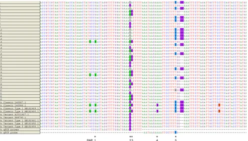
(Fig. 1) revealed SNPs to 3 loci to which the forward primer and 2 loci to which the probe hybridized (Fig. 1). The “V” on SNP 2 was represented by nucleotides (nt) A, C, and G. This “C” is a new nucleotide observed in this study and was included in recently
submitted GenBank sequences (KC633224.1, KC633226.1, and
KC866363.1). It is also observed only inP. ovale curtisi(classic) type.
We therefore modified the currently usedP. ovale forward
primer and probe sequences based on these polymorphisms and
designed the new degenerateP. ovaleforward primer D2 andP.
ovaleprobe D.
Performance of new duplexP. malariae/P. ovaleqPCR
as-say. Modified P. ovale forward primer and probe sequences
(Table 4) were tested by performing qPCR onP. falciparum,P.
[image:2.585.298.544.77.173.2]vivax,P. malariae, andP. ovale18S rRNA clones. The optimized
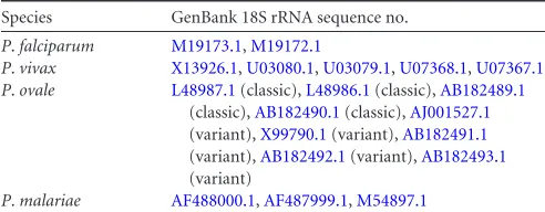
TABLE 1GenBank sequences used for sequence alignments
Species GenBank 18S rRNA sequence no.
P.falciparum M19173.1,M19172.1
P.vivax X13926.1,U03080.1,U03079.1,U07368.1,U07367.1
P.ovale L48987.1(classic),L48986.1(classic),AB182489.1
(classic),AB182490.1(classic),AJ001527.1 (variant),X99790.1(variant),AB182491.1 (variant),AB182492.1(variant),AB182493.1 (variant)
P.malariae AF488000.1,AF487999.1,M54897.1
on May 16, 2020 by guest
http://jcm.asm.org/
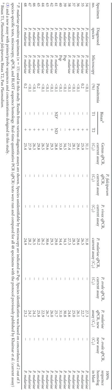
TABLE 2 Specimens positive for Plasmodium ovale a Specimen no. Diagnosed species Microscopy analysis result Parasitemia (%) Binax b Genus qPCR, current assay ( CT ) P. falciparum qPCR, current assay ( CT ) P. vivax qPCR, current assay ( CT ) P. malariae qPCR, current assay ( CT ) P. ovale qPCR, current assay ( CT ) P. malariae qPCR, new assay ( CT ) P. ovale qPCR, new assay ( CT )
100% Sequence homolog in
blast P. ovale subspecies yr of importation T1 T2 1 P. ovale P. ovale ⬍ 0.1 ⫺⫹ 27.6 ⫺⫺ ⫺ 26.4 ⫺ 24.5 P. ovale curtisi 2007 2 P. ovale P. ovale 0.3 ⫺⫹ 23.8 ⫺⫺ ⫺ 28.3 ⫺ 23.5 P. ovale wallikeri 2008 3 P. ovale P. ovale 0.3 ⫺⫹ 26.1 ⫺⫺ ⫺ 24.4 ⫺ 22.6 P. ovale curtisi 2008 4 P. ovale P. ovale ⬍ 0.1 ND c ND 26.9 ⫺⫺ ⫺ 33.7 ⫺ 26.6 P. ovale wallikeri 2009 5 P. ovale P. ovale ⬍ 0.1 ND ND ⫺⫺ ⫺ ⫺ 36.5 ⫺ 29.6 P. ovale wallikeri 2009 6 P. ovale P. ovale ⬍ 0.1 ⫺⫺ 26.0 ⫺⫺ ⫺ 25.4 ⫺ 23.9 P. ovale curtisi 2009 7 P. ovale P. ovale ⬍ 0.1 ⫺⫺ 31.0 ⫺⫺ ⫺ 30.1 ⫺ 28.2 P. ovale curtisi 2009 8 P. ovale P . malariae ⬍ 0.1 ⫺⫺ 27.2 ⫺⫺ ⫺ ⫺ ⫺ 27.5 P. ovale wallikeri 2009 9 P. ovale Psp ⬍ 0.1 ⫺⫹ 32.3 ⫺⫺ ⫺ ⫺ ⫺ 24.1 P. ovale wallikeri 2010 10 P. ovale P. ovale ⬍ 0.1 ⫺⫺ 31.1 ⫺⫺ ⫺ 30.1 ⫺ 29.9 P. ovale curtisi 2010 11 P. ovale P. ovale ⬍ 0.1 ⫺⫺ 31.4 ⫺⫺ ⫺ 31.3 ⫺ 28.1 P. ovale curtisi 2010 12 P. ovale P. ovale ⬍ 0.1 ⫺⫺ 29.9 ⫺⫺ ⫺ 28.8 ⫺ 27.7 P. ovale curtisi 2010 13 P. ovale P. ovale ⬍ 0.1 ⫺⫺ 29.7 ⫺⫺ ⫺ 27.4 ⫺ 27.7 P. ovale curtisi 2010 14 P. ovale P. ovale ⬍ 0.1 ⫺⫺ 27.2 ⫺⫺ ⫺ 34.8 ⫺ 28.3 P. ovale wallikeri 2011 15 P. ovale P. ovale 0.1 ⫺⫹ 26.4 ⫺⫺ ⫺ 25.5 ⫺ 24.6 P. ovale curtisi 2011 16 P. ovale P. ovale 0.1 ⫺⫺ 34.7 ⫺⫺ ⫺ ⫺ ⫺ 27.5 P. ovale wallikeri 2011 17 P. ovale P. ovale 0.5 ⫺⫺ 32.7 ⫺⫺ ⫺ 30.9 ⫺ 23.8 P. ovale wallikeri 2011 18 P. ovale P. ovale ⬍ 0.1 ⫺⫺ 33.3 ⫺⫺ ⫺ 32.7 ⫺ 31.7 P. ovale curtisi 2011 19 P. ovale P. ovale 0.3 ⫺⫺ 24.4 ⫺⫺ ⫺ 34.5 ⫺ 26.8 P. ovale wallikeri 2011 20 P. ovale Psp 0.5 ⫺⫹ 30.9 ⫺⫺ ⫺ ⫺ ⫺ 23.0 P. ovale wallikeri 2011 21 P. ovale P. ovale 0.3 ⫺⫹ 28.4 ⫺⫺ ⫺ 27.0 ⫺ 22.4 P. ovale wallikeri 2012 22 P. ovale P. ovale 0.3 ⫺⫹ 32.2 ⫺⫺ ⫺ 29.5 ⫺ 25.4 P. ovale wallikeri 2012 23 P. ovale P. ovale 0.2 ⫺⫹ 26.8 ⫺⫺ ⫺ 26.4 ⫺ 24.4 P. ovale curtisi 2012 24 P. ovale P. ovale ⬍ 0.1 ⫺⫺ 27.7 ⫺⫺ ⫺ ⫺ ⫺ 28.2 P. ovale wallikeri 2012 25 P. ovale P. ovale ⬍ 0.1 ⫺⫺ 28.4 34.1 ⫺⫺ 35.8 ⫺ 29.8 P. ovale wallikeri 2012 26 P. ovale P. ovale ⬍ 0.1 ⫺⫺ 33.6 ⫺⫺ ⫺ ⫺ ⫺ 31.6 P. ovale curtisi 2012 27 P. ovale P. ovale 0.3 ⫺⫹ 22.9 ⫺⫺ ⫺ 28.5 ⫺ 24.0 P. ovale wallikeri 2012 28 P. ovale Psp ⬍ 0.1 ⫺⫺ ⫺ ⫺ ⫺ ⫺ 32.5 ⫺ 27.5 P. ovale wallikeri 2012 29 P. ovale P. ovale ⬍ 0.1 ⫺⫺ 34.7 ⫺⫺ ⫺ 34.1 ⫺ 28.1 P. ovale wallikeri 2013 30 P. ovale P. ovale ⬍ 0.1 ⫺⫺ 30.1 ⫺⫺ ⫺ 29.3 ⫺ 26.7 P. ovale curtisi 2013 31 P. ovale P. ovale ⬍ 0.1 ⫺⫺ 29.8 ⫺⫺ ⫺ ⫺ ⫺ 33.1 P. ovale wallikeri 2013 32 P. ovale P. ovale 0.1 ⫺⫹ 26.2 ⫺⫺ ⫺ ⫺ ⫺ 29.7 P. ovale wallikeri 2013 33 P. ovale P. ovale ⬍ 0.1 ⫺⫺ 29.0 ⫺⫺ ⫺ ⫺ ⫺ 33.1 P. ovale wallikeri 2013 aP. ovale -positive specimens ( n ⫽ 33) used in this study. Results from various diagnostic assays are shown. Species unidentifiable by microscopy are indicated as Psp. Species identific ation was based on concordance of 2 out of 3 species-specific tests, microscopy, qPCR, and BLAST sequence homology. Real-time quantitative PCR (qPCR) tests were run and compared for all 48 spec imens with the protocol previously published by Khairnar et al. (current assay) ( 3 ) and a new assay with primer/probe sequences and concentrations designed in this study. Based on sequence analysis, 11 P. ovale curtisi (classic type) and 18 P. ovale wallikeri (variant type) cases were identified. Specimen numbers 5, 11, 13, and 22 were duplicate samples from the same patient above and thus were not counted as a case. bBinax T1, Plasmodium falciparum ;Binax T2, Pan Plasmodium . cND, not determined
on May 16, 2020 by guest
http://jcm.asm.org/
primer/probe mix in the new assay had the following
concentra-tions: 200 nM (P. malariaeforward primer), 200 nM (P. ovale
forward primer D2), 200 nM (Plasmodiumreverse primer), 160
nM (P. ovaleprobe D), and 80 nM (P. malariaeprobe) (Table 4). TheP. malariaeforward primer was increased in concentration from our current assay with the aim to maintain or improve sitivity. These concentrations were found to have the highest sen-sitivity without cross-reactivity. The new assay was further
vali-dated against the same set of P. malariae-positive, P. ovale
-positive,P. falciparum-positive,P. vivax-positive, and negative
specimens. AllP. ovale-positive specimens (n⫽33) were positive
with the new assay, improving sensitivity to 100% from 72.7% while maintaining 100% specificity. Using the current assay, the
LOD forP. malariaewas 115 copies/reaction, while the LOD forP.
ovalewas 452 copies/reaction. With the new assay, the LOD forP. malariaewas 84 copies/reaction and the LOD forP. ovalewas 477
copies/reaction. Therefore, the LOD forP. malariae improved
with the new assay, while the LOD for the current and new assays
were comparable forP. ovale.
Classification ofP. ovaleasP. ovale curtisiorP. ovale
wal-likeri.Based on GenBank sequences, we identified 3 loci that can
distinguishP. ovale curtisifromP. ovale wallikeridenoted by SNP
5 and the double-G deletions three nucleotides downstream (Fig. 1). Using these sequence differences, we classified 13 specimens
(11 cases) asP. ovale curtisiand 20 specimens (18 cases) asP. ovale
wallikeri(Table 2).
DISCUSSION
The diagnosis of malaria remains challenging in settings of non-endemicity, yet, rapid and accurate diagnosis with species identi-fication are required to inform treatment and are critical to
posi-tive patient outcomes (8,9). Although microscopic examination
of thick and thin blood smears remains the diagnostic gold stan-dard, this technique requires considerable technologist training and ongoing internal and external proficiency assessments, and it is insensitive at very low parasitemia. RDTs have the advantage of rapid turnaround, but their excellent performance characteristics
are limited toP. falciparum, with suboptimal diagnostic sensitivity
for non-falciparumspecies, especiallyP. ovaleandP. malariae, as
reported in this study and other studies (10,11). Molecular
meth-ods do not require highly specialized technologists and have the advantage of sensitivity and rapidity; however, their practicality is limited to well-resourced reference centers, typically in settings of nonendemicity.
In our center, PCR is used to resolve cases difficult to speciate by microscopy, those with discordant or unusual RDT patterns, and those in which clinical suspicion is high, but microscopy and RDT are noncontributory. We noted several cases of microscopy and qPCR discordance and sought to optimize our molecular
de-tection assays forP. malariaeandP. ovale. Our new assay
demon-strated superior sensitivity forP. ovaleand improved LOD forP.
malariae, without compromising specificity. With the recognition
of 2 subspecies ofP. ovale, both of which were imported to Ontario
over our enrollment period, using a diagnostic assay that can re-liably detect both strains is imperative to appropriate clinical management (6). In addition, using an assay that can distinguish the 2 subspecies may become epidemiologically important as well, given new published information regarding their differing laten-cies (12).
In summary, we improved our molecular detection
algo-3 Specimens positive for Plasmodium malariae a Diagnosed species Microscopy Parasitemia (%) Binax b Genus qPCR, current assay ( C T ) P. falciparum qPCR, current assay ( C T ) P. vivax qPCR, current assay ( C T ) P. malariae qPCR, current assay ( C T ) P. ovale qPCR, current assay ( C T ) P. malariae qPCR, new assay ( C T ) P. ovale qPCR, new assay ( C T ) 100% sequence homolog in blast T1 T2 P. malariae P. malariae ⬍ 0.1 ⫺⫹ 31.1 ⫺⫺ 31.8 ⫺ 27.3 ⫺ P. malariae P. malariae P. malariae 0.2 ⫺⫹ 25.9 ⫺⫺ 26.5 ⫺ 22.7 ⫺ P. malariae P. malariae P. malariae ⬍ 0.1 ⫺⫹ 29.6 ⫺⫺ 29.6 ⫺ 26.1 ⫺ P. malariae P. malariae P. malariae ⬍ 0.1 ⫺⫹ 28.1 ⫺⫺ 28.3 ⫺ 25.0 ⫺ P. malariae P. malariae P. malariae ⬍ 0.1 ⫺⫹ 27.4 ⫺⫺ 27.5 ⫺ 24.6 ⫺ P. malariae P. malariae Psp ⬍ 0.1 ⫺⫺ 34.5 ⫺⫺ 34.3 ⫺ 30.8 ⫺ P. malariae P. malariae Psp ⬍ 0.1 ⫺⫺ 33.8 ⫺⫺ 34.4 ⫺ 31.0 ⫺ P. malariae P. malariae P. malariae ⬍ 0.1 ND c ND 28.8 ⫺⫺ 29.0 ⫺ 25.7 ⫺ P. malariae P. malariae P. malariae ⬍ 0.1 ⫺⫹ 29.3 ⫺⫺ 29.3 ⫺ 26.0 ⫺ P. malariae P. malariae P. malariae ⬍ 0.1 ⫺⫺ 29.6 ⫺⫺ 30.6 ⫺ 26.7 ⫺ P. malariae P. malariae P. malariae ⬍ 0.1 ⫺⫹ 26.5 ⫺⫺ 26.2 ⫺ 22.9 ⫺ P. malariae P. malariae P. malariae 0.2 ⫺⫹ 29.8 ⫺⫺ 30.2 ⫺ 25.8 ⫺ P. malariae P. malariae P. malariae ⬍ 0.1 ⫺⫹ 26.3 ⫺⫺ 26.5 ⫺ 24.2 ⫺ P. malariae P. malariae P. malariae ⬍ 0.1 ⫺⫹ 27.9 ⫺⫺ 28.2 ⫺ 24.7 ⫺ P. malariae P. malariae P. malariae 0.2 ⫺⫹ 25.4 ⫺⫺ 24.9 ⫺ 23.2 ⫺ P. malariae P. malariae -positive specimens ( n ⫽ 15) used in this study. Results from various diagnostic assays are shown. Species unidentifiable by microscopy are indicated as Psp. Species identific ation was based on concordance of 2 out of 3 tests, microscopy, qPCR, and BLAST sequence homology. Real-time quantitative PCR (qPCR) tests were run and compared for all 48 spec imens with the protocol previously published by Khairnar et al. (current assay) ) and a new assay with primer/probe sequences and concentrations designed in this study. Binax T1, Plasmodium falciparum ;Binax T2, Pan Plasmodium . ND, not determined.
on May 16, 2020 by guest
http://jcm.asm.org/
[image:4.585.67.258.63.725.2]rithm forP. malariaeandP. ovaleby optimizing primers and probe concentrations and developing a new degenerate primer
and probe set forP. ovalebased on sequence analysis of the 18S
region and accommodation of known and newly identified SNPs.
ACKNOWLEDGMENTS
We thank MR4 for providing us withPlasmodiumsmall-subunit rRNA clones contributed by Peter A. Zimmerman.
This work was funded by Public Health Ontario.
REFERENCES
1.Obare P, Ogutu B, Adams M, Odera JS, Lilley K, Dosoo D, Adhiambo C,
Owusu-Agyei S, Binka F, Wanja E, Johnson J.2013. Misclassification of
Plas-modiuminfections by conventional microscopy and the impact of
reme-dial training on the proficiency of laboratory technicians in species iden-tification. Malar. J.12:113.http://dx.doi.org/10.1186/1475-2875-12-113.
2.Collins WE, Jeffery GM.2007.Plasmodium malariae: parasite and disease. Clin.
Microbiol. Rev.20:579–592.http://dx.doi.org/10.1128/CMR.00027-07.
3.Khairnar K, Martin D, Lau R, Ralevski F, Pillai DR.2009. Multiplex
real-time quantitative PCR, microscopy and rapid diagnostic immuno-chromatographic tests for the detection ofPlasmodiumspp: performance, limit of detection analysis and quality assurance. Malar. J.8:284.http://dx .doi.org/10.1186/1475-2875-8-284.
4.Shokoples SE, Ndao M, Kowalewska-Grochowska K, Yanow SK.2009.
Multiplexed real-time PCR assay for discrimination ofPlasmodium spe-cies with improved sensitivity for mixed infections. J. Clin. Microbiol. 47:975–980.http://dx.doi.org/10.1128/JCM.01858-08.
5.Cohen R, Feghali K, Alemayehu S, Komisar J, Hang J, Weina PJ,
Coggeshall P, Kamau E, Zapor M. 2013. Use of qPCR and genomic
sequencing to diagnosePlasmodium ovale wallikerimalaria in a returned soldier in the setting of a negative rapid diagnostic assay. Am. J. Trop. Med. Hyg.89:501–506.http://dx.doi.org/10.4269/ajtmh.12-0724.
6.Bauffe F, Desplans J, Fraisier C, Parzy D.2012. Real-time PCR assay for
discrimination ofPlasmodium ovale curtisiandPlasmodium ovale wallikeri
FIG 1The 18S rRNA gene was sequenced and aligned with 4 classic and 5 variant type sequences from GenBank for 33 specimens positive forP. ovale.
P. ovaleforward primer and probe (nucleotide positions 1112 to 1139 and 1141 to 1159, respectively, of GenBank sequenceL48987.1) sequences of the
[image:5.585.45.544.66.350.2]current assay were shown. The new primer and probe designed in this study with degenerate nucleotides bind to the same location. Single nucleotide polymorphisms (denoted by * and labeled as SNPs 1 to 5) were identified on 3 loci to which the primer and 2 loci to which the probe hybridized. SNP 5, along with the double-G deletions three nucleotides downstream, was used to differentiate between classic and variant types. In specimen number 30, in the SNP 5 position, although a dominant C nt was observed in the chromatogram, a minor A was also evident (denoted by an M). The dominant nt of the SNPs were indicated here; however, analysis of the peak heights of chromatograms revealed that the other polymorphic nt was also evident in SNPs 1 to 3, suggesting coinfection of more than one strain ofP. ovale.
TABLE 4P. malariaeandP. ovaleduplex qPCR primer/probe sequences
and final concentrationsa
Primer/probe Sequenceb
Final concn (nM)
P. malariaeforward
primerc
CCGACTAGGTGTTGGATGATAGAG TAAA
200
P. malariaeprobec 6FAM-CTATCTAAAAGAAACACTC
AT-MGBNFQ
80
P. ovaleforward
primer D2
CCRACTAGGTTTTGGATGAAAVR TTTTT
200
P. ovaleprobe D
VIC-CRAAAGGAATTYTCTTATT-MGBNFQ
160
Plasmodiumreverse
primerc
AACCCAAAGACTTTGATTTCTCA TAA
200
aP. ovaledegenerate (D) primer and probe were designed based on sequence heterogeneity found in this study.
bR⫽A or G; Y⫽C or T; V⫽A or C or G. Underlining indicates degenerate
nucleotides.
cSequences as previously published (4).
on May 16, 2020 by guest
http://jcm.asm.org/
[image:5.585.40.287.552.679.2]in the Ivory Coast and in the Comoros Islands. Malar. J.11:307.http://dx .doi.org/10.1186/1475-2875-11-307.
7.Tamura K, Peterson D, Peterson N, Stecher G, Nei M, Kumar S. 2011.
MEGA5: molecular evolutionary genetics analysis using maximum likelihood, evolutionary distance, and maximum parsimony methods. Mol. Biol. Evol.28: 2731–2739.http://dx.doi.org/10.1093/molbev/msr121.
8.Kain KC, MacPherson DW, Kelton T, Keystone JS, Mendelson J,
MacLean JD.2001. Malaria deaths in visitors to Canada and in Canadian
travellers: a case series. CMAJ164:654 – 659.
9.Suh KN, Kain KC, Keystone JS.2004. Malaria. CMAJ.170:1693–1702.
http://dx.doi.org/10.1503/cmaj.1030418.
10. Maltha J, Gillet P, Jacobs J. 2013. Malaria rapid diagnostic tests in
endemic settings. Clin. Micro. Infect.19:399 – 407.http://dx.doi.org/10 .1111/1469-0691.12151.
11. Maltha J, Gillet P, Jacobs J.2013. Malaria rapid diagnostic tests in travel
medicine. Clin. Micro. Infect. 19:408 – 415. http://dx.doi.org/10.1111 /1469-0691.12152.
12. Nolder D, Oguike MC, Maxwell-Scott H, Niyazi HA, Smith V, Chiodini
PL, Sutherland CJ.2013. An observational study of malaria in British
travellers:Plasmodium ovale wallikeriandPlasmodium ovale curtisidiffer significantly in the duration of latency. BMJ Open3:e002711.http://dx .doi.org/10.1136/bmjopen-2013-002711.