comment
reviews
reports
deposited research
interactions
information
refereed research
Protein family review
The synucleins
Julia M George
Address: Department of Cell and Structural Biology, University of Illinois, Urbana, IL 61801, USA. E-mail: [email protected]
Summary
Synucleins are small, soluble proteins expressed primarily in neural tissue and in certain tumors. The
family includes three known proteins: D-synuclein, E-synuclein, and J-synuclein. All synucleins have in
common a highly conserved D-helical lipid-binding motif with similarity to the class-A2 lipid-binding
domains of the exchangeable apolipoproteins. Synuclein family members are not found outside vertebrates, although they have some conserved structural similarity with plant
‘late-embryo-abundant’ proteins. The D- and E-synuclein proteins are found primarily in brain tissue, where they are
seen mainly in presynaptic terminals. The J-synuclein protein is found primarily in the peripheral
nervous system and retina, but its expression in breast tumors is a marker for tumor progression. Normal cellular functions have not been determined for any of the synuclein proteins, although some
data suggest a role in the regulation of membrane stability and/or turnover. Mutations in D-synuclein
are associated with rare familial cases of early-onset Parkinson’s disease, and the protein accumulates abnormally in Parkinson’s disease, Alzheimer’s disease, and several other neurodegenerative illnesses. The current challenge is to understand the normal cellular function of these proteins and how they might contribute to the development of human disease.
Published: 20 December 2001
GenomeBiology2001, 3(1):reviews3002.1–3002.6
The electronic version of this article is the complete one and can be found online at http://genomebiology.com/2001/3/1/reviews/3002 © BioMed Central Ltd (Print ISSN 1465-6906; Online ISSN 1465-6914)
Gene organization and evolutionary history
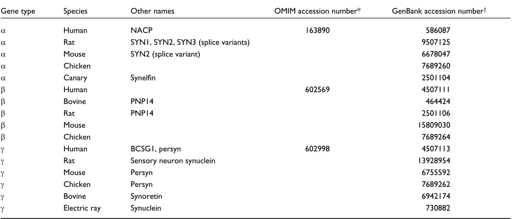
The synuclein family consists of three distinct genes, D -synu-clein, E-synuclein, and J-synuclein, which have so far been described only in vertebrates. Table 1 catalogs the unique members of the synuclein family that are currently listed in GenBank [1]; these 16 sequences encode the orthologs of each of the three synucleins in the species in which they have been described. The sequences are shown aligned in Figure 1a and their estimated relationships are indicated by the dendro-gram in Figure 1b. The D-synuclein gene has been mapped to human chromosome 4q21.3-q22 [2], E-synuclein to human chromosome 5q35 [3], and J-synuclein to human chromo-some 10q23.2-q23.3 [4]. The D-synuclein gene is organized as 7 exons, 5 of which are protein-coding, while the E -synu-clein gene has 6 exons (5 protein-coding) and theJ-synuclein gene has 5 exons (all protein-coding) (reviewed in [5]).Characteristic structural features
All synuclein protein sequences consist of a highly conserved amino-terminal domain that includes a variable number of
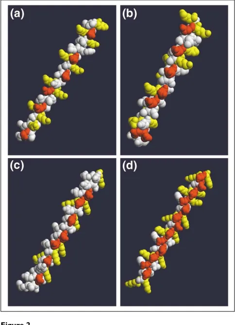
11-residue repeats and a less-conserved carboxy-terminal domain that includes a preponderance of acidic residues. The only significant divergences within the repeat domain are the deletion of 11 amino acids (residues 53-63) in all E-synucleins and the addition of a repeat after residue 32 in the J-synuclein of the electric ray Torpedo californica (Figure 1a). The 11-mer repeats make up a conserved apolipoprotein-like class-A2helix (Figure 2a,b), which medi-ates binding to phospholipid vesicles; lipid binding is accom-panied by a large shift in protein secondary structure, from around 3% to over 70% D-helix [6].
conformation [7], and are hypothesized to associate with and stabilize cellular membranes against desiccation stress. A Caenorhabditis elegansLEA homolog has been reported [8] that also shares this structural motif (Figure 2d). Thus, despite the low degree of primary sequence similarity, further scrutiny of the LEA proteins’ potential functional relationships to the synucleins is warranted.
Localization and function
The first synuclein was identified in 1988 by Maroteaux et al. [9], who screened an expression library with an antiserum raised against purified cholinergic vesicles from the electric organ of the Pacific electric ray Torpedo californica. This initial cDNA clone (encoding electric-ray J-synuclein; Table 1) was used to isolate a rat clone encoding a 140-amino-acid protein (rat D-synuclein, Table 1). The product of the E-synuclein gene was first isolated as a bovine brain-specific phosphoprotein (phosphoneuroprotein 14 kDa or PNP14), and its sequence was first described in 1993 [10].
The D- and E-synuclein proteins are predominately expressed in brain, particularly in the neocortex, hippocam-pus, striatum, thalamus, and cerebellum; protein immunoreactivity is enriched at presynaptic terminals [11,12]. Although their normal physiological functions are unknown, several lines of evidence suggest a role in mem-brane-associated processes at the presynaptic terminal: D-synuclein is specifically upregulated in a discrete popula-tion of presynaptic terminals of the songbird brain during a period of song-acquisition-related synaptic rearrangement [13]; D- and E-synuclein proteins were biochemically
purified from bovine brain as constitutive inhibitors of phos-pholipase D2 [14], an enzyme that catalyzes the hydrolysis of phosphatidylcholine to phosphatidic acid and appears to play a role in cytoskeletal reorganization and/or endocytosis at the plasma membrane [15]; D-synuclein knockout mice have enhanced dopamine release at nigrostriatal terminals in response to paired electrical stimuli, suggesting that D-synuclein is an activity-dependent negative regulator of dopamine neurotransmission [16]; and, finally, depletion of D-synuclein from cultured primary hippocampal neurons by treatment with antisense oligonucleotides results in a decrease in the distal pool of presynaptic vesicles, as visual-ized by electron microscopy [17].
Mammalian J-synuclein was first identified as breast cancer-specific gene 1 (BCSG1) in a high-throughput direct differen-tial-cDNA-sequencing screen for markers of breast cancer [18]. The protein is expressed in the peripheral nervous system (in primary sensory neurons, sympathetic neurons, and motor neurons) [18] and is also detected in brain [19], ovarian tumors [20], and in the olfactory epithelium [21]. A sequence dubbed synoretin was independently isolated from ocular tissues in a screen for novel proteins regulating pho-totransduction and is now thought to represent the bovine ortholog of J-synuclein [22]. The normal cellular function of J-synuclein is likewise unknown, but exogenous expression of the protein increases the invasive and metastatic potential of breast tumors [23].
Synucleins in neurodegenerative disease
In 1993, Ueda et al. reported [24] that a short peptide (non-amyloid component, NAC) derived from purified (non-amyloid
D Rat SYN1, SYN2, SYN3 (splice variants) 9507125
D Mouse SYN2 (splice variant) 6678047
D Chicken 7689260
D Canary Synelfin 2501104
E Human 602569 4507111
E Bovine PNP14 464424
E Rat PNP14 2501106
E Mouse 15809030
E Chicken 7689264
J Human BCSG1, persyn 602998 4507113
J Rat Sensory neuron synuclein 13928954
J Mouse Persyn 6755592
J Chicken Persyn 7689262
J Bovine Synoretin 6942174
J Electric ray Synuclein 730882
[image:2.609.56.561.118.333.2]comment reviews reports deposited research interactions information refereed research
plaques from the brains of people with Alzheimer’s disease was derived from a larger precursor protein, non-amyloid component precursor (NACP), which is now known to be identical to human D-synuclein. Then, in 1997, Poly-meropoulos and colleagues reported [25] that a missense mutation (A53T) in D-synuclein was genetically linked to early-onset, familial Parkinson’s disease. Description of a second linked mutation (A30P) followed in 1998 [26]. Anti-bodies to D-synuclein protein were used to show that the protein accumulates in the ubiquitinated protein inclusions and dystrophic neurites (Lewy bodies and Lewy neurites) that are the hallmark pathological features of the disease; this localization was observed even in cases of sporadic Parkin-son’s disease, which are not associated with D-synuclein muta-tions [27]. Indeed, D-synuclein is the primary fibrillar component of Lewy bodies, and D-synuclein lesions are also observed in cases of dementia with Lewy bodies and multiple system atrophy, and in some cases of Alzheimer’s disease, the parkinsonism-dementia complex of Guam, and Hallervorden-Spatz disease (reviewed in [28]). Although the accumulation of fibrillar D-synuclein in neurodegenerative disease can be interpreted as evidence that the fibrils are themselves toxic, an equally plausible view is that the fibrils are inert or even pro-tective. Conway et al.[29] recently observed that the neuro-transmitter dopamine forms adducts with D-synuclein that
stabilize a protofibrillar form of the protein (at the expense of fibril formation); the authors proposed that the accumulation of toxic D-synuclein protofibrils could account for the selective loss of dopamine-containing neurons in Parkinson’s disease.
The detection of D-synuclein in ubiquitinated inclusions raises the issue of whether D-synuclein is normally targeted for turnover by the ubiquitin-proteasome machinery. Although the evidence for D-synuclein turnover by the proteasome is equivocal [30-32], proteasomal inhibitors do not appear to cause accumulation of poly-ubiquitinated D-synuclein. The D-synuclein binding partner synphilin-1 was, however, recently shown to be ubiquitinated and targeted for protea-somal turnover by parkin, a ubiquitin ligase, mutation of which is itself a risk factor for familial Parkinson’s disease. This may provide a common pathological mechanism linking familial mutations in D-synuclein and parkin via their common interactions with synphilin-1 [33].
[image:3.609.54.555.86.375.2]The E- and J-synuclein proteins are not found in Lewy bodies, but both are associated with hippocampal axon pathology in Parkinson’s disease and dementia with Lewy bodies [34]. A change in the expression of J-synuclein has also been specifically observed in the retina of patients with Alzheimer’s disease [22].
Figure 1
Alignment and relationships of the 16 known synuclein sequences. There are about 80 synuclein sequences in GenBank [1], which can be further sorted into 16 unique groups, each representing a single protein-coding sequence orthologous to one of the three synucleins (summarized in Table 1). (a) The resulting 16 synuclein sequences were aligned with the Multalin program [37]. Shading indicates identity with rat D-synuclein; blue bars represent the 11-residue repeats. (b)A dendrogram of synuclein relationships, generated with ClustalW [38] and displayed using TreeView [39].
Rat MDVFMKGLSKAKEGVVAAAEKTKQGVAEAA---GKTKEGVLYVGSKTKEGVVHGVTTVAEKTKEQVTNVGGA Mouse MDVFMKGLSKAKEGVVAAAEKTKQGVAEAA---GKTKEGVLYVGSKTKEGVVHGVTTVAEKTKEQVTNVGGA Human MDVFMKGLSKAKEGVVAAAEKTKQGVAEAA---GKTKEGVLYVGSKTKEGVVHGVATVAEKTKEQVTNVGGA Chicken MDVFMKGLNKAKEGVVAAAEKTKQGVAEAA---GKTKEGVLYVGSRTKEGVVHGVTTVAEKTKEQVSNVGGA Canary MDVFMKGLSKAKEGVVAAAEKTKQGVAEAA---GKTKEGVLYVGSRTKEGVVHGVTTVAEKTKEQVSNVGGA Rat MDVFMKGLSMAKEGVVAAAEKTKQGVTEAA---EKTKEGVLYVGSKTKEGVVQGVASVAEKTKEQASHLGGA Mouse MDVFMKGLSMAKEGVVAAAEKTKQGVTEAA---EKTKEGVLYVGSKT-SGVVQGVASVAEKTKEQASHLGGA Human MDVFMKGLSMAKEGVVAAAEKTKQGVTEAA---EKTKEGVLYVGSKTREGVVQGVASVAEKTKEQASHLGGA Bovine MDVFMKGLSMAKEGVVAAAEKTKQGVTEAA---EKTKEGVLYVGSKTKEGVVQGVASVAEKTKEQASHLGGA Chicken MEVFMKGLSKAKEGVVAAAEKTKQGVAEAA---EKTKEGVLYVGSKT-QGVVQGVTSVAEKAKEQASQLGEA Bovine MDVFKKGFSIAKEGVVGAVEKTKQGVTEAA---EKTKEGVMYVGAKTKEGVVQSVTSVAEKTKEQANAVSEA Human MDVFKKGFSIAKEGVVGAVEKTKQGVTEAA---EKTKEGVMYVGAKTKENVVQSVTSVAEKTKEQANAVSEA Rat MDVFKKGFSIAREGVVGAVEKTTQGVTEAA---EKTKEGVMYVGAKTKGERGTSVTSVAEKTKEQANAVSEA Mouse MDVFKKGFSIAKEGVVGAVEKTKQGVTEAA---EKTKEGVMYVGTKTKENVVQSVTSVAEKTKEQANAVSEA Chicken MDVFKKGFSIAKEGVVAAAEKTKQGVTEAA---EKTKEGVMYVGTKTKEGVVQSVTSVAEKTKEQANVVGEA Electric ray MDVLKKGFSFAKEGVVAAAEKTKQGVQDAAEKTKQGVQDAAEKTKEGVMYVGTKTKEGVVQSVNTVTEKTKEQANVVGGA
Rat VVTGVTAVAQKTVEGAGNIAAATGFVKKDQMGK--GEEGYPQEGILE---DMPVDPSSEAYEMPSEEGYQDYEPEA Mouse VVTGVTAVAQKTVEGAGNIAAATGFVKKDQMGK--GEEGYPQEGILE---DMPVDPGSEAYEMPSEEGYQDYEPEA Human VVTGVTAVAQKTVEGAGSIAAATGFVKKDQLGK--NEEGAPQEGILE---DMPVDPDNEAYEMPSEEGYQDYEPEA Chicken VVTGVTAVAQKTVEGAGNIAAATGLVKKDQLAKQ-NEEGFLQEGMVNN-TDIPVDPENEAYEMPPEEEYQDYEPEA Canary VVTGVTAVAQKTVEGAGNIAAATGLVKKDQLAKQ-NEEGFLQEGMVNN-TGAAVDPDNEAYEMPPEEEYQDYEPEA Rat VFS---GAGNIAAATGLVKKEEFPTDLKPEEVAQEAAEEPLIEPLMEPEGESYEDSPQEEYQEYEPEAKGP Mouse VFS---GAGNIAAATGLVKKEEFPTDLKPEEVAQEAAEEPLIEPLMEPEGESYEDSPQEEYQEYEPEA Human VFS---GAGNIAAATGLVKREEFPTDLKPEEVAQEAAEEPLIEPLMEPEGESYEDPPQEEYQEYEPEA Bovine VFS---GAGNIAAATGLVKKEEFPTDLKPEEVAQEAAEEPLIEPLMEPEGESYEEQPQEEYQEYEPEA Chicken AFS---GAGNIAAATGLVKKEEFPADLKAEEVAQEAVEEPLVEPLLEPEGESYEESPQEEYQEYEPEA Bovine VVSSVNTVATKTVEEVENIAVTSGVVHKEALKQPVPSQED---EAAKA-EEQVAEETKSGGD Human VVSSVNTVATKTVEEAENIAVTSGVVRKEDLRPSAPQQEG---VASKE-KEEVAEEAQSGGD Rat VVSSVNTVATETVEEAENIVVTTGVVRKEDLEPPAQDQE---AKE-QEE-GEEAKSGGD Mouse VVSSVNTVANKTVEEAENIVVTTGVVRKEDLEPPAQDQE---AKE-QEE-NEEAKSGED Chicken VVASVNTVANKTVEGAETIVATTGVVKKEDLAPQQPAAEG---EAAIPGSTEGGGEGENEGN Electric ray VVAGVNTVASKTVEGVENVAAASGVVKLDEHGREIPAEQV---AEGKQTTQEPLVEATEATEETGK
β Rat
β Mouse
β Human
β Bovine
β Chicken
α Human
α Rat
α Mouse
α Chicken
α Canary
γ Electric ray
γ Chicken
γ Human
γ Bovine
γ Rat
γ Mouse
Frontiers
The association of synucleins with human disease has focused a great deal of interest on this protein family. The question of what the synucleins do still remains, however. Most of the experimental evidence available with regard to function is gleaned from experiments with D-synuclein, but the conser-vation of an extended, class-A2amphipathic Dhelix compris-ing more than half of each of the protein sequences indicates a common biochemical mechanism among all synucleins, probably involving lipid binding. Perrin et al.[35] report irre-versible multimerization of D-, E-, and J-synuclein upon exposure to polyunsaturated phospholipid species, cellular membrane components that are the most susceptible to oxidative damage. This specific interaction might serve to
maintain cellular membrane integrity. But much work remains to be done to elucidate the normal cellular functions of these unusually conserved proteins and to determine how they contribute to diverse disease processes spanning neuro-degenerative disease and cancer.
Acknowledgements
The author is supported by NIH grant 1RO1 AG13762 from the National Institute on Aging.
Additional data files
Additional data files available with the online version of this article include animated versions of Figure 2a, Figure 2b, Figure 2c and Figure 2d, which can be viewed with Quick-Time Player.
References
1. Searching GenBank
[http://www.ncbi.nlm.nih.gov/Genbank/GenbankSearch.html]
An annotated sequence database maintained by the National Institutes of Health.
2. Chen X, de Silva HA, Pettenati MJ, Rao PN, St George-Hyslop P, Roses AD, Xia Y, Horsburgh K, Ueda K, Saitoh T: The human NACP/alpha-synuclein gene: chromosome assignment to 4q21.3-q22 and TaqI RFLP analysis.Genomics1995, 26:425-427.
Chromosomal mapping of the D-synuclein gene.
3. Spillantini MG, Divane A, Goedert M: Assignment of human alpha-synuclein (SNCA) and beta-synuclein (SNCB) genes to chromosomes 4q21 and 5q35.Genomics1995, 27:379-381.
Chromosomal mapping of the D- and E-synuclein genes.
4. Ninkina NN, Alimova-Kost MV, Paterson JW, Delaney L, Cohen BB, Imreh S, Gnuchev NV, Davies AM, Buchman VL: Organization, expression and polymorphism of the human persyn gene. Hum Mol Genet1998, 7:1417-1424.
Chromosomal mapping of the J-synuclein gene.
5. Lavedan C: The synuclein family.Genome Res1998, 8:871-880.
A review of the common properties of the synuclein protein family, including gene organization, tissue distribution, and the relationship of protein family members to human disease.
6. Perrin RJ, Woods WS, Clayton DF, George JM: Interaction of human alpha-synuclein and Parkinson’s disease variants with phospholipids. Structural analysis using site-directed mutagenesis.J Biol Chem2000, 275:34393-34398.
The D-helical lipid-binding domain of D-synuclein was mapped to exons 3, 4, and 5 using site directed mutagenesis.
7. Wolkers WF, McCready S, Brandt WF, Lindsey GG, Hoekstra FA: Isolation and characterization of a D-7 LEA protein from pollen that stabilizes glasses in vitro.Biochim Biophys Acta2001, 1544:196-206.
The authors describe a shift in secondary structure from random coil to Dhelix by a plant LEA protein upon fast drying of the protein. The shift is thought to contribute to stabilization of the dehydrating cyto-plasm.
8. Entrez Nucleotide entry for Caenorhabditis elegans LEA [http://www.ncbi.nlm.nih.gov/entrez/viewer.cgi?val=AF016513.1]
[image:4.609.56.298.88.422.2]A C. eleganssequence with homology to plant late embryo abundant (LEA) proteins.
Figure 2
Comparison of the amphipathic D-helical domains of D-synuclein and related proteins. Sequences of interest were imported into Swiss-PdbViewer [40], assigned an ideal D-helical structure, and exported as .pdb files. Models were then formatted and exported with RASMol [41], and animations (available with the complete version of this article, online) were compiled with QuickTime Pro. (a)Human D-synuclein residues 1-50, modeled as an D-helix. The initial frame shown here shows just the hydrophilic face of the helix, with acidic residues confined to the center (red) and basic residues at each interface (yellow); the opposite
9. Maroteaux L, Campanelli JT, Scheller RH: Synuclein: a neuron-specific protein localized to the nucleus and presynaptic nerve terminal.J Neurosci1988, 8:2804-2815.
The first synuclein family member was identified in the Pacific electric ray Torpedo californica.
10. Nakajo S, Tsukada K, Omata K, Nakamura Y, Nakaya K: A new brain-specific 14-kDa protein is a phosphoprotein. Its complete amino acid sequence and evidence for phosphory-lation.Eur J Biochem1993, 217:1057-1063.
The first E-synuclein was directly isolated from bovine brain.
11. Iwai A, Masliah E, Yoshimoto M, Ge N, Flanagan L, de Silva HA, Kittel A, Saitoh T: The precursor protein of non-A beta component of Alzheimer’s disease amyloid is a presynaptic protein of the central nervous system.Neuron1995, 14:467-475.
This study maps the distribution of D-synuclein protein in rat brain and demonstrates localization to presynaptic terminals.
12. Nakajo S, Shioda S, Nakai Y, Nakaya K: Localization of phospho-neuroprotein 14 (PNP 14) and its mRNA expression in rat brain determined by immunocytochemistry and in situ hybridization.Brain Res Mol Brain Res1994, 27:81-86.
The authors map the distribution of E-synuclein protein and mRNA in rat brain.
13. George JM, Jin H, Woods WS, Clayton DF: Characterization of a novel protein regulated during the critical period for song learning in the zebra finch.Neuron1995, 15:361-372.
Zebra finch D-synuclein protein is transiently expressed in a brain area associated with song acquisition during the critical period for song learning; expression declines concomitantly with song stabilization.
14. Jenco JM, Rawlingson A, Daniels B, Morris AJ: Regulation of phos-pholipase D2: selective inhibition of mammalian phospholi-pase D isoenzymes by alpha- and beta-synucleins.Biochemistry 1998, 37:4901-4909.
The activity of phospholipase D2 (PLD2) is thought to be constitutively repressed in vivo. Purification of an endogenous inhibitor of PLD2 yielded D- and E-synuclein proteins.
15. Colley WC, Sung TC, Roll R, Jenco J, Hammond SM, Altshuller Y, Bar-Sagi D, Morris AJ, Frohman MA: Phospholipase D2, a distinct phospholipase D isoform with novel regulatory properties that provokes cytoskeletal reorganization. Curr Biol 1997, 7:191-201.
Summarizes the enzymatic activity and potential physiological roles of PLD2.
16. Abeliovich A, Schmitz Y, Farinas I, Choi-Lundberg D, Ho WH, Castillo PE, Shinsky N, Verdugo JM, Armanini M, Ryan A,et al.: Mice lacking alpha-synuclein display functional deficits in the nigrostriatal dopamine system.Neuron2000, 25:239-252.
Electrophysiological characterization of D-synuclein null mice demon-strates altered dopamine release and a reduction in striatal dopamine.
17. Murphy DD, Rueter SM, Trojanowski JQ, Lee VM: Synucleins are developmentally expressed, and alpha-synuclein regulates the size of the presynaptic vesicular pool in primary hip-pocampal neurons.J Neurosci2000, 20:3214-3220.
Antisense depletion of D-synuclein from cultured primary hippocampal neurons causes a decline in a discrete population of presynaptic vesicles.
18. Ji H, Liu YE, Jia T, Wang M, Liu J, Xiao G, Joseph BK, Rosen C, Shi YE: Identification of a breast cancer-specific gene, BCSG1, by direct differential cDNA sequencing.Cancer Res1997, 57: 759-764.
The first mammalian member of the J-synuclein subfamily, breast-cancer-specific gene 1 (BCSG1), was identified in a screen for markers of breast cancer.
19. Buchman VL, Adu J, Pinon LG, Ninkina NN, Davies AM: Persyn, a member of the synuclein family, has a distinct pattern of expression in the developing nervous system.J Neurosci1998, 18:9335-9341.
Mapping of the expression of J-synuclein protein within the brain and peripheral nervous system.
20. Lavedan C, Leroy E, Dehejia A, Buchholtz S, Dutra A, Nussbaum RL, Polymeropoulos MH: Identification, localization and charac-terization of the human gamma-synuclein gene.Hum Genet 1998, 103:106-112.
Reports the organization and chromosomal location of the J-synuclein gene and also maps the tissue distribution of the protein.
21. Duda JE, Shah U, Arnold SE, Lee VM, Trojanowski JQ: The expres-sion of alpha-, beta-, and gamma-synucleins in olfactory
mucosa from patients with and without neurodegenerative diseases.Exp Neurol1999, 160:515-522.
Documentation of the expression of J-synuclein protein in the olfac-tory epithelium.
22. Surguchov A, McMahan B, Masliah E, Surgucheva I: Synucleins in ocular tissues.J Neurosci Res2001, 65:68-77.
Mapping of the distributions of D-, E-, and J-synuclein proteins in retina and optic nerve.
23. Jia T, Liu YE, Liu J, Shi YE: Stimulation of breast cancer invasion and metastasis by synuclein gamma.Cancer Res1999, 59:742-747.
Demonstrates that overexpression of J-synuclein in breast-cancer cells increases motility and invasiveness in vitroand metastatic potential in vivo.
24. Ueda K, Fukushima H, Masliah E, Xia Y, Iwai A, Yoshimoto M, Otero DA, Kondo J, Ihara Y, Saitoh T: Molecular cloning of cDNA encoding an unrecognized component of amyloid in Alzheimer disease.Proc Natl Acad Sci USA 1993, 90: 11282-11286.
A fragment of D-synuclein was isolated from plaque material from patients with Alzheimer’s disease. This is the first report of an associa-tion of D-synuclein with neurodegenerative disease.
25. Polymeropoulos MH, Lavedan C, Leroy E, Ide SE, Dehejia A, Dutra A, Pike B, Root H, Rubenstein J, Boyer R,et al.: Mutation in the alpha-synuclein gene identified in families with Parkinson’s disease.Science1997, 276:2045-2047.
Identification of the first D-synuclein mutation linked to familial early-onset Parkinson’s disease.
26. Kruger R, Kuhn W, Muller T, Woitalla D, Graeber M, Kosel S, Przuntek H, Epplen JT, Schols L, Riess O: Ala30Pro mutation in the gene encoding alpha-synuclein in Parkinson’s disease. Nat Genet1998, 18:106-108.
Description of the second D-synuclein mutation linked to Parkinson’s disease.
27. Irizarry MC, Growdon W, Gomez-Isla T, Newell K, George JM, Clayton DF, Hyman BT: Nigral and cortical Lewy bodies and dystrophic nigral neurites in Parkinson’s disease and cortical Lewy body disease contain alpha-synuclein immunoreactiv-ity.J Neuropathol Exp Neurol1998, 57:334-337.
Identification of D-synuclein as an excellent immunological marker for Lewy bodies and Lewy neurites.
28. Goedert M: Alpha-synuclein and neurodegenerative diseases. Nat Rev Neurosci2001, 2:492-501.
A comprehensive review of the relationship between D-synuclein accu-mulation and human neurodegenerative disease.
29. Conway KA, Rochet JC, Bieganski RM, Lansbury PT: Kinetic stabi-lization of the DD-synuclein protofibril by a dopamine-DD -synu-clein adduct.Science2001, 294:1346-1349.
A screen for compounds inhibiting D-synuclein fibrilization yielded pri-marily catecholamines related to dopamine.
30. Bennett MC, Bishop JF, Leng Y, Chock PB, Chase TN, Mouradian MM: Degradation of alpha-synuclein by proteasome. J Biol Chem1999, 274:33855-33858.
The authors report that A53T D-synuclein has a longer half-life than wild-type D-synuclein in transfected SY5Y cells. Turnover of both iso-forms was blocked by the selective proteasomal inhibitor beta-lactone.
31. Shimura H, Schlossmacher MG, Hattori N, Frosch MP, Trocken-bacher A, Schneider R, Mizuno Y, Kosik KS, Selkoe DJ: Ubiquitina-tion of a new form of alpha-synuclein by parkin from human brain: implications for Parkinson’s disease. Science 2001, 293:263-269.
This paper reports the identification of a novel O-glycosylated form of
D-synuclein (alphaSp22), which is a target for ubiquitination by parkin, an E3 ubiquitin ligase implicated in Parkinson’s disease.
32. Paxinou E, Chen Q, Weisse M, Giasson BI, Norris EH, Rueter SM, Trojanowski JQ, Lee VM, Ischiropoulos H: Induction of alpha-synuclein aggregation by intracellular nitrative insult. J Neurosci2001, 21:8053-8061.
The authors conclude that inhibition of the proteasome in HEK-293 cells does not alter the steady-state levels of transfected D-synuclein.
33. Chung KK, Zhang Y, Lim KL, Tanaka Y, Huang H, Gao J, Ross CA, Dawson VL, Dawson TM: Parkin ubiquitinates the alpha-synu-clein-interacting protein, synphilin-1: implications for Lewy-body formation in Parkinson disease. Nat Med 2001, 7:1144-1150.
Demonstrates ubiquitination of the D-synuclein-associated protein syn-philin by the E3 ubiquitin ligase parkin, and shows that mutations in
Parkinson’s disease and dementia with Lewy bodies.
35. Perrin RJ, Woods WS, Clayton DF, George JM: Exposure to long-chain polyunsaturated fatty acids triggers rapid multimer-ization of synucleins.J Biol Chem2001, 276:41958-41962.
This paper reports rapid and irreversible multimerization of each of the synuclein family members when exposed to very long-chain polyunsat-urated fatty acids such as the second-messenger arachidonic acid.
36. Online Mendelian Inheritance in Man (OMIM) [http://www.ncbi.nlm.nih.gov/entrez/query.fcgi?db=OMIM]
A catalog of human genes and genetic disorders at the National Center for Biotechnology Information.
37. Corpet F: Multiple sequence alignment with hierarchical clus-tering.Nucleic Acids Res1988, 16:10881-10890.
The Multalin program is useful for creating multiple alignments and for-matting them for display.
38. Thompson J, Higgins D, Gibson T: CLUSTAL W: improving the sensitivity of progressive multiple sequence alignment through sequence weighting, position-specific gap penalties and weight matrix choice.Nucleic Acids Res1994, 22:4673-4680.
This program calculates the optimal alignment between multiple sequences; output can be as a multiple alignment or as a phylogenetic tree.
39. Page RD: TreeView: an application to display phylogenetic trees on personal computers.Comput Appl Biosci1996, 12: 357-358.
This application is useful for displaying the output of ClustalW.
40. Guex N, Peitsch MC: SWISS-MODEL and the Swiss-Pdb-Viewer: an environment for comparative protein modeling. Electrophoresis1997, 18:2714-2723.
This program can be used to render molecular coordinates into three-dimensional images; it can also be used to assign an idealized secondary structure to a sequence and then generate the corresponding molecu-lar coordinates.
41. Sayle RA, Milner-White EJ: RASMOL: biomolecular graphics for all.Trends Biochem Sci1995, 20:374.
![Figure 1Alignment and relationships of the 16 known synuclein sequences. There are about 80 synuclein sequences in GenBank [1], which can be further sortedinto 16 unique groups, each representing a single protein-coding sequence orthologous to one of the t](https://thumb-us.123doks.com/thumbv2/123dok_us/8646278.866318/3.609.54.555.86.375/alignment-relationships-synuclein-sequences-synuclein-sortedinto-representing-orthologous.webp)