Heparan sulfate proteoglycans of the
cardiovascular system. Specific structures
emerge but how is synthesis regulated?
R D Rosenberg, … , J J Schwartz, L Zhang
J Clin Invest.
1997;
99(9)
:2062-2070.
https://doi.org/10.1172/JCI119377
.
Perspective
Find the latest version:
J. Clin. Invest.
© The American Society for Clinical Investigation, Inc. 0021-9738/97/05/2062/09 $2.00
Volume 99, Number 9, May 1997, 2062–2070
Perspectives Series:
Cell Adhesion in Vascular Biology
Introduction
The cell surfaces and surrounding extracellular matrix of the cardiovascular system possess large quantities of heparan sul-fate proteoglycans (HSPGs)1 (1). These highly charged
macro-molecules consist of different core proteins with covalently linked heparan sulfate chains (HS) of varying monosaccharide sequence which serve as critical mediators of biologic pro-cesses (2, 3). For example, these components are involved in regulating mesodermal cell fate, positioning of the heart, vas-culogenesis and angiogenesis after ischemic injury, interac-tions of cells with adhesive proteins and blood vessels, prolifer-ation of smooth muscle cells during atherogenesis, metabolism of lipoproteins, nonthrombogenic characteristics of endothe-lial cells, etc. (4–9). Detailed investigations over the past de-cade have defined the structures of HSPGs, uncovered the mo-lecular mechanisms by which these components carry out their diverse functions, and revealed that specific monosaccharide sequences of HS are required for interaction with biologic tar-gets. In this review, we outline the current state of our knowl-edge about the structure and the biosynthesis of HSPGs as well as describe interactions of these components with growth factors, enzymes, and protease inhibitors. These observations provide a conceptual framework for elucidating the roles of HSPGs in other biologic systems.
Structure and biosynthesis of HSPGs
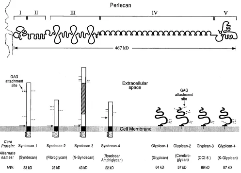
The syndecan, glypican, and perlecan core protein families constitute the major HSPGs generated within the cardiovascu-lar system (Fig. 1). The syndecan core protein family contains four human or murine members as well as a Drosophila homo-logue. Syndecan family members exhibit an extracellular re-gion with multiple glycosaminoglycan (GAG) attachment sites and a protease dibasic cleavage sequence, a homologous mem-brane spanning region, and a short highly conserved cytoplas-mic tail with four tyrosine residues at fixed positions (for re-view see reference 10). The sequences of the extracellular regions of syndecan family members are quite divergent ex-cept for the GAG attachment and putative cleavage sites, sug-gesting that this domain may function as a potentially cleav-able protein scaffold onto which GAGs are attached. The residues of the extracellular and transmembrane regions of syndecan family members allow homo-oligomerization which can permit the cytoplasmic tail to interact with and activate protein kinase C (11, 12). The cytoplasmic tails of syndecan family members also interact with either intracellular microfil-aments or focal adhesion, depending upon the core protein (13). In the former instance, a specific tyrosine residue appears to be involved in regulating this association (14). The phosphor-ylation of intracytoplasmic serine residues and possibly ty-rosine residues may play a critical role in the above process (15). Syndecan family core proteins exhibit cell type–specific distributions with vascular endothelial cells/smooth muscle cells expressing syndecans-1, -2, and -4 with predominant tar-geting to basolateral surfaces. The glypican core protein family is comprised of four human or murine members as well as the
Drosophila homologue dally. (Recent data reveal the glypican family to be even larger [Saunders, S., S. Paine-Saunders, and A.D. Lander, manuscript submitted for publication].) Glypi-can family members posses an extracellular region with GAG attachment sites, 14 invariant cysteine residues, which stabilize a highly compact tertiary structure, and a COOH-terminal GPI anchor. The extracellular regions of the different family mem-bers are quite similar, which suggests that these areas may be involved in important cellular functions such as binding to ligands or interaction with Golgi components to direct glyca-nation. Glypican family core proteins are selectively expressed on different cell types with only glypican-1 present on vascular endothelial cells/smooth muscle cells (for review see reference 16). These core proteins are mainly targeted to apical surfaces which are partially dependent upon the extent of glycanation (17). Perlecan represents the final class of core protein to be considered with only a single human or murine family mem-ber, but multiple splice variants are possible (18). The intact Address correspondence to Robert D. Rosenberg, Massachusetts
In-stitute of Technology, Bldg. 68-480, 77 Massachusetts Ave., Cam-bridge, MA 02139. Phone: 617-253-8804; FAX: 617-258-6553; E-mail: [email protected]
Accepted for publication 31 March 1997.
1. Abbreviations used in this paper: AT, antithrombin; CS, chon-droitin sulfate; FGFs, fibroblast growth factors; GAG, glycosami-noglycan; GlcA, glucuronic acid; GlcA 2S, 2-O-sulfated glucuronic acid; GlcNAc, N-acetylglucosamine; GlcN(Ac/S), N-acetyl or N -sul-fated glucosamine; GlcNAc 6S, 6-O-sulfated N-acetylglucosamine; GlcNS 3S, 3-O-sulfated N-sulfated glucosamine; HexA, unspecified uronic acid, either GlcA or IdoA; HS, heparan sulfate chains; HSPGs, heparan sulfate proteoglycans; IdoA, iduronic acid; IdoA 2S, 2-O-sulfated iduronic acid; LPL, lipoprotein lipase; NST, N -deacety-lase/N-sulfotransferase; 6-OST, 6-O-sulfotransferase. For all se-quences given, the linkages and configurations are →4-d-GlcApb1→,
→4-d-GlcNp(Ac/S)a1→, and →4-l-IdoApa1→.
Heparan Sulfate Proteoglycans of the Cardiovascular System
Specific Structures Emerge But How Is Synthesis Regulated?
Robert D. Rosenberg,*‡ Nicholas W. Shworak,*‡ Jian Liu,* John J. Schwartz,* and Lijuan Zhang*
*Department of Biology, Massachusetts Institute of Technology, Cambridge, Massachusetts 02139; and ‡Department of Medicine, Harvard
core protein contains five separate regions with a variety of in-triguing structural motifs decorated by posttranslational modi-fications such as GAG chains, O-linked nonsulfated oligosac-charides as well as long chain fatty acids. Perlecan is secreted by multiple cell types including vascular endothelial cells/ smooth muscle cells, and interacts with collagens, laminin, and other components within the basement membrane (19).
The biosynthetic mechanism of core protein glycanation has been extensively investigated over the past decade, but only partially defined. The glycanation of core proteins is initi-ated by four specific enzymes which generate a unique linkage region tetrasaccharide (20). The tetrasaccharide acceptor site is represented by a Ser-Gly (Ala)-X-Gly (Ala) sequence with subsequent attachment of HS, rather than chondroitin sulfate (CS), favored by multiple acidic amino acids at a distance of seven to nine residues, the occurrence of tryptophan residues in close proximity, and the presence of adjacent tracts of Ser-Gly repeats (for review see reference 21). However, the rela-tive intracellular concentrations of metabolic intermediates required for HS versus CS biosynthesis, and the overall
struc-tures of core proteins also play a critical role in specifying the linkage of a particular GAG. The attachment of HS to glypican family members is unusually selective, frequently ap-proaching 100%; the linkage of HS to the NH2 terminus of
perlecan is highly specific frequently approximating 80% with CS linked to the remaining sites, whereas the coupling of HS to syndecan family members is favored averaging 60% with the remaining sites decorated with CS (22–24). Under certain conditions, occasional acceptor sites in the above core proteins may be unsubstituted.
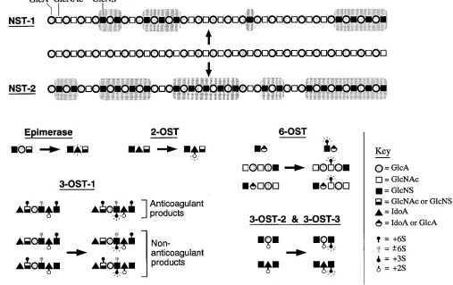
[image:3.612.60.557.63.419.2]spreading in both directions to generate modified domains of z 5 disaccharide units separated by relatively unmodified re-gions of z 18 disaccharide units. The C5 glucuronosyl/idu-ronosyl epimerase then catalyzes transformation of occasional
d-GlcA residues to l-iduronic acid (IdoA) residues if N
-sul-fate groups are present at the immediate upstream position. The equilibrium distribution of this reaction lies far in the di-rection of GlcA but epimerization is favored by sulfation of a IdoA. The iduronosyl 2-O-sulfotransferase (2-OST) then sulfates selected IdoA residues (creates IdoA 2S) provided that N -sul-fated glucosamine (GlcNS) groups are located at the immedi-ate upstream position and 6-O-sulfated glucosamine [GlcN (Ac/S) 6S] groups are absent from the immediate downstream position. The glucuronosyl 2-O-sulfotransferase also sulfates rare GlcA residues (creates GlcA 2S) which prevent subse-quent epimerization to IdoA. The glucosaminyl 6-O -sulfo-transferase (6-OST) then frequently sulfates GlcN(Ac/S) resi-dues provided that N-sulfate groups are present at either the immediate upstream or downstream positions (extensively re-viewed in reference 25). Finally, the glucosaminyl 3-O -sul-fotransferase (3-OST-1) sulfates occasional GlcNS and GlcNS 6S residues (creates GlcNS 3S and GlcNS 3S 6S, respectively) predominantly when →HexA (GlcA or IdoA)→GlcN(Ac/S)66S 6S→GlcA→ is present in the immediate upstream position and the downstream location is filled by an →IdoA 2S→ GlcNS→. Thus, the above enzymatic reactions which generate
distinct HS fine structures represent, at the very minimum, a partially ordered biochemical pathway with clear cut precur-sor/product relationships.
Until recently, all biosynthetic enzymes were postulated entities based upon the biochemical activities of cell lysates. During the past 4 yr, virtually all of these proteins have been purified and several have been molecularly cloned (26–32). The available data on the initial and final sulfation enzymes in the biosynthetic pathway provide intriguing clues about poten-tial mechanisms for generating HS with regions of defined monosaccharide sequence. The primary structures and bio-chemical specificities of murine/human NSTs and 3-OSTs have been determined. We note that NST-1 and NST-2 were cloned from hepatic cells and mast cells, respectively, but are also ex-pressed in other cell types (30–32). The two enzymes exhibit alternate specificities that initially lead to a varying extent of
[image:4.612.48.555.66.385.2]3-OSTs have been expressed, and the sites of sulfation have been determined. The results demonstrate that 3-OST-1 sul-fates GlcNS residues when the upstream uronic acid is GlcA, whereas 3-OST-2 and 3-OST-3 sulfate GlcNS residues when the upstream uronic acid is IdoA 2S or GlcA 2S (Fig. 2). The specificities of these enzymes are also dependent upon additional residues in the neighborhood of the site of sulfation. Based upon these data, we speculate that isoforms of biosynthetic en-zyme exist in different cell types which sulfate HS at specific sites based upon the surrounding monosaccharide sequence and hence are capable of producing GAGs with regions of de-fined structure. The regulation of the concentrations of these enzymes, provided that they serve as limiting components in a biosynthetic pathway, would establish particular levels of HS with regions of defined monosaccharide sequence.
Growth factors and HSPGs
Fibroblast growth factors (FGFs), vascular endothelial growth factor, heparin binding EGF, the Wnts, interleukin-3, GM-CSF, and IFN-gamma form tight complexes with HSPGs (for review see reference 33). These mitogens, differentiation fac-tors, and cytokines are critically involved in early develop-ment, angiogenesis, thrombosis, and atherosclerosis of the car-diovascular system. In the section below, we describe the regulation of FGF signaling which serves as a paradigm for elucidating the role of HSPGs in modulating the function of other growth factors/cytokines.
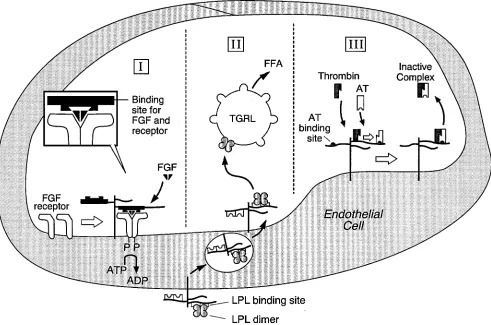
The HSPGs are required for high-affinity binding of FGFs to their receptors (34, 35) (Fig. 3). Syndecans-1, -2, and -4 as well as glypican-1, possess HS which carry out this function. The crystal structures of FGF-2 and heparin oligosaccharides reveal specific interactions between asp28, arg121, lys126, and gln135 of
[image:5.612.62.553.61.386.2]signaling sequence must be present on a dodecasaccharide to induce mitogenesis with no activity noted if the same se-quences are located on a decasaccharide. The two sese-quences appear to be necessary for binding to both growth factor and receptor.
Based upon the above data, it is surmised that HSPGs bearing specific binding and signaling sequences complex with FGF-2 to induce growth factor dimer formation and also in-teract directly with FGFR1 (34, 35, 39, 40). The end result is transient dimerization of the receptor which facilitates phos-phorylation of cytoplasmic tails, assembly of intracellular com-ponents, and mitogenesis (46, 47). Additional monosaccharide sequences may also be required to generate stably phosphory-lated higher order receptor complexes that lead to an in-creased strength of cell signaling (46). The regulation of FGFs by cell surface HSPGs is also likely to be modulated by extra-cellular and intraextra-cellular events. On the one hand, the FGFs interact with basement membrane perlecan which reduces the quantities of growth factor binding to receptor but also pro-vides a ready reservoir for rapid mobilization (48). On the other hand, the binding of the cytoplasmic tails of syndecans to cytoskeletal elements and protein kinase C may modulate the response to FGF signaling (11, 12). Finally, we note that the structures of HS required to initiate a mitogenic response with FGF-1 and FGF-4 are different from those described above (45). Thus, it appears likely that specific monosaccharide se-quences of HS may differentially regulate the biologic effects of FGF family members within the cardiovascular system.
Surprising results have emerged from genetic screens in
Drosophila that underscore the importance of growth factor regulation by HSPGs at the whole animal level. The Wnts are growth factors known to generate complexes with HS and play an important role in development. The seven membrane span-ning Frizzled gene family has been identified recently as the long-sought Wnt receptors, and in vitro depletion of HS from
Drosophila cells inhibits binding of Wnt 1 to Frizzled as well as suppresses cell signaling (49, 50). A genetic screen to identify new components in the Wnt signaling pathway generated mutant flies with deletions of NST that exhibit a severe devel-opmental malfunction of this system (51). Using a similar ap-proach, saturation mutagenesis was used to identify new effec-tors involved in cell division patterning in Drosophila, which uncovered dally mutants with abnormalities in M phase pro-gression (52). The sequencing of wild-type dally cDNA reveals an open reading frame extremely homologous to glypican with 14 cysteine residues and several GAG attachment sites as well as a putative GPI anchor. It appears likely that dally is a core protein that bears HS and is required for the binding of growth factor needed for normal cell division.
Lipoprotein metabolism and HSPGs
The metabolism of lipoproteins is partially regulated by HSPGs through interactions with lipoprotein lipase (LPL) and apolipoproteins B and E (apoB and apoE). LPL is the rate-limiting enzyme for hydrolysis of triglycerides in very low den-sity lipoproteins as well as chylomicrons and therefore controls the delivery of fatty acids to tissues. In the section below, we describe the modulation of this pathway by HSPGs which pro-vide a conceptual framework for considering the effects of GAGs on other cell surface bound enzymes in the cardiovas-cular system.
The primary site of action of LPL is the luminal surface of capillary endothelial cells where the enzyme is anchored to HSPGs (53) (Fig. 3). The core proteins involved in this interac-tion have not been completely defined but the available data are compatible with a major role for syndecan 1 (54). The en-zymatically active dimeric LPL binds 6,000-fold more tightly than inactive monomeric enzyme to HSGPs (55). This interac-tion allows dimeric LPL to be localized on the endothelial cell surface whereas monomeric enzyme enters the blood and is cleared by the liver. The binding of LPL to HSPGs fails to ap-preciably alter the conformation of the enzyme or its ability to interact with lipoproteins. Thus, LPL is targeted to the appro-priate locale to carry out its biologic role without perturbing structural features needed for optimal function. The localiza-tion of LPL to the endothelial cell surface may also involve a 116-kD NH2-terminal region processed form of apoB (NTAB)
which contains separate binding domains for LPL and HS. The endothelial cell expression of NTAB may alter the interactions of LPL with HSPGs or apoB containing lipoproteins. The api-cal anchoring of LPL exposes the enzyme to circulating triglyc-eride-rich lipoproteins, and the presence of apoE may facili-tate binding to endothelial cell HSGPs. The surface bound LPL attaches to lipoproteins, possibly by complexing to apoB, with the cofactor apoCII activating the enzyme. During lipoly-sis, LPL can dissociate from cell surfaces which makes it un-clear whether hydrolysis of triglycerides takes place at the cell surface or within the capillary lumen (for an extensive review see reference 56). Surprisingly, LPL is synthesized predomi-nantly in myocytes and adipocytes, and presumably exchanges between HSPGs of the extracellular matrix to reach the baso-lateral surface of endothelial cells. At this latter site, the en-zyme bound to carrier HSPGs is internalized and transcellu-larly transported to the apical surface (57). LPL complexed with HSPGs of cell surfaces and extracellular matrix can simul-taneously interact with lipoproteins, and such nonenzymatic bridging may affect lipoprotein localization and metabolism. On the one hand, retention of LDL and VLDL within the sub-endothelial space is dependent on the presence of both HSPGs and LPL (58). On the other hand, LPL-dependent degradation of lipoproteins is initiated through uptake by several distinct receptors and, in specific cell types, HSPGs may constitute a major, functionally independent internalization pathway (59–61).
The interaction of HS with dimeric LPL leads to the forma-tion of a 1:1 stoichiometric complex which depends upon spe-cific groups of the enzyme and GAG. Based upon the crystal-lographic structure of the homologous pancreatic lipase, the front surface of LPL is thought to contain the active site, whereas the back surface is believed to possess four regions of clustered basic residues (62). In particular residues 279–282 and 292–304 (human LPL numeration) show homology to the heparin-binding consensus sequences -X-B-B-X-B-X- and -X-B-B-B-X-X-B-X-, which are comprised of basic (B) and small neutral (X) amino acid residues (63). Mutational analysis demonstrates that Arg279, Arg280, Arg282, Arg296, Arg297, as well
de-casaccharide that represented 2% of the initial HS mass. These decasaccharides constitute the minimum size capable of bind-ing dimeric enzyme as efficiently as HS, and are represented by the sequences (IdoA 2S→GlcNS 6S→)5 and (IdoA 2S→
GlcNS 6S→)3 GlcA→GlcNS 6S→IdoA 2S→GlcNS 6S (67,
68). Detailed biochemical investigations reveal that ionic bonding to LPL only requires 10 of the 20 available negative groups, with nonelectrostatic interactions accounting for 40% of the total binding energy (55). Thus, more in depth structural analyses are required to establish the exact set of ionic and nonionic groups involved in the interaction of HS with LPL.
Blood coagulation and HSPGs
The blood coagulation cascade consists of a series of linked in-active precursor–serine protease transformations which gener-ate thrombin, thereby activating plgener-atelets as well as producing fibrin. The various natural anticoagulant mechanisms of the blood vessel wall oppose the action of the blood coagulation cascade and hence prevent thrombotic events. In the section below, we describe the endothelial cell anticoagulant heparan sulfate pathway which accelerates inhibition of coagulation proteases by a circulating plasma protease inhibitor and pro-vides a framework for considering how HSPGs with regions of defined monosaccharide sequence might be generated in other biologic systems.
The plasma protease inhibitor antithrombin (AT) slowly inactivates thrombin (T), Factor Xa, and other coagulation proteases by formation of enzyme–AT complexes (69) (Fig. 3). The binding of the active center of coagulation proteases to the reactive site of AT induces a partial insertion of the amino terminal of the protease inhibitor into its interior, which arrests cleavage of the reactive site bond and traps the enzyme in a stable complex. Heparin induces a conformational alteration in AT by binding to two sets of positively charged residues, and in certain cases augments interactions of protease inhibi-tor with free enzyme as well as enhances enzyme reactivity. These molecular events dramatically accelerate enzyme–AT complex generation. Once generation of the interaction prod-uct is completed, heparin dissociates and then catalyzes addi-tional rounds of enzyme neutralization by plasma inhibitor (69–71; for reviews see references 72 and 73). The affinity frac-tionation of heparin by AT revealed a small subpopulation of GAG with the unique sequence: →HexA→GlcN(Ac/S) 6S→ GlcA→GlcNS 3S 6S→IdoA 2S→GlcNS 6S→ which is mainly responsible for binding protease inhibitor and accelerating Factor Xa inhibition (74–79). The evaluation of individual res-idues demonstrate that the 6-O-sulfate group on residue 2 and the 3-O-sulfate group on residue 4 act in a concerted fashion to bind AT and accelerate Factor Xa neutralization (78, 79). The downstream flanking disaccharide, →IdoA 2S→GlcNS 6S→, is also of importance in this process (80). Two additional hep-arin domains of unknown structure are required for neutral-ization of thrombin, as well as the remaining coagulation pro-teases other than Factor Xa. Cultured cloned endothelial cells synthesize small amounts of anticoagulant HSPGs with co-valently linked HS possessing the AT binding site, which accel-erates enzyme–inhibitor complex formation. The physiologic role of endothelial cell anticoagulant HSPGs has been defined by perfusing rat hindlimbs with T and AT, and showing that T–AT complex generation is dramatically accelerated by a heparan sulfate vessel wall component. The in vivo location of anticoagulant HSPGs was ascertained by light and EM level
autoradiography which revealed small amounts of anticoagu-lant HSPGs on the luminal surface of endothelial cells with much larger quantities deposited in the subendothelial space (for review see reference 81). Thus, it appears likely that coag-ulation enzyme–AT interactions are dramatically accelerated in normal and damaged blood vessels by anticoagulant HSPGs. The HS biosynthetic pathway generates limiting amounts of anticoagulant HSPGs with regions of defined structure that contain the AT binding site, and also produces the more abun-dant nonanticoagulant HSPGs with regions of varying struc-ture that carry out other biologic functions. It has been argued that the amounts of anticoagulant HSPGs generated could not be formed by a completely random process and that the anti-coagulant HS biosynthetic mechanism must be ordered. How-ever, the anticoagulant HSPGs are produced by a complex pathway with multiple enzymatic components, and until re-cently regulation of this process remained obscure. The analy-ses of cell mutants, created by overexpression of the syndecan-4 core protein or chemical exposure, revealed that anticoagulant HS generation requires a pathway-specific component present in limiting amounts (82, 83). The rate-limiting step was identi-fied by establishing a cell-free system using microsomal con-version activity from wild-type microsomes, and radiolabeled HSPG precursor from microsomes of mutants blocked in anti-coagulant HSPG generation (84). The addition of these two extracts results in the production of large amounts of anticoag-ulant HSPG. Further investigation demonstrated that the con-centrations of microsomal conversion activity in many differ-ent cell types predicts the cellular levels of anticoagulant HSPG generated. The treatment of radiolabeled wild-type mi-crosomal and cell surface HSPGs with excess mimi-crosomal con-version activity transforms a maximum of 35% of total HSPGs into anticoagulant HSPG. This extent of conversion should be contrasted to the existing levels of cell surface anticoagulant HSPG, which average z 1–5% of total HSPGs (84). These in-vestigations provide convincing evidence that microsomal con-version activity represents the limiting intracellular component that transforms small amounts of an HSPG precursor popula-tion into anticoagulant HSPGs.
anticoagulant HS is controlled by the intracellular levels of a 3-OST isoform which sulfates specific sites in precursor GAGs depending upon the neighboring monosaccharide sequence. We note that 3-OST-2 or 3-OST-3, neither of which appears to be present in endothelial cells, sulfates precursor HS at other sites and hence produces GAGs with potential interaction sites for biologic targets other than AT. It is of interest that HS modified by the latter 3-OST isoforms has been identified in the glomeru-lar basement membrane where it has been hypothesized to be involved in regulating permeability to proteins (85, 86).
Conclusions
We have summarized recent advances in our knowledge of HSGPs, which demonstrate that covalently linked HS with regions of defined monosaccharide sequence interact with re-ceptors and proteins to regulate different biologic functions. Indeed, given the various cellular processes thought to be con-trolled by GAGs, one should anticipate the existence of nu-merous discrete HS sequences. However, it remains unclear how the biosynthetic mechanism is able to generate and specif-ically regulate the concentrations of each of these components. Based upon available data, we speculate that the various classes of sulfotransferases exist as multiple isoforms, each able to recognize and modify slightly different monosaccharide sequences. Cell type–specific expression of these isoforms would then dictate the synthesis of a particular array of GAGs. The actual levels of a given HS population with regions of defined monosaccharide sequence could then be regulated by controlling the concentrations/activities of key enzymes present in limiting amount. The validation of this hypothesis, or the development of alternative models, will require im-proved approaches for separating HS into functionally discrete components, more powerful methods for sequencing GAGs, and the availability of enzyme cDNAs to allow genetic manip-ulation of biosynthetic pathways. Recent progress in these three areas suggests that the mechanisms for controlling the di-versity of HSPGs will be delineated over the next few years. Success in this endeavor should provide us with novel insights into the regulation of different biologic functions, and lead to the development of new therapeutic approaches for cardiovas-cular disorders.
Acknowledgments
Apologies to all our colleagues whose experiments were not cited for lack of space.
The studies cited from this laboratory were supported by the Na-tional Institutes of Health (HL41484).
References
1. Simionescu, M., N. Simionescu, J.E. Sibert, and G.E. Palade. 1981. Dif-ferentiated microdomains on the luminal surface of the capillary endothelium.
II. Partial characterization of their anionic sites. J. Cell Biol. 90:614–621.
2. Conrad, H.E. 1989. Structure of heparan sulfate and dermatan sulfate. Ann. NY Acad. Sci. 556:18–28.
3. Yanagishita, M., and V.C. Hascall. 1992. Cell surface heparan sulfate
pro-teoglycans. J. Biol. Chem. 267:9451–9454.
4. Nathan, A., M.A. Nugent, and E.R. Edelman. 1995. Tissue engineered
perivascular endothelial cell implants regulate vascular injury. Proc. Natl. Acad.
Sci. USA. 92:8130–8134.
5. Yost, H.J. 1992. Regulation of vertebrate left-right symmetries by
extra-cellular matrix. Nature (Lond.). 357:158–161.
6. Sanderson, R.D., J.E. Turnbull, J.T. Gallagher, and A.D. Lander. 1994. Structure of heparan sulfate regulates proteoglycan function and cell behavior.
J. Biol. Chem. 269:13100–13106.
7. Guyton, J.R., R.D. Rosenberg, A.W. Clowes, and M.J. Karnovsky. 1980. Inhibition of rat arterial smooth muscle cell proliferation by heparin. In vivo
studies with anticoagulant and nonanticoagulant heparin. Circ. Res. 46:625–634.
8. Itoh, K., and S.Y. Sokol. 1994. Heparan sulfate proteoglycans are
re-quired for mesoderm formation in Xenopus embryos. Development (Camb.).
120:2703–2711.
9. Nikkari, S.T., H.T. Jarvelainen, T.N. Wight, M. Ferguson, and A.W. Clowes. 1994. Smooth muscle cell expression of extracellular matrix genes after
arterial injury. Am. J. Pathol. 144:1348–1356.
10. Bernfield, M., R. Kokenyesi, M. Kato, M.T. Hinkes, J. Spring, R.L. Gallo, and E.J. Lose. 1992. Biology of the syndecans: a family of
transmem-brane heparan sulfate proteoglycans. Annu. Rev. Cell Biol. 8:365–393.
11. Asundi, V.K., and D.J. Carey. 1995. Self-association of N-syndecan
(syndecan-3) core protein is mediated by a novel structural motif in the
trans-membrane domain and ectodomain flanking region. J. Biol. Chem. 270:26404–
26410.
12. Oh, E.-S., A. Woods, and J.R. Couchman. 1997. Multimerization of the cytoplasmic domain of syndecan-4 is required for its ability to activate protein
kinase C. J. Biol. Chem. In press.
13. Baciu, P.C., and P.F. Goetinck. 1995. Protein kinase C regulates the
re-cruitment of syndecan-4 into focal contacts. Mol. Biol. Cell. 6:1503–1513.
14. Carey, D.J., K.M. Bendt, and R.C. Stahl. 1996. The cytoplasmic domain of syndecan-1 is required for cytoskeleton association but not detergent
insolu-bility. Identification of essential cytoplasmic domain residues. J. Biol. Chem.
271:15253–15260.
15. Itano, N., K. Oguri, Y. Nagayasu, Y. Kusano, H. Nakanishi, G. David, and M. Okayama. 1996. Phosphorylation of a membrane-intercalated proteo-glycan, syndecan-2, expressed in a stroma-inducing clone from a mouse Lewis
lung carcinoma. Biochem. J. 315:925–930.
16. Weksberg, R., J.A. Squire, and D.M. Templeton. 1996. Glypicans: a
growing trend. Nat. Genet. 12:225–227.
17. Mertens, G., B. Van der Schueren, H. van den Berghe, and G. David. 1996. Heparan sulfate expression in polarized epithelial cells. The apical sorting of glypican (GPI-anchored proteoglycan) is inversely related to its heparan
sul-fate content. J. Cell Biol. 132:487–497.
18. Noonan, D.M., A. Fulle, P. Valente, S. Cai, E. Horigan, M. Sasaki, Y. Yamada, and J.R. Hassell. 1991. The complete sequence of perlecan, a base-ment membrane heparan sulfate proteoglycan, reveals extensive similarity with laminin A chain, low density lipoprotein-receptor, and the neural cell adhesin
molecule. J. Biol. Chem. 266:22939–22947.
19. Murdoch, A.D., G.R. Dodge, I. Cohen, R.S. Tuan, and R.V. Iozzo. 1992. Primary structure of the human heparan sulfate proteoglycan from base-ment membrane (HSPG2/perlecan). A chimeric molecule with multiple do-mains homologous to the low density lipoprotein receptor, laminin, neural cell
adhesion molecules, and epidermal growth factor. J. Biol. Chem. 267:8544–
8557.
20. Sugahara, K., H. Tsuda, K. Yoshida, S. Yamada, T. de Beer, and J.F. Vliegenthart. 1995. Structure determination of the octa- and decasaccharide se-quences isolated from the carbohydrate-protein linkage region of porcine
intes-tinal heparin. J. Biol. Chem. 270:22914–22923.
21. Esko, J.D., and L. Zhang. 1996. Influence of core protein sequence on
glycosaminoglycan assembly. Curr. Opin. Struct. Biol. 6:663–670.
22. David, G., V. Lories, B. Decock, P. Marynen, J.-J. Cassiman, and H. Van den Berghe. 1990. Molecular cloning of a phosphatidylinositol-anchored
membrane heparan sulfate proteoglycan from human lung fibroblasts. J. Cell
Biol. 111:3165–3176.
23. Dolan, M., T. Horchar, B. Rigatti, and J.R. Hassell. 1997. Identification
of sites in domain I of perlecan that regulate heparan sulfate synthesis. J. Biol.
Chem. 272:4316–4322.
24. Shworak, N.W., M. Shirakawa, R.C. Mulligan, and R.D. Rosenberg.
1994. Characterization of ryudocan glycosaminoglycan acceptor sites. J. Biol.
Chem. 269:21204–21214.
25. Lindahl, U. 1989. Biosynthesis of heparin and related polysaccharides.
In Heparin, Chemical and Biological Properties: Clinical Applications. D.A.
Lane and U. Lindahl, editors. Edward Arnold, London. 159–191.
26. Lind, T., U. Lindahl, and K. Lidholt. 1993. Biosynthesis of heparin/
heparan sulfate. Identification of a 70-kDa protein catalyzing both the d
-glucu-ronosyl- and the N-acetyl-d-glucosaminyltransferase reactions. J. Biol. Chem.
268:20706–20708.
27. Kobayashi, M., H. Habuchi, O. Habuchi, M. Saito, and K. Kimata. 1996. Purification and characterization of heparan sulfate 2-sulfotransferase from
cul-tured Chinese hamster ovary cells. J. Biol. Chem. 271:7645–7653.
28. Habuchi, H., O. Habuchi, and K. Kimata. 1995. Purification and charac-terization of heparan sulfate 6-sulfotransferase from the culture medium of
Chinese hamster ovary cells. J. Biol. Chem. 270:4172–4179.
29. Liu, J., N.W. Shworak, L.M.S. Fritze, J.M. Edelberg, and R.D.
Rosen-berg. 1996. Purification of heparan sulfate d-glucosaminyl 3-O-sulfotransferase.
J. Biol. Chem. 271:27072–27082.
30. Orellana, A., C.B. Hirschberg, Z. Wei, S.J. Swiedler, and M. Ishihara.
1994. Molecular cloning and expression of a glycosaminoglycan N
J. Biol. Chem. 269:2270–2276.
31. Hashimoto, Y., A. Orellana, G. Gil, and C.B. Hirschberg. 1992.
Molecu-lar cloning and expression of rat liver N-heparan sulfate sulfotransferase. J.
Biol. Chem. 267:15744–15750.
32. Eriksson, I., D. Sandbäck, B. Ek, U. Lindahl, and L. Kjellén. 1994.
cDNA cloning and sequencing of mouse mastocytoma glucosaminyl N
-deacety-lase/N-sulfotransferase, an enzyme involved in the biosynthesis of heparin. J.
Biol. Chem. 269:10438–10443.
33. Nelson, R.M., A. Venot, M.P. Bevilacqua, R.J. Linhardt, and I.
Stamen-kovic. 1995. Carbohydrate-protein interactions in vascular biology. Annu. Rev.
Cell Dev. Biol. 11:601–631.
34. Olwin, B.B., and A. Rapraeger. 1992. Repression of myogenic
differen-tiation by aFGF, bFGF, and K-FGF is dependent on cellular heparan sulfate. J.
Cell Biol. 118:631–639.
35. Ornitz, D.M., and P. Leder. 1992. Ligand specificity and heparin
depen-dence of fibroblast growth factor receptors 1 and 3. J. Biol. Chem. 267:16305–
16311.
36. Faham, S., R.E. Hileman, J.R. Fromm, R.J. Linhardt, and D.C. Rees. 1996. Heparin structure and interactions with basic fibroblast growth factor. Science (Wash. DC). 271:1116–1120.
37. Ornitz, D.M., A.B. Herr, M. Nilsson, J. Westman, C.-M. Svahn, and G. Waksman. 1995. FGF binding and FGF receptor activation by synthetic
hepa-ran-derived di- and trisaccharides. Science (Wash. DC). 268:432–436.
38. Yayon, A., M. Klagsbrun, J.D. Esko, P. Leder, and D.M. Ornitz. 1991. Cell surface, heparin-like molecules are required for binding of basic fibroblast
growth factor to its high affinity receptor. Cell. 64:841–848.
39. Kan, M., F. Wang, J. Xu, J.W. Crabb, J. Hou, and W.L. McKeehan. 1993. An essential heparin-binding domain in the fibroblast growth factor
re-ceptor kinase. Science (Wash. DC). 259:1918–1921.
40. Brickman, Y.G., M.D. Ford, D.H. Small, P.F. Bartlet, and V. Nur-combe. 1995. Heparan sulfates mediate the binding of basic fibroblast growth
factor to a specific receptor on neural precursor cells. J. Biol. Chem. 270:24941–
24948.
41. Habuchi, H., S. Suzuki, T. Saito, T. Tamura, T. Harada, K. Yoshida, and K. Kimata. 1992. Structure of a heparan sulphate oligosaccharide that binds to
basic fibroblast growth factor. Biochem. J. 285:805–813.
42. Maccarana, M., B. Casu, and U. Lindahl. 1993. Minimal sequence in
he-parin/heparan sulfate required for binding of basic fibroblast growth factor. J.
Biol. Chem. 268:23898–23905.
43. Turnbull, J.E., D.G. Fernig, Y. Ke, M.C. Wilkinson, and J.T. Gallagher. 1992. Identification of the basic fibroblast growth factor binding sequence in
fi-broblast heparan sulfate. J. Biol. Chem. 267:10337–10341.
44. Bai, X., and J.D. Esko. 1996. Mutant defective in heparan sulfate
hexu-ronic acid 2-O-sulfation. J. Biol. Chem. 271:17711–17717.
45. Guimond, S., M. Maccarana, B.B. Olwin, U. Lindahl, and A.C. Raprae-ger. 1993. Activating and inhibitory heparin sequences for FGF-2 (basic FGF).
Distinct requirements for FGF-1, FGF-2, and FGF-4. J. Biol. Chem. 268:23906–
23914.
46. Krufka, A., S. Guimond, and A.C. Rapraeger. 1996. Two hierarchies of FGF-2 signaling in heparin: mitogenic stimulation and high-affinity
binding/re-ceptor transphosphorylation. Biochemistry. 35:11131–11141.
47. Ueno, H., M. Gunn, K. Dell, A.J. Tseng, and L. Williams. 1992. A trun-cated form of fibroblast growth factor receptor 1 inhibits signal transduction by
multiple types of fibroblast growth factor receptor. J. Biol. Chem. 267:1470–
1476.
48. Aviezer, D., D. Hecht, M. Safran, M. Eisinger, G. David, and A. Yayon. 1994. Perlecan, basal lamina proteoglycan, promotes basic fibroblast growth
factor-receptor binding, mitogenesis, and angiogenesis. Cell. 79:1005–1013.
49. Perrimon, N. 1996. Serpentine proteins slither into the wingless and
hedgehog fields. Cell. 86:513–516.
50. Reichsman, F., L. Smith, and S. Cumberledge. 1996.
Glycosaminogly-cans can modulate extracellular localization of the wingless protein and
pro-mote signal transduction. J. Cell Biol. 135:819–827.
51. Lin, X., U. Haecker, and N. Perrimon. 1996. Mutations in the synthesis
of heparan sulfate result in defects of wg signaling. Wnt meeting 1996, Stanford
University.
52. Nakato, H., T.A. Futch, and S.B. Selleck. 1995. The division abnormally
delayed (dally) gene: a putative integral membrane proteoglycan required for cell division patterning during postembryonic development of the nervous
sys-tem in Drosophila. Development (Camb.). 121:3687–3702.
53. Cheng, C.F., A. Oosta, A. Bensadoun, and R.D. Rosenberg. 1981.
Bind-ing of lipoprotein lipase to endothelial cells in culture. J. Biol. Chem. 256:
12893–12896.
54. Saxena, U., M.G. Klein, and I.J. Goldberg. 1991. Identification and
characterization of the endothelial cell surface lipoprotein lipase receptor. J.
Biol. Chem. 266:17516–17521.
55. Lookene, A., O. Chevreuil, P. Ostergaard, and G. Olivecrona. 1996. In-teraction of lipoprotein lipase with heparin fragments and with heparan sulfate:
stoichiometry, stabilization, and kinetics. Biochemistry. 35:12155–12163.
56. Goldberg, I.J. 1996. Lipoprotein lipase and lipolysis: central roles in
li-poprotein metabolism and atherogenesis. J. Lipid Res. 37:693–707.
57. Saxena, U., M.G. Klein, and I.J. Goldberg. 1991. Transport of
lipopro-tein lipase across endothelial cells. Proc. Natl. Acad. Sci. USA. 88:2254–2258.
58. Eisenberg, S., E. Sehayek, T. Olivecrona, and I. Vlodavsky. 1992. Lipo-protein lipase enhances binding of lipoLipo-proteins to heparan sulfate on cell
sur-faces and extracellular matrix. J. Clin. Invest. 90:2013–2021.
59. Ji, Z.S., S. Fazio, and R.W. Mahley. 1994. Variable heparan sulfate pro-teoglycan binding of apolipoprotein E variants may modulate the expression of
type III hyperlipoproteinemia. J. Biol. Chem. 269:13421–13428.
60. Obunike, J.C., I.J. Edwards, S.C. Rumsey, L.K. Curtiss, W.D. Wagner, R.J. Deckelbaum, and I.J. Goldberg. 1994. Cellular differences in lipoprotein
li-pase-mediated uptake of low density lipoproteins. J. Biol. Chem. 269:13129–
13135.
61. Fernandez-Borja, M., D. Bellido, E. Vilella, G. Olivecrona, and S. Vilaro. 1996. Lipoprotein lipase-mediated uptake of lipoprotein in human fi-broblasts: evidence for an LDL receptor-independent internalization pathway. J. Lipid Res. 37:464–481.
62. van Tilbeurgh, H., A. Roussel, J.M. Lalouel, and C. Cambillau. 1994. Li-poprotein lipase. Molecular model based on the pancreatic lipase x-ray
struc-ture: consequences for heparin binding and catalysis. J. Biol. Chem. 269:4626–
4633.
63. Cardin, A.C., and H.R.J. Weintraub. 1989. Molecular modeling of
pro-tein-glycosaminoglycan interactions. Arteriosclerosis. 9:21–32.
64. Hata, A., D.N. Ridinger, S. Sutherland, M. Emi, Z. Shuhua, R.L. Myers, K. Ren, T. Cheng, I. Inoue, D.E. Wilson, P.-H. Iverius, and J.-M. Lalouel. 1993. Binding of lipoprotein lipase to heparin. Identification of five critical residues
in two distinct segments of the amino-terminal domain. J. Biol. Chem. 268:
8447–8457.
65. Nielsen, M.S., J. Brejning, R. García, H. Zhang, M.R. Hayden, S. Vilaró, and J. Gliemann. 1997. Segments in the C-terminal folding domain of lipopro-tein lipase important for binding to the low density lipoprolipopro-tein receptor-related
protein and to heparan sulfate proteoglycans. J. Biol. Chem. 272:5821–5827.
66. Hoogewerf, A.J., L.A. Cisar, D.C. Evans, and A. Bensadoun. 1991. Ef-fect of chlorate on the sulfation of lipoprotein lipase and heparan sulfate pro-teoglycans. Sulfation of heparan sulfate proteoglycans affects lipoprotein lipase
degradation. J. Biol. Chem. 266:16564–16571.
67. Parthasarathy, N., I.J. Goldberg, P. Sivaram, B. Mulloy, D.M. Flory, and W.D. Wagner. 1994. Oligosaccharide sequences of endothelial cell surface
heparan sulfate proteoglycan with affinity for lipoprotein lipase. J. Biol. Chem.
269:22391–22396.
68. Larnkjær, A., A. Nykjær, G. Olivecrona, H. Thogersen, and P.B. Øster-gaard. 1995. Structure of heparin fragments with high affinity for lipoprotein lipase and inhibition of lipoprotein lipase binding to alpha 2-macroglobulin-receptor/low-density-lipoprotein-receptor-related protein by heparin
frag-ments. Biochem. J. 307:205–214.
69. Rosenberg, R.D., and P.S. Damus. 1973. The purification and
mecha-nism of action of human antithrombin-heparin cofactor. J. Biol. Chem. 248:
6490–6505.
70. Carrell, R.W., P.E. Stein, G. Fermi, and M.R. Wardell. 1994. Biological
implications of a 3 Å structure of dimeric antithrombin. Structure (Lond.). 2:
257–270.
71. Gettins, P.G.W., B. Fan, B.C. Crews, and I.V. Turko. 1993. Transmis-sion of conformational change from the heparin binding site to the reactive
cen-ter of antithrombin. Biochemistry. 32:8385–8389.
72. Shworak, N.W., and R.D. Rosenberg. 1995. Antithrombotic defence
po-tential of the blood vessel wall: the heparan sulfate-antithrombin pathway. In
The Endothelial Cell in Health and Disease. J.R. Vane, G.V.R. Born, and D. Welzel, editors. Schattauer, Stuttgart. 119–146.
73. Olson, S.T., and I. Bjork. 1994. Regulation of thrombin activity by
anti-thrombin and heparin. Semin. Thromb. Hemostasis. 20:373–409.
74. Lam, L.H., J.E. Silbert, and R.D. Rosenberg. 1976. The separation of
active and inactive forms of heparin. Biochem. Biophys. Res. Commun. 69:
570–577.
75. Rosenberg, R.D., and L. Lam. 1979. Correlation between structure and
function of heparin. Proc. Natl. Acad. Sci. USA. 76:1218–1222.
76. Lindahl, U., G. Bäckström, L. Thunberg, and I.G. Leder. 1980.
Evi-dence for a 3-O-sulfated d-glucosamine residue in the antithrombin-binding
se-quence of heparin. Proc. Natl. Acad. Sci. USA. 77:6551–6555.
77. Choay, J., M. Petitou, J.C. Lormeau, P. Sinaÿ, B. Casu, and G. Gatti. 1983. Structure-activity relationship in heparin: a synthetic pentasaccharide with high affinity for antithrombin III and eliciting high anti-factor Xa activity. Biochem. Biophys. Res. Commun. 116:492–499.
78. Atha, D.H., J.C. Lormeau, M. Petitou, R.D. Rosenberg, and J. Choay. 1985. Contribution of monosaccharide residues in heparin binding to
anti-thrombin III. Biochemistry. 24:6723–6729.
79. Atha, D.H., J.C. Lormeau, M. Petitou, R.D. Rosenberg, and J. Choay.
1987. Contribution of 3-O- and 6-O-sulfated glucosamine residues in the
hep-arin-induced conformational change in antithrombin III. Biochemistry. 26:
6454–6461.
80. Casu, B. 1985. Structure and biological activity of heparin. Adv.
Carbo-hydr. Chem. Biochem. 43:51–134.
81. Marcum, J.A., and R.D. Rosenberg. 1989. The biochemistry, cell biol-ogy, and pathophysiology of anticoagulantly active heparin-like molecules of
Appli-cations. D.A. Lane and U. Lindahl, editors. Edward Arnold, London. 275–294. 82. Shworak, N.W., M. Shirakawa, S. Colliec-Jouault, J. Liu, R.C. Mulligan, L.K. Birinyi, and R.D. Rosenberg. 1994. Pathway-specific regulation of the
syn-thesis of anticoagulantly active heparan sulfate. J. Biol. Chem. 269:24941–
24952.
83. Colliec-Jouault, S., N.W. Shworak, J. Liu, A.I. de Agostini, and R.D. Rosenberg. 1994. Characterization of a cell mutant specifically defective in the
synthesis of anticoagulantly active heparan sulfate. J. Biol. Chem. 269:24953–
24958.
84. Shworak, N.W., L.M.S. Fritze, J. Liu, L.D. Butler, and R.D. Rosenberg. 1996. Cell-free synthesis of anticoagulant heparan sulfate reveals a limiting
ac-tivity which modifies a nonlimiting precursor pool. J. Biol. Chem. 271:27063–
27071.
85. Edge, A.S., and R.G. Spiro. 1990. Characterization of novel sequences
containing 3-O-sulfated glucosamine in glomerular basement membrane
hepa-ran sulfate and localization of sulfated disaccharides to a peripheral domain. J.
Biol. Chem. 265:15874–15881.
86. Kanwar, Y.S., A. Linker, and M.G. Farquhar. 1980. Increased perme-ability of the glomerular basement membrane to ferritin after removal of
gly-cosaminoglycans (heparan sulfate) by enzyme digestion. J. Cell Biol. 86:688–
693.
87. Ishihara, M., Y. Guo, Z. Wei, Z. Yang, S.J. Swiedler, A. Orellana, and C.B. Hirschberg. 1993. Regulation of biosynthesis of the basic fibroblast growth
factor binding domains of heparan sulfate by heparan sulfate-N-deacetylase/N
-sulfotransferase expression. J. Biol. Chem. 268:20091–20095.
88. Cheung, W.-F., I. Eriksson, M. Kusche-Gullberg, U. Lindahl, and L.
Kjellén. 1996. Expression of the mouse mastocytoma glucosaminyl N
-deacety-lase/N-sulfotransferase in human kidney 293 cells results in increased N
-sulfa-tion of heparan sulfate. Biochemistry. 35:5250–5256.
“Cell Adhesion In Vascular Biology”
Series Editors, Mark H. Ginsberg, Zaverio M. Ruggeri, and Ajit P. Varki
October 15, 1996 Adhesion and signaling in vascular cell–cell interactions ... Guy Zimmerman, Tom McIntyre, and
Stephen Prescott
November 1, 1996 Endothelial adherens junctions: implications in the control of vascular
permeability and angiogenesis ... Elisabetta Dejana
November 15, 1996 Genetic manipulation of vascular adhesion molecules in mice ...Richard O. Hynes and Denisa D. Wagner
December 1, 1996 The extracellular matrix as a cell cycle control element in
atherosclerosis and restenosis ... Richard K. Assoian and Eugene E. Marcantonio
December 15, 1996 Effects of fluid dynamic forces on vascular cell adhesion... Konstantinos Konstantopoulos and
Larry V. McIntire January 1, 1997 The biology of PECAM-1 ... Peter J. Newman January 15, 1997 Selectin ligands: Will the real ones please stand up?... Ajit Varki February 1, 1997 Cell adhesion and angiogenesis... Joyce Bischoff February 15, 1997 von Willebrand Factor... Zaverio Ruggeri
March 1, 1997 Therapeutic inhibition of carbohydrate–protein interactions in vivo ... John B. Lowe and Peter A. Ward
March 15, 1997 Integrins and vascular matrix assembly... Erkki Ruoslahti
April 1, 1997 Platelet GPIIb/IIIa antagonists: The first anti-integrin receptor therapeutics... Barry S. Coller
April 15, 1997 Biomechanical activation: An emerging paradigm in endothelial
adhesion biology... Michael A. Gimbrone, Jr., Tobi Nagel, and James N. Topper
May 1, 1997 Heparan sulfate proteoglycans of the cardiovascular system. Specific
structures emerge but how is synthesis regulated?... Robert D. Rosenberg, Nicholas W. Shworak, Jian Liu, John J. Schwartz, and Lijuan Zhang
May 15, 1997 New insights into integrin-ligand interaction ... Robert Liddington and Joseph Loftus
June 1, 1997 Adhesive interactions of Sickle erythrocytes with endothelium ... Robert Hebbel
June 15, 1997 Cell migration in vascular biology ... Stephen Schwartz
July 1, 1997 Integrin signaling in vascular biology ... Sanford Shattil and Mark Ginsberg
July 15, 1997 Multi-step mechanisms of leukocyte homing ... Eugene Butcher