Development 140, 2365-2376 (2013) doi:10.1242/dev.083865 © 2013. Published by The Company of Biologists Ltd
INTRODUCTION
In mice, lymphatic vascular development initiates around embryonic day (E) 9.75 with the expression of Prox1 in a subset of endothelial cells (ECs) of the cardinal vein (CV) (Oliver et al., 1993; Oliver and Harvey, 2002). Prox1, a homeobox transcription factor, functions as the master regulator of lymphatic endothelial cell (LEC) specification and maintains LEC fate (Wigle et al., 2002). In vitrostudies suggest that Prox1 functions in LEC progenitors to promote differentiation and upregulate LEC genes, such as those encoding SLC (Ccl21), neuropilin 2 (Nrp2), podoplanin and α9 integrin, and downregulate blood endothelial cell (BEC) genes, such as those that encode CD34, neuropilin 1 (Nrp1) and endoglin (Petrova et al., 2002; Hong and Detmar, 2003). Prox1deletion in mice results in a loss of LEC progenitor cells in veins and blocks lymphatic vascular development (Wigle and Oliver, 1999; Wigle et al., 2002; Johnson et al., 2008). By contrast, ectopic Prox1 expression in the blood endothelium results in enlarged lymphatic sacs (LSs) and edema (Kim et al., 2010). Loss of Prox1after completion of lymphatic development leads to a loss of LEC-specific proteins and misexpression of BEC markers, demonstrating that Prox1 is required to maintain LEC identity (Johnson et al., 2008).
At E9.75, expression of Prox1 in the CV requires Coup-TFII (Nr2f2 – Mouse Genome Informatics) and Sox18 transcription factors (Srinivasan et al., 2007; François et al., 2008; Lin et al., 2010). After E13.5, maintenance of Prox1 expression no longer requires Coup-TFII or Sox18, and maintenance of Prox1 transcription occurs by different mechanisms (Srinivasan et al., 2007; Srinivasan et al., 2010). Coup-TFII further cooperates with Prox1 to drive the expression of LEC genes, such as Nrp2and Vegfr3(Flt4) (Lee et al., 2009; Lin et al., 2010). Recently, it has been shown that Prox1 and Coup-TFII function together to regulate lymphovenous valve formation (Srinivasan and Oliver, 2011).
Migration of Prox1-positive LEC progenitors from the veins and formation of LSs require the activity of vascular endothelial growth factor C (VEGFC) (Karkkainen et al., 2004). VEGFR3 is a tyrosine kinase receptor that, in complex with the co-receptor Nrp2, binds VEGFC (Favier et al., 2006; Kärpänen et al., 2006; Xu et al., 2010). VEGFC through VEGFR3 induces LEC proliferation, survival and migration in culture (Mäkinen et al., 2001).
Notch signaling modulates cell-fate decisions via direct cell-cell contact (Andersson et al., 2011). The Notch pathway comprises a family of four transmembrane receptors (Notch 1-4) and five membrane-bound ligands (delta-like 1, delta-like 3, delta-like 4, jagged 1 and jagged 2). Receptor-ligand interactions release the intracellular domain via proteolysis that translocates to the nucleus, where it binds to CBF1/Su(H)/LAG1 (CSL) (Borggrefe and Liefke, 2012). The intracellular Notch peptide recruits a transcriptional activating complex to CSL converting it from a repressor to an activator of transcription, inducing multiple downstream targets such as the transcriptional repressors, HES and Hey.
In the blood vasculature, Notch signaling regulates arterial/ venous specification and sprouting angiogenesis. In the arterial endothelium, high delta-like 4 (Dll4)/Notch1 signaling maintains 1Department of Obstetrics and Gynecology, Columbia University Medical Center,
New York, NY 10032, USA. 2Department of Biosciences, Viikki Biocenter, University
of Helsinki, Helsinki, FI-00014, Finland. 3Department of Pharmacology, Columbia
University Medical Center, New York, NY 10032, USA. 4Pathology, Columbia
University Medical Center, New York, NY 10032, USA. 5Herbert Irving
Comprehensive Cancer Center, Columbia University Medical Center, New York, NY 10032, USA.
*These authors contributed equally to this work
‡These authors contributed equally to this work §Author for correspondence ([email protected])
Accepted 26 March 2013 SUMMARY
In development, lymphatic endothelial cells originate within veins and differentiate via a process requiring Prox1. Notch signaling regulates cell-fate decisions, and expression studies suggested that Jag1/Notch1 signaling functions in veins during lymphatic endothelial specification. Using an inducible lymphatic endothelial Prox1CreERT2driver, Notch signaling was suppressed by deleting Notch1or expressing dominant-negative Mastermind-like in Prox1+ endothelial cells. Either loss of Notch1or reduced Notch signaling increased Prox1+ lymphatic endothelial progenitor cell numbers in the veins, leading to incomplete separation of venous and lymphatic vessels. Notch loss of function resulted in excessive Prox1+ lymphatic cells emerging from the cardinal vein and significant lymphatic overgrowth. Moreover, loss of one allele of Notch1in Prox1heterozygous mice rescued embryonic lethality due to Prox1 haploinsufficiency and significantly increased Prox1+ lymphatic endothelial progenitor cell numbers. Expression of a constitutively active Notch1 protein in Prox1+ cells suppressed endothelial Prox1 from E9.75 to E13.5, resulting in misspecified lymphatic endothelial cells based upon reduced expression of podoplanin, LYVE1 and VEGFR3. Notch activation resulted in the appearance of blood endothelial cells in peripheral lymphatic vessels. Activation of Notch signaling in the venous endothelium at E10.5 did not arterialize the cardinal vein, suggesting that Notch can no longer promote arterialization in the cardinal vein during this developmental stage. We report a novel role for Notch1 in limiting the number of lymphatic endothelial cells that differentiate from the veins to assure proper lymphatic specification.
KEY WORDS: Notch1, Lymphatic endothelial cells, Prox1, Mouse
Notch1 functions as a negative regulator of lymphatic
endothelial cell differentiation in the venous endothelium
Aino Murtomaki1,2,*, Minji K. Uh1,3,*, Yun K. Choi1, Christopher Kitajewski1, Valeriya Borisenko1,Jan Kitajewski1,4,5,‡and Carrie J. Shawber1,‡,§
D
E
V
E
LO
P
M
E
N
arterial identity and inhibits venous EC differentiation (Gridley, 2010). In sprouting angiogenesis, Dll4 in tip cells signals to Notch1 in adjacent stalk cells to restrict the number of tip cells and vascular density (Jakobsson et al., 2009; Tung et al., 2012). Dll4/Notch1 signaling functions in lymphatic vascular development of zebrafish, where Dll4 promotes thoracic duct morphogenesis (Geudens et al., 2010). Dll4 expressed on the intersomitic arteries guides secondary lymphangiogenic sprouting from the posterior CV. In mice, Dll4/Notch1 signaling has been shown to function in sprouting lymphangiogenesis, by acting to control lymphatic tip cell differentiation (Niessen et al., 2011; Zheng et al., 2011). Consistent with this finding, Notch1 and Notch4 are expressed in dermal lymphatic endothelium, when the embryonic lymphatics are actively remodeling (Shawber et al., 2007).
We demonstrate a novel role for Notch in lymphatic vascular development: that of regulating the choice between venous and lymphatic endothelial identity during murine development. Using the Prox1CreERT2-inducible driver (Srinivasan et al., 2007), Notch
activity was manipulated in Prox1+ LEC progenitor cells in veins. Conditional loss of Notch1 or inhibition of Notch/CSL transcriptional activation resulted in precocious and excessive LEC differentiation that correlated with misexpression of Prox1 in veins and dilation of lymphatics. Ectopic Notch1 activation in Prox1+ ECs resulted in misspecified LECs, leading to severe edema, blood-filled lymphatics and incorporation of BECs into the peripheral lymphatics.
MATERIALS AND METHODS Constructs/cell culture
Early passage HdMVECs (Lonza) were infected with 25 pfu/cell adenoviruses encoding the intracellular domain of human NOTCH1 (N1IC), or lacZ, as previously described (Shawber et al., 2007). HdLECs were isolated from human foreskins (Exemption AAAB-1700) using CD31 conjugated Dynabeads (Invitrogen). Passage 1 cells were sorted by fluorescence-activated cell sorting (FACS) for podoplanin (Angiobio), then negatively sorted by FACS for CD34 (Pharmingen). HdLEC were maintained in EGM-2MV BulletKit (Lonza) with 10 ng/ml VEGFC (RnD). HdLEC lines were generated by lentiviral infection using pCCL.pkg.wpre vector encoding N1IC, Hey1, Hey2 or GFP (Tung et al., 2009). Transgene expression was confirmed by quantitative (q) RT-PCR and western blotting. For activation of endogenous Notch signaling, HDLEC grown to confluency on fibronectin-coated dishes were treated overnight with 200 nM Compound E, a gamma secretase inhibitor (GSI). The next day, GSI was removed and cells treated with 10 mM EDTA/PBS at 37C for up to 15 minutes. Medium was then replaced with EGM-2 MV containing 10 ng/ml VEGFC (R&D Systems).
RT-PCR/western analyses
RNA (RNeasy Mini Kit; Qiagen) and protein were isolated 24 hours after adenoviral infections and 48 hours after lentiviral infection (Shawber et al., 2007). RT-PCR was performed (Funahashi et al., 2010), and western blotting performed with antibodies against Prox1 (Millipore), or α-tubulin (Sigma). cDNAs were synthesized and qPCR performed with SYBR Green PCR Master Mix (ABI) and 7300 Real-Time PCR System (ABI). PCR amplicons for genes cloned into pDrive (Stratagene) served as reference standards. β-actin qRT-PCR was used to normalize samples. Primers are described in supplementary material Table S1.
Mice
Notch1+/−(Krebs et al., 2000), Prox1CreERT2(Srinivasan et al., 2007), Prox1GFPcre(Srinivasan et al., 2010; Srinivasan and Oliver, 2011), NI1C
(Buonamici et al., 2009), Notch1fl/fl(Yang et al., 2004), and DNMAML(Tu
et al., 2005) mice are described. Notch1fl/fland ROSA:lacZfl/flmice were
purchased (Jax Labs). Tamoxifen (5 mg/40 g or 7 mg/40 g) dissolved in corn oil was injected intraperitoneally at E9.75, E10.5, E13.5 and E14.5 for
N1ICand Notch1fl/flstudies. Tamoxifen (5 mg/40 g) was administered orally
at E9.75 for DNMAMLstudies. Two to seven litters were evaluated for each time point and are described in supplementary material Table S2.
Immunohistochemistry
Immunohistochemistry on 5 μm fixed-frozen/paraffin-embedded and 30 μm fixed-frozen sections was performed (Shawber et al., 2007). Whole mounts were performed as described (Lohela et al., 2008). Antibodies are listed in supplementary material Table S3. A M.O.M. kit (Vector Labs) was used with mouse monoclonal antibodies. Secondary antibodies were from Invitrogen (Alexa Fluor 488, Alexa Fluor 594) and Vector Labs (biotinylated anti-goat, biotinylated anti-hamster). Colorimetric staining was Hematoxylin counterstained. Nuclei visualized with DAPI in immunofluorescent staining. Images captured using a Nikon SMZ-U Zoom 1:10 microscope and Nikon 4500 digital camera, or Nikon ECLIPSE E800 microscope, Nikon DXM 1200 digital camera, and Image ProPlus software. Confocal microscopy performed with a Zeiss LSM 510 META confocal microscope and the LSM software.
Quantitation/statistical analyses
ImageJ software (NIH) was used for quantitative analyses of images. Number of embryos or section analyzed is summarized in supplementary material Table S4. Prox1+ cells within the CV or LS were normalized by vessel/sac circumference and emerging from CV by area analyzed. LYVE1 vessels were normalized by area analyzed to determine lymphatic vessel density. Significance between two groups determined by a t-test.
RESULTS
Notch1 activation suppresses lymphatic endothelial gene expression in microvascular endothelial cells
To determine whether Notch regulates LEC-specific gene expression, a constitutively active Notch1 protein (N1IC) was ectopically expressed in human neonatal dermal microvascular endothelial cells (HdMVECs) by adenoviral infection and human dermal lymphatic endothelial cells (HdLECs) by lentiviral infection. HdMVECs consist of BECs and LECs, and a subset of HdMVECs express LEC genes, Prox1, LYVE1 and podoplanin. Notch1 activation suppressed Prox1, podoplanin and LYVE1 transcripts in both HdMVECs and HdLECs, and Prox1 protein in HdMVECs (Fig. 1A,B). Notch activation did not affect the expression of Sox18 in HdLECs (supplementary material Fig. S1A). In HdLECs, ectopic expression of the Notch effectors, Hey1 or Hey2, reduced expression of LEC genes Prox1, LYVE1, and podoplanin (Fig. 1B). Hey2 suppression of Prox1 and LYVE1 was stronger relative to Hey1. Notch1- and Hey2-mediated downregulation of Prox1 and podoplanin in cultured LECs is consistent with previous observations (Kang et al., 2010).
Notch regulates the expression of Vascular endothelial growth factor receptors (VEGFRs) in BECs. Notch downregulates VEGFR2 (Taylor et al., 2002; Shawber et al., 2007; Funahashi et al., 2010). Notch either induces or suppresses VEGFR3, depending on the cellular context in vivo(Shawber et al., 2007; Tammela et al., 2008). The effect of activating Notch1 or ectopic Hey1 or Hey2 on VEGFR2/3 was evaluated in HdLECs. N1IC, Hey1 or Hey2 suppressed VEGFR2 transcript levels (Fig. 1C,D). N1IC induced VEGFR3, whereas ectopic Hey1 or Hey2 suppressed VEGFR3 (Fig. 1C,D), suggesting that Notch dynamically regulates VEGFR3 expression.
We previously established that Notch directly induces VEGFR3 via Notch/CSL transactivation of the VEGFR3promoter (Shawber et al., 2007). We hypothesize that VEGFR3 may be downregulated, secondary to Notch/CSL induction, by the action of Hey proteins, as part of a negative feedback loop. To evaluate this possibility, we determined the effect of activating endogenous Notch on the timing
D
E
V
E
LO
P
M
E
N
of VEGFR3 and Hey1/2 induction. HdLECs express Notch1, Notch4, jagged 1 and Dll4 (supplementary material Fig. S1B). Confluent HdLECs were grown overnight with the GSI Compound E to suppress Notch signaling. The following day GSI was removed, endogenous Notch was activated by EDTA, and RNA was collected at different time points post-Notch activation. Significant induction of VEGFR3 was observed after 5 minutes and reached maximum expression at 10 minutes (Fig. 1E). At 20 minutes, VEGFR3 induction was significantly downregulated, which followed low-level Hey1/2 induction, observed at 5 and 10 minutes. After Hey1/2 levels went back to baseline at 20 minutes, VEGFR3 was significantly up at 30 minutes. Strong induction of Hey1/2 observed at 60 minutes correlated with VEGFR3 downregulation. These data suggest that VEGFR3 is an immediate response gene downstream of Notch activation and downregulated by the Notch effectors Hey1 and Hey2.
Notch1 expression and activity in the CV during venous/lymphatic endothelial specification
LEC differentiation is a dynamic process that initiates around E9.75 with the onset of Prox1 expression and progresses from anterior to posterior along the CVs (Oliver et al., 1993; François et al., 2011). As Notch1 suppressed Prox1 in vitro(Fig. 1A,B), we hypothesized
that Notch acts in venous endothelium to restrict the differentiation of Prox1+ progenitor cells. Expression of Notch proteins (Notch 1-4) and Notch ligands Dll4 and jagged 1, relative to endothelial Prox1, was determined in murine E9.75 and E10.5 wild-type and Notch reporter (TNR) embryos. The transgenic TNR mouse line expresses green fluorescent protein (GFP) in response to Notch signal activation (Duncan et al., 2005). We found that the venous endothelium primarily expressed Notch1 and jagged 1 during this developmental period (Fig. 1F-H; supplementary material Fig. S1C-E).
[image:3.612.51.368.57.466.2]Analysis of E9.75 transverse sections anterior to the common atrial chamber identified a region of the CV in which Notch1 expression was the opposite of polarized Prox1 (Fig. 1F). This pattern differed from the aorta, where high Notch1 expression was observed throughout the aortic endothelium (Fig. 1F), suggesting distinct expression patterns in arterial versus venous vessels. The Notch ligands Dll4 and jagged 1 were both expressed in the aortic endothelium at E9.75 (supplementary material Fig. S1C). Notch activity was detected using a GFP antibody in a subset of Notch1-expressing CV ECs (Fig. 1F), when weak punctate jagged 1 expression was first observed in the E9.75 CV (supplementary material Fig. S1C). Thus, jagged 1 may function as the ligand for Notch1 in venous endothelium.
Fig. 1. Notch1 regulation of LEC-specific genes in
vitro, and Notch1 expression/activity in E9.75/E10.5 veins. (A) Prox1, LYVE1 and β-actin
RT-PCR and Prox1 and α-tubulin westerns of lacZ
(β-gal)- and N1IC-expressing HMVECs. (B-D) Prox1,
LYVE1, podoplanin, VEGFR2 and VEGFR3 qRT-PCR of N1IC-, Hey1-, Hey2- and GFP-expressing HdLECs.
(E) VEGFR3, Hey1 and Hey2 qRT-PCR on RNA isolated
at different times post EDTA-induced Notch activation. Data represented as means.e.m. *P<0.05, **P<0.05, ***P<0.005. (F) Endomucin (BEC marker), Prox1 (yellow arrowheads), and Notch1 staining of serial E9.75 transverse sections (original
magnification 20×). Endomucin or Notch1 (N1)/GFP staining of E9.75 TNR transverse sections to determine Notch activation (white arrowheads;
original magnification 50×). (G) Endomucin/Notch1,
endomucin (endo)/Prox1 or Notch1/Prox1 staining of serial E10.5 sagittal thick sections. Boxed areas enlarged on right (original magnification 20×).
(H) Notch1/Prox1 staining of E10.5 transverse thick
sections. White arrowheads highlight Prox1–/N1+ ECs, yellow arrowheads Prox1+/N1– ECs, blue arrowheads, Prox1+/N1+ within the CV (original
magnification 20×). (I) Endomucin/GFP staining of
E10.5 TNR transverse section. White arrowheads highlight ECs with Notch activity. ao, aorta; ba, brachial arch; cv, cardinal vein; d, dorsal; dao, descending aorta; jv, jugular vein; pcv, posterior cardinal vein; v, ventral.
D
E
V
E
LO
P
M
E
N
Analysis of thick sagittal sections of E10.5 embryos demonstrated that Notch1 expression was discontinuous in the CV and jugular vein (JV) with punctate, weak Notch1 expression observed dorsally (Fig. 1G). In serial sections, Prox1+ progenitors resided in the dorsal JV, where Notch1 was sometimes co-expressed with Prox1 (Fig. 1G). Dorsally migrating Prox1+ progenitors were negative for Notch1. In thick E10.5 transverse sections, Notch1 and Prox1 had both distinct and overlapping expression in the posterior CV (PCV; Fig. 1H). At this posterior position of the PCV, Prox1+ progenitors were evenly distributed, not yet polarized to one side. Three distinct expression patterns were observed for Prox1 and Notch1: PCV cells that expressed high Prox1 and low Notch1, cells that expressed low Prox1 and high Notch1 and cells that co-expressed both proteins (Fig. 1H). At E10.5, jagged 1 expression was polarized medially in the anterior CV (supplementary material Fig. S1D); this region displayed punctate Notch activation (GFP) (Fig. 1I; supplementary material Fig. S2A).
The level of Notch signaling in the CV was much lower than that observed in the aortic endothelium and vascular smooth muscle cells at E10.5 and E11.5 (supplementary material Fig. S2A,C), which correlated with the level of Notch1 expression (supplementary material Fig. S2B). Notch1 expression was evaluated in mesenteric vasculature that consists of arterial, venous and lymphatic vessels (supplementary material Fig. S2D). At E16.5 and E17.5, Notch1 expression was strongest in arteries, weaker in veins and weakest in lymphatics (supplementary material Fig. S2D,E). Quantification showed that Notch1 expression in the veins was 51% and 43% of that in the arteries at E16.5 and E17.5, respectively (supplementary material Fig. S2E). In the lymphatic vessels, Notch1 was expressed at 22% (E16.5) and 13% (E17.5) levels relative to the artery (supplementary material Fig. S2E). These studies show that Notch1 expression and activity is found in arterial, venous and lymphatic endothelium, but differs depending on the vessel type and developmental stage.
Loss of Notch signaling in Prox1-positive endothelial cells caused ectopic lymphatic endothelial cell specification and enlarged lymph sacs
To determine the effect of deleting Notch in the Prox1+ LEC progenitors residing in veins, we used the inducible Prox1-CreERT2
driver (Johnson et al., 2008). The Prox1-CreERT2 line drives
recombination in Prox1+ ECs in response to tamoxifen without disrupting the endogenous Prox1gene. Prox1CreERT2drivers were
crossed with mice carrying a floxed allele of Notch1(N1fl/fl) (Yang et
al., 2004), or a dominant-negative isoform of Mastermind-like (DNMAML) (Tu et al., 2005). DNMAML forms an inactive transcriptional complex with Notch on CSL without disrupting CSL repressor activity. Prox1CreERT2;N1fl/+males were crossed
with N1fl/+ females. Prox1CreERT2 females were crossed with
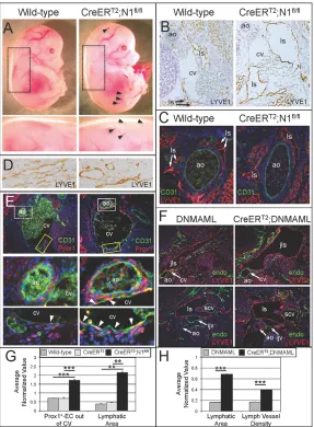
homozygous DNMAML males. Tamoxifen was administered at E9.75, a time when Prox1 expression initiates and Notch1 and Prox1 are co-expressed in the venous endothelium (Fig. 1F-H). E14.5 Prox1CreERT2;N1fl/fl (Fig. 2A) and Prox1CreERT2;DNMAML
embryos (data not shown) displayed mild edema and small foci of blood filled dermal lymphatics. Prox1CreERT2;N1fl/+,Prox1CreERT2
and N1fl/+control embryos were indistinguishable from wild-type
littermates (data not shown).
Immunohistochemistry on E14.5 wild-type and Prox1CreERT2;
N1fl/fltransverse sections was performed using antibodies against
BEC- (CD31/endomucin) and LEC- (Prox1/LYVE1) specific proteins. Compared with wild-type littermates, Prox1CreERT2;N1fl/fl
embryos had enlarged LSs surrounding the CV at the level of the aortic arch (Fig. 2B), descending thoracic aorta (Fig. 2C), and dermal lymphatic vessels (Fig. 2D). Prox1CreERT2;N1fl/flLS luminal area
was 5.7-fold greater than that in wild-type littermates and 4.7-fold greater than in Prox1CreERT2littermates (Fig. 2G). The increase in
LS size in Prox1CreERT2;N1fl/flembryos correlated with a 2.5-fold
increase in Prox1+ LECs emerging from veins relative to control embryos (Fig. 2E,G), indicating increased LEC differentiation. Similarly, Prox1CreERT2;DNMAMLlymphatic vessels were enlarged
4.2-fold and lymphatic vessel density increased 2.3-fold relative to DNMAMLcontrols (Fig. 2F,H). These data suggest that the increase in Prox1+ progenitors emerging from veins in Notch-deficient embryos contributed to lymphatic sac enlargement.
As VEGFC signaling is necessary for LEC progenitors to migrate from the CV (Favier et al., 2006; Kärpänen et al., 2006), expression of the VEGFC receptors VEGFR2 and VEGFR3 was evaluated. Conditional deletion of Notch1 in the Prox1+ ECs resulted in increased VEGFR2 expression in the venous endothelium, but not the lymphatic endothelium (supplementary material Fig. S3A), and is consistent with a loss of Notch-mediated suppression of VEGFR2 (Taylor et al., 2002; Shawber et al., 2007). VEGFR3 expression was relatively unaffected (supplementary material Fig. S3B). The increase in migrating Prox1+ cells may be due to changes in LEC VEGFR2 expression.
Loss of Notch in Prox1+ EC progenitors resulted LECs incorporation into veins
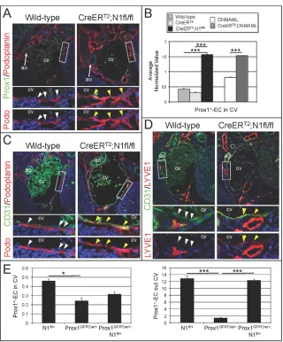
As expected, Prox1 expression was restricted to the lymphatic endothelium at E14.5 and rarely observed in the CV of control embryos (Fig. 2E, Fig. 3A). Prox1CreERT2;N1fl/fltissues had a
significant 3.75- and 5.32-fold increase in the number of Prox1+ cells in the CV relative to wild-type and Prox1CreERT2embryos,
respectively (Fig. 3B). Prox1 expression was observed throughout the CV of Prox1CreERT2;N1fl/fltissues at E14.5. This phenotype
was also observed in Prox1CreERT2;DNMAMLembryos that had a
1.89-fold increase in Prox1+ cells in the CV (Fig. 3B). Similar to conditional Notch loss-of-function mice, Prox1+ ECs residing in the CV was increased 1.58-fold in N1+/−heterozygotes at E10,
relative to wild-type littermates (supplementary material Fig. S4A,B). Thus, Notch1 activity is necessary to assure that Prox1 is suppressed in venous endothelium.
The presence of Prox1+ ECs in the CV of Notch mutant embryos suggested a specification defect in the CV endothelium. To explore this, we determined whether the CV expressed LYVE1 and podoplanin, proteins normally restricted to the lymphatic endothelium at E14.5 (Fig. 3A,C,D). In Prox1CreERT2;N1fl/fl
embryos, LYVE1 and podoplanin were misexpressed in the CV endothelium (Fig. 3A,C,D). Staining for podoplanin revealed that lymphatic vessels surrounding the CV of Prox1CreERT2;N1fl/fl
embryos were sometimes merged with the CV (Fig. 3A,C), indicating a failure to segregate lymphatic and venous vessels. LYVE1 was also misexpressed in the CV of E10.0 N1+/−embryos
relative to their wild-type littermates (supplementary material Fig. S4C). These data indicate that reduced Notch1activity resulted in an increase of venous Prox1+ ECs coincident with the expression of several key lymphatic proteins within veins, well beyond the time when LECs should have segregated from venous endothelium.
We examined whether Notch1loss could rescue the embryonic lethality of Prox1 haploinsufficiency, as might be predicted if Notch1 downregulates Prox1. In a mixed C57Bl and NMRI background, Prox1GFPCre/+mice, in which one allele of Prox1is
disrupted with a GFPCre cassette (Srinivasan et al., 2010; Srinivasan
D
E
V
E
LO
P
M
E
N
and Oliver, 2011), are not viable (Table 1). Prox1GFPCre/+mice were
crossed with N1fl/+mice to generate Prox1GFPCre/+;N1fl/+mice where
one copy of Notch1is lost in Prox1+ ECs. Prox1GFPCre/+;N1fl/+were
viable and observed at predicted frequency. Endomucin and Prox1 staining of E10.5 Prox1GFPCre/+;N1fl/+, Prox1GFPCre/+and N1fl/+
embryos was carried out to determine if improved viability correlated with rescued lymphatic endothelial specification. Prox1+ LECs were reduced 1.9-fold in the CV and 3.35-fold emerging out of the CV in Prox1GFPCre/+tissues relative to N1fl/+(Fig. 3E). Prox1+
LEC numbers were not significantly different in the CV of Prox1GFPCre/+;N1fl/+tissues relative to N1fl/+tissues. Prox1+ LECs
emerging from the CV were significantly increased 2.41-fold in Prox1GFPCre/+;N1fl/+tissues compared with Prox1GFPCre/+tissues.
Thus, losing one copy of Notch1in LEC progenitors rescued the LEC specification defect within the CV and viability of Prox1 heterozygous mice.
Notch1 activation in Prox1+ endothelial cells resulted in edema and blood-filled lymphatics To conditionally activate Notch1 within Prox1+ ECs, the Prox1CreERT2 driver was crossed with mice carrying a
Cre-responsive, constitutively activated NOTCH1 (N1IC) transgene downstream (N1IC) (Buonamici et al., 2009). Tamoxifen was administered at E9.75 or E10.5. Prox1CreERT2;N1ICembryos died
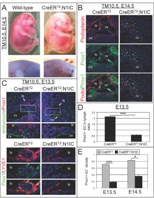
before E15.5. When analyzed at E14.5, Prox1CreERT2;N1IC
embryos displayed severe edema, and extensive networks of blood-filled superficial lymphatics (Fig. 4A; supplementary material Fig. S5A). A blood-filled jugular LS was observed in Prox1CreERT2;N1IC embryos (Fig. 4A). When tamoxifen was
administered at E10.5 and embryos isolated at E13.5, Prox1CreERT2;N1ICembryos displayed mild edema and appeared
to undergo normal blood vascular development (supplementary material Fig. S5A). Thus, the severe lymphatic defects arose between E13.5 and E14.5, when flow begins in the embryonic lymphatics. Staining for the intracellular domain of Notch1 confirmed that the N1IC transgene was expressed in the lymphatic endothelium of Prox1CreERT2;N1IC tissues (supplementary
material Fig. S5B). The Prox1CreERT2driver was crossed with a
mouse carrying a conditional lacZtransgene downstream of the ROSA26promoter to generate Prox1CreERT2;lacZfl/flmice that were
crossed with N1IC mice. Prox1CreERT2;lacZfl/+;N1ICembryos
displayed severe lymphatic vascular defects at E14.5. Staining for β-gal and LYVE1 confirmed recombination occurred in ECs incorporated into the LSs and nearby lymphatic vessels in control Prox1CreERT2;lacZfl/+, and Prox1CreERT2;lacZfl/+;N1ICtissues
[image:5.612.50.337.61.451.2](supplementary material Fig. S5C). Thus, Notch1 activation in Prox1+ ECs led to severe edema and embryonic lethality, probably related to venous/lymphatic defects.
Fig. 2. E9.75 conditional loss of Notch in Prox1+ ECs increased LEC differentiation at E14.5.(A) Prox1CreERT2;N1fl/fl
(CreERT2;N1fl/fl) embryos displayed mild edema and occasional
blood-filled dermal lymphatics (black arrowheads). Boxed
areas enlarged below. (B) LYVE1 staining of transverse sections
at the level of the aortic arch (original magnification 20×).
(C) CD31/LYVE1 staining of transverse sections through the
descending thoracic aorta (original magnification 20×).
(D) LYVE1 staining of dermal cross-sections (original
magnification 50×) (E) CD31/Prox1 staining of transverse
sections at the level of the aortic arch (original magnification 20×). White boxes enlarged below highlight Prox1+ ECs surrounding the aorta. Yellow boxes enlarged below highlight Prox1+ ECs residing in (white arrowheads) and emerging from
CV. (F) Endomucin/LYVE1 staining of E14.5 DNMAMLand
Prox1CreERT2;DNMAML(CreERT2;DNMAML) transverse sections through the level of the brachial arch (upper) and aortic arch
(lower; original magnification 10×). (G) Quantitation of Prox1+
EC density surrounding the CV (out), and LS luminal area in
Prox1CreERT2;N1fl/fland control tissues normalized by area.
(H) Quantitation of LS luminal area and lymphatic vessel
density in Prox1CreERT2;DNMAMLand DNMAMLtissues
normalized by area. (G,H) Data represented as means.e.m.
*P<0.05, ***P<0.0005. ao, aorta; cv, cardinal vein; ijv, internal jugular vein; jls, jugular lymph sac; ls, lymph sac; scv, subclavian vein.
D
E
V
E
LO
P
M
E
N
We hypothesized that the Prox1CreERT2;N1ICphenotype was
due to aberrant lymphatic endothelial specification. The lymphatic vascular phenotype was determined by evaluating Prox1, podoplanin, and LYVE1. In wild-type tissues, Prox1 expression was absent from the CV endothelium and restricted to the LSs and peripheral lymphatic vasculature at E13.5 and E14.5 (Fig. 4B,C). In E14.5 Prox1CreERT2;N1IC embryos, expression of Prox1 and
podoplanin was strongly reduced in the presumptive LSs (Fig. 4B). The presumptive LSs of E14.5 Prox1CreERT2;N1ICconsisted of
numerous small disorganized blood-filled channels (Fig. 4B; supplementary material Fig. S5D). These abnormal lymphatic channels had spotty Prox1 and weak podoplanin expression (Fig. 4B; supplementary material Fig. S5D). Unlike Notch loss-of-function models, the presumptive LSs clearly segregated from the
CV in Prox1CreERT2;N1IC embryos. A less severe lymphatic
phenotype was observed in E13.5 Prox1CreERT2;N1ICembryos
and correlated with a 3.72-fold decrease in Prox1+ ECs in the lymphatic endothelium, where LYVE1 expression was also reduced (Fig. 4C,D). The reduction in Prox1+ ECs was associated with a significant 3.8-fold and 2.31-fold decrease in Prox1+ EC density in Prox1CreERT2;N1ICtissues relative to control tissues at E13.5 and
E14.5, respectively (Fig. 4E). To determine if this phenotype was due to an early migration defect, the number of Prox1+ progenitor cells within and emerging from the CV was determined in E10.5 Prox1CreERT2;N1IC and control embryos with tamoxifen
administered at E9.75. Prox1+ progenitors numbers did not significantly differ between the two groups (supplementary material Fig. S6). Thus, activation of Notch1 in the Prox1+ progenitor cells suppressed lymphatic specification, opposite to that observed when Notch signaling was inhibited in the lymphatic progenitor ECs.
Notch1 inhibition of Prox1 was restricted to the initiation and early maintenance phase of lymphatic vascular development
[image:6.612.53.373.57.443.2]The transcription factors Sox18 and Coup-TFII cooperate to regulate Prox1 expression in lymphatic progenitor ECs between E9.75 and E13.5 in mice (François et al., 2008; Lee et al., 2009; Srinivasan et al., 2010). In blood vasculature, Coup-TFII and Notch function reciprocally to inhibit one another’s expression and
Fig. 3. Loss of Notch signaling in Prox1+ ECs resulted in lymphatic specification within the CV at E14.5, and rescued the Prox1
heterozygous phenotype at E10.5.
(A) Prox1/podoplanin staining of transverse
sections at the level of the aortic arch (original
magnification 20×). (B) Quantitation of Prox1+ EC
density in the CV. (C) CD31/podoplanin staining of
transverse sections at the level of the aortic arch. (A,C) Podoplanin expression (yellow arrowheads) and non-expression (white arrowheads) was observed in the CV Prox1CreERT2;N1fl/fland
wild-type tissues, respectively. (D) CD31/LYVE1 staining
of transverse sections at the level of the aortic arch. LYVE1 expression (yellow arrowheads) and non-expression (white arrowheads) was observed in the CV. (A,C,D) Original magnification 20×. Boxed areas
are enlarged below. (E) Quantitation of Prox1+ EC
density in and emerging from CV of N1fl/+, Prox1GFPCre/+and Prox1GFPCre/+;N1fl/+. (B,E) Prox1+ EC
[image:6.612.48.301.666.725.2]in CV normalized by CV circumference. Data represented as means.e.m. *P<0.05, **P<0.005., ***P<0.0005. ao, aorta; cv, cardinal vein.
Table 1. Viability of mice from Prox1CreGFP/+and Notch1fl/+ crosses
Genotype Predicted (%) Observed (%)
Wild-type 25 14.81
Notch1fl/+ 25 69.96
Prox1CreGFP/+ 25 0.0*
Prox1CreGFP/+;Notch1fl/+ 25 22.22
n=27; *P<0.001.
D
E
V
E
LO
P
M
E
N
maintain venous and arterial EC identity, respectively (You et al., 2005; Diez et al., 2007), suggesting that Notch downregulates TFII. In fact, Notch via Hey1 and Hey2 directly suppresses Coup-TFII expression in cultured HdLECs (Kang et al., 2010).
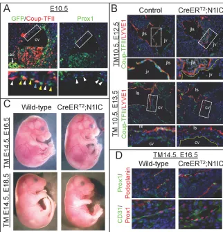
As Notch1 did not suppress Sox18 in HdLECs (supplementary material Fig. S1B), we analyzed Notch regulation of Coup-TFII during lymphatic endothelial specification. On the side of the CV where Prox1+ progenitors resided, Notch activity and Coup-TFII expression did not overlap, but they were observed in neighboring cells at E10.5 (Fig. 5A), consistent with Notch functioning to suppress Coup-TFII. To determine whether activation of Notch1 in Prox1+ ECs influenced Coup-TFII expression in vivo, E12.5 and E13.5 wild-type and Prox1CreERT2;N1ICtissues were stained for
Coup-TFII. In the wild-type CV, Coup-TFII was expressed throughout the venous and lymphatic endothelium (Fig. 5B). By contrast, Coup-TFII was strongly suppressed in the venous and LS ECs of Prox1CreERT2;N1ICembryos. The loss of Coup-TFII in the
Prox1CreERT2;N1IC embryos preceded Prox1 downregulation
(Fig. 4C), suggesting the loss of Prox1 may be secondary to that of Coup-TFII.
As Coup-TFII is not required to maintain Prox1 expression in LECs after E13.5 (Srinivasan et al., 2010), tamoxifen was administered at E13.5 and E14.5 to Prox1CreERT2and N1ICcrosses
and embryos isolated at E16.5 and E18.5. Tamoxifen administration at E14.5 resulted in Prox1CreERT2;N1ICembryos indistinguishable
from their wild-type and heterozygote littermates (Fig. 5C). Expression of Prox1 and podoplanin was unchanged in lymphatic endothelium of E16.5 Prox1CreERT2;N1ICtissues compared to
wild-type tissues (Fig. 5D). When recombination was initiated at E13.5, ~40% of E18.5 Prox1CreERT2;N1IC embryos displayed
mild lymphatic defects (supplementary material Fig. S7), including small blood-filled subcutaneous lymphatics around the base of the head. Thus, a developmental window exists from E9.75 to E13.5 during which Notch1 functions to suppress endothelial Prox1 in veins, most likely via its suppression of Coup-TFII.
Notch1 activation suppressed VEGFR2 and VEGFR3, but not Nrp2
Expression of multiple lymphatic-specific proteins was reduced in LEC progenitors in Prox1CreERT2;N1IC embryos; however,
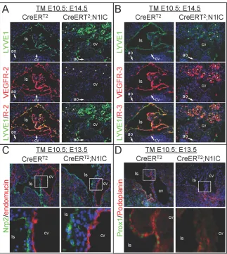
[image:7.612.52.354.58.445.2]misspecified LECs still segregated from veins and coalesced at sites of LS formation. LEC migration from veins requires VEGFC/VEGFR3/Nrp2 signaling (Karkkainen et al., 2004; Kärpänen et al., 2006; Xu et al., 2010). Expression of the VEGFC receptor complexes, VEGFR2, VEGFR3 and the VEGFR3 co-receptor, Nrp2, was evaluated. At E14.5, VEGFR2 was expressed in the CV and LS endothelium, and VEGFR3 expressed in the LS of control tissues (Fig. 6A,B; supplementary material Fig. S8A). By contrast, VEGFR2 and VEGFR3 expression was strongly reduced in E14.5 Prox1CreERT2;N1IC CV and LS. At E13.5, reduced
Fig. 4. Notch1 activation in Prox1+ LEC progenitors resulted in severe edema, blood-filled lymphatics and loss of Prox1.(A) E14.5 Prox1CreERT2;N1ICembryos
displayed severe edema (red arrowheads), blood-filled superficial lymphatics and jugular LS (blue arrowhead).
(B) Podoplanin/Prox1 staining of transverse sections at
the level of the aortic arch. Asterisks mark lymph sac-like structures (original magnification 20×).
(C) Endomucin/Prox1 (original magnification 20×) or
Prox1/LYVE1 (original magnification 50×) staining of transverse sections at the level of the aortic arch. Boxed
areas enlarged below. (D) Quantitation of Prox1+ ECs in
lymphatic sacs normalized by LS circumference.
(E) Quantitation of Prox1+ EC density at E13.5 and E14.5
normalized by area. (D,E) Data represented as
means.e.m. *P<0.05, ***P<0.0005. ao, aorta; cv, cardinal vein; ls, lymph sac.
D
E
V
E
LO
P
M
E
N
VEGFR2 expression was observed in LYVE1+ lymphatic vessels of Prox1CreERT2;N1IC tissues, whereas VEGFR3 expression
was absent in the Prox1CreERT2;N1IC lymphatic vessels
(supplementary material Fig. S8B,C). A reduction in VEGFR3 was observed in limb bud peripheral lymphatics of gain-of-function tissues (supplementary material Fig. S9A). Nrp2 expression within the LS endothelium was unaffected in Prox1CreERT2;N1IC
presumptive lymphatics (Fig. 6C), even though Prox1 and podoplanin expression was reduced (Fig. 6D). VEGFR2 and VEGFR3 were both strongly downregulated by Notch activation in Prox1+ LEC progenitors, whereas Nrp2 expression was maintained.
Notch1 activation in Prox1+ endothelial cells resulted in abnormal thoracic duct and peripheral lymphatic morphogenesis
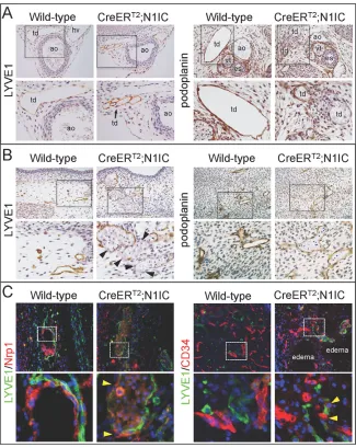
We examined the morphology of the thoracic duct and the peripheral lymphatics in embryos with ectopic Notch1 activation. Staining for LYVE1 or podoplanin revealed a defect in thoracic duct morphogenesis at the level of the descending thoracic aorta at E14.5. Instead of a well-defined thoracic duct observed in wild-type embryos, the thoracic duct of Prox1CreERT2;N1IC embryos
consisted of numerous small vessels (Fig. 7A). This phenotype correlated with discontinuous expression of podoplanin. The dermal lymphatics were more numerous in Prox1CreERT2;N1ICtissues,
with discontinuous LYVE1 expression and reduced podoplanin levels (Fig. 7B). As podoplanin is necessary to maintain the segregation between BEC and LECs, we evaluated expression of the blood endothelial markers, CD34 and Nrp1, in the peripheral lymphatics. CD34+ and Nrp1+ ECs were found in the enlarged peripheral lymphatic vessels of Prox1CreERT2;N1ICembryos, a
phenotype not seen in control tissue (Fig. 7C). LYVE1+ ECs were not observed in the peripheral blood vasculature, indicating that defects were restricted to the lymphatic vasculature. The ectopic appearance of Nrp1 and CD34 in peripheral lymphatics seen upon Notch1 activation could be interpreted as a defect in separation of blood and LECs during development, which might lead to abnormal blood-filled lymphatic vasculature.
Presumptive LS of Prox1CreERT2;N1ICembryos did
not express blood endothelial cell markers
Prox1 downregulates the expression BEC genes (Petrova et al., 2002; Hong and Detmar, 2003; Johnson et al., 2008). We determined the expression of the BEC markers, Nrp1 and CD34, both repressed by Prox1 (Wigle et al., 2002; Johnson et al., 2008). In E13.5 and E14.5 Prox1CreERT2;N1ICtissues, Nrp1 and CD34
expression was identical to controls (supplementary material Fig. S10A,B). Neither Nrp1, nor CD34 was misexpressed in LS endothelium, demonstrating that the LS endothelium of Prox1CreERT2;N1ICembryos did not misexpress BEC-specific
proteins.
Notch1 activation in Prox1+ endothelium did not induce arterial endothelial cell proteins
[image:8.612.52.374.60.395.2]Global or endothelial loss of Notch signaling results in venous gene expression in the arterial endothelium, whereas ectopic Notch activation leads to misexpression of arterial genes in the venous endothelium (Gridley, 2010). CV expression of arterial endothelial-specific proteins, CD34 and ephrinB2, and vascular smooth muscle cell recruitment was determined in the gain-of-function model. CD34 expression was unaltered in Prox1CreERT2;N1ICtissues
Fig. 5. Notch activation downregulated venous and lymphatic endothelial Coup-TFII.
(A) GFP/Coup-TFII or Prox1 staining of E10.5 TNR
transverse sections at the level of the heart. Boxed area enlarged below and readjusted to highlight expression pattern. White arrowheads highlight Coup–TFII+/GFP– ECs, yellow arrowheads Coup-TFII–/GFP+ ECs, and blue arrowheads Coup-TFII+/GFP+ ECs (original
magnification 20×). (B) Coup-TFII/LYVE1 staining
of E12.5 transverse sections at the level of jugular vein/LS and E13.5 transverse sections at the level of the aortic arch (original magnification 20×). E12.5 wild-type, E13.5
control-Prox1CreERT2. (C) E16.5 and E18.5 wild-type and Prox1CreERT2;N1IC(CreERT2;N1IC) embryos.
(D) Prox1/podoplanin or CD31/Prox1 staining of
E16.5 of peripheral lymphatic vessels (original magnification 50×). Tamoxifen was administered at E10.5 (B) and E14.5 (C,D). Boxed areas enlarged below. ao, aorta; cv, cardinal vein; jls, jugular lymph sac; jv, jugular vein; ls, lymph sac.
D
E
V
E
LO
P
M
E
N
(supplementary material Fig. S10B). EphrinB2 expression was restricted to the aortic endothelium, and SMA was expressed in aortic vascular smooth muscle cells of Prox1CreERT2;N1ICand
wild-type embryos (supplementary material Fig. S11A,B). Weak punctate expression of ephrinB2 observed in wild-type LS was not seen in Prox1CreERT2;N1IC LSs (supplementary material Fig.
S11A). Thus, mid-gestational Notch activation in the venous endothelium did not promote arterial endothelial differentiation, suggesting that there is a limited time window before E9.75 in which Notch functions to regulate arterial/venous endothelial identity.
DISCUSSION
We report a novel role for jagged 1/Notch1 signaling in the venous endothelium during lymphatic endothelial specification, whereby Notch suppresses lymphatic endothelial differentiation to maintain venous cell identity. We demonstrated that Notch1 inhibited Prox1 expression within the CV, most likely via suppression of Coup-TFII. Loss of Notch signaling, via either loss of Notch1or expression of DNMAML, in the LEC progenitors resulted in Prox1+ ECs in the CV and disrupted lymphatic and CV separation. Loss of Notch signaling led to over-commitment of LEC progenitors, resulting in enlarged lymphatic sacs and vessels. Loss of one allele of Notch1 also rescued embryonic lethality due to Prox1haploinsufficiency and resulted in a significant increase in Prox1+ EC progenitor cells at E10.5. By contrast, ectopic Notch1 signaling suppressed endothelial expression of Prox1, resulting in perturbed LEC
differentiation. These misspecified LECs failed to express LEC-specific proteins, podoplanin, LYVE1 and VEGFR3, but still emerged from veins and formed disorganized lymphatic sac-like structures. In gain-of-function embryos, BECs were found incorporated into the endothelium of blood-filled peripheral lymphatics, suggesting a failure in either segregation of BEC and LEC types or maintenance of LEC fate. Taken together, our results demonstrate an essential role for Notch1 in limiting the number of LECs that differentiate from the embryonic veins.
Our studies suggest that Notch has a distinct role in veins relative to arterial endothelium. Unlike the aorta, where Notch1, Dll4 and jagged 1 are strongly and uniformly expressed, Notch1 and jagged 1 expression was weak and discontinuous in the E9.75 and E10.5 veins, when Prox1+ LEC progenitors arise. Consistent with low levels of ligand and receptor expression, Notch activity was weaker in the CV compared with the aorta. In leukemogenesis and T-cell development, different levels of Notch signaling transactivate distinct target genes (Liu et al., 2010). Thus, the low levels of Notch signaling in the venous endothelium may induce unique target genes from those in arteries. In fact, activation of Notch in the Prox1+ progenitors did not lead to the expression of arterial-specific proteins in the CV. The ability of Notch to drive arterial specification may be limited to a specific developmental time window. Alternatively, Prox1+ progenitors may be sufficiently committed to the lymphatic fate to override Notch-driven arterialization.
[image:9.612.53.376.60.421.2]The genetic studies demonstrate that Notch1 functions in the venous endothelium to suppress lymphatic endothelial
Fig. 6. Notch activation downregulated VEGFR2 and VEGFR3.(A) LYVE1/VEGFR2,
(B) LYVE1/VEGFR3 staining of E14.5 transverse
sections. Asterisks mark lymph sac-like structures.
(C) Nrp2/endomucin, (D) Prox1/podoplanin
staining of E13.5 transverse serial section. Boxed areas are enlarged below. (A-D) Images at the level of the aortic arch (original magnification 20×). ao, aorta; cv, cardinal vein; ls, lymph sac.
D
E
V
E
LO
P
M
E
N
differentiation. Reduction of Notch signaling in Prox1+ venous cells resulted in precocious Prox1 expression in the CV. The extent of Prox1 misexpression in the CV of Notch loss-of-function embryos was much more extensive than that observed for either podoplanin or LYVE1. Therefore, Prox1 expression in the venous endothelium was not sufficient to drive LEC differentiation, and an additional factor with polarized expression may cooperate with Prox1 in LEC specification. One such potential factor, Sox18, is necessary for Prox1 expression and LEC specification (François et al., 2008). We found that Notch activation did not alter Sox18 in cultured HdLECs, although the effect of altering Notch1 signaling on Sox18 expression in vivoremains to be determined.
The ability of Notch1 to suppress endothelial Prox1 was limited to E9.75-E13.5. This developmental window overlaps with the requirement of Coup-TFII for Prox1 expression (Srinivasan et al., 2010), suggesting that Notch1 inhibited Prox1 via Coup-TFII repression. In HdLECs, Notch via its induction Hey1 and Hey2, inhibited Coup-TFII reporters (Kang et al., 2010). In the E10.5 CV, Notch activity and Coup-TFII expression was in neighboring cells consistent with Notch inhibiting Coup-TFII via lateral inhibition. In fact, ectopic Notch activation in Prox1+ progenitors strongly suppressed Coup-TFII expression in both the venous and lymphatic endothelium. Loss of Coup-TFII was observed at E12.5, 2 days
before strong Prox1 downregulation observed at E14.5. Taken together, the data suggest that jagged 1/Notch1 signaling in a subset of venous ECs downregulates Coup-TFII to suppress LEC differentiation and, by default, maintain venous identity. In cells with little to no Notch signaling, Coup-TFII is upregulated to drive Prox1 in LEC progenitors and repress Notch1 signaling. Thus, we propose that Notch1 regulates venous/lymphatic specification via suppression of the Coup-TFII/Prox1 signaling axis. Whether Notch signaling more directly represses Prox1, possibly at the level of transcription, remains unknown.
Loss and activation of Notch1 signaling both led to a failure to separate blood and lymphatic vasculature, but by distinct mechanisms. In Prox1CreERT2;N1fl/flembryos, lymphatic vessels
sometimes failed to separate from the CV, most likely because of the persistence of LECs within the CV. By contrast, BECs were incorporated in the affected peripheral lymphatic vessels of Prox1CreERT2;N1ICembryos. This gain-of-function phenotype
may arise from the reduced podoplanin expression, as podoplanin is necessary to maintain the segregation of blood and lymphatic vessels (Bertozzi et al., 2010). In support of this hypothesis, Notch1 signaling via the induction of Hey1 and Hey2 suppressed podoplanin in cultured HdLECs (Fig. 1) (Kang et al., 2010). In murine dermal lymphatics, Notch1 and podoplanin have
non-Fig. 7. Thoracic duct morphogenesis and BEC/LEC segregation defects in
Prox1CreERT2;N1ICembryos.(A) LYVE1 or
podoplanin staining of E14.5 transverse sections at the level of the descending aorta (left) and
esophagus (right). (B) LYVE1 or podoplanin
staining of dermal cross-sections. Black arrowheads highlight discontinuous LYVE1 in
affected dermal lymphatics. (C) Nrp1/LYVE1 or
CD34/LYVE1 staining of peripheral lymphatics. Yellow arrowheads mark Nrp1 and CD34 BECs incorporated into LYVE1+ vessels. (A-C) Boxed areas are enlarged below (original magnification 20×). ao, aorta; cv, cardinal vein; es, esophagus; hv, hemiazygos vein; ls, lymph sac; td, thoracic duct; vt, vagal trunk.
D
E
V
E
LO
P
M
E
N
[image:10.612.53.378.61.470.2]overlapping expression (Kang et al., 2010). The reduction of podoplanin in Prox1CreERT2;N1ICembryos was most likely the
result of a loss of Prox1, as podoplanin is induced by Prox1 (Johnson et al., 2008; Kim et al., 2010). Indeed, we found that loss of podoplanin expression was coincident with the loss of Prox1.
Loss of Notch signaling resulted in an increased number of LECs that emerged from the CV that correlated with lymphatic vessel enlargement and increased lymphatic density. Unexpectedly, misspecified LECs in Prox1CreERT2;N1ICembryos still separated
from veins and formed disorganized lymph sac-like structures, although Notch1 activation suppressed VEGFR3 in this model. This downregulation of VEGFR3 may occur via Notch induction of Hey1/2. Alternatively, it may be secondary to Notch suppression of the Coup-TFII/Prox1 axis, as VEGFR3 is a transcriptional target of both Coup-TFII and Prox1 (Lin et al., 2010; Srinivasan and Oliver, 2011). VEGFR3 and its co-receptor Nrp2 have been shown to be necessary to promote LEC migration towards VEGFC (Karkkainen et al., 2004; Xu et al., 2010). It is unknown if alternative signaling pathways can promote lymphatic endothelial migration independent of VEGFR3. The expression of VEGFR3 co-receptor, Nrp2, was unaffected in the LS and lymphatic vessel endothelium of the gain-of-function embryos. Nrp2 can form complexes with plexin to bind semaphorin3F in cultured LECs (Coma et al., 2011). Thus, Nrp2 may provide signals for migration away from the CV via different ligands, despite the reduced VEGFR3 levels. Alternatively, our in vitrostudies suggest that Notch transiently induces VEGFR3 in HdLECs, and this may be sufficient for the misspecified LECs to migrate away from veins. In fact, the number of Prox1+ progenitors emerging from the CV did not differ between control and Prox1CreERT2;N1IC embryos at E10.5. The level of VEGFR3
necessary for LEC migration may also be below the level of antibody detection and still functional in our model. Finally, a population of Nrp2high/LYVE1low LECs has been identified to migrate directly off the CV, but do not incorporate into lymph sacs and instead go on to form peripheral lymphatic vessels (François et al., 2011). In the Notch gain-of-function model, lymphatic channels that arise at lymph sac sites may arise from the migratory Nrp2high/LYVE1lowLECs. Thus, the disorganized lymph sac-like structures consist of numerous misspecified lymphatic vessels in Prox1CreERT2;N1ICembryos. Whether this Nrp2high/LYVE1low population also expresses VEGFR3 is unknown.
Our study of venous/lymphatic differentiation in mammals uncovered a unique role for Notch in limiting lymphatic endothelial differentiation in veins. Recent studies in zebrafish suggest that blood endothelial Dll4 signals to adjacent lymphatic endothelium to regulate lymphatic sprouting angiogenesis and formation of the thoracic duct (Geudens et al., 2010). Our studies do not exclude the possibility that Notch has a role in lymphatic sprouting or functions in guiding lymphatic growth along arterial vasculature. We observed that Notch signal activation no longer suppressed Prox1 after E14.5, and thus this would allow for Notch to function in the Prox1+ lymphatic endothelium later in development. Using inhibitors of Notch1 or Dll4, it was recently shown that Notch regulates postnatal and pathological lymphatic sprouting angiogenesis (Niessen et al., 2011), and that Notch has a role in restricting in lymphatic tip cells (Zheng et al., 2011). Consistent with these findings, Notch1 and Notch4 are expressed in postnatal dermal lymphatic vessels and tumor lymphatic vasculature (Shawber et al., 2007). Thus, Notch may have a distinct role in the postnatal lymphatic endothelium.
We conclude that venous to lymphatic differentiation represents a unique Notch-regulated cell-fate decision. Once the fate of lymphatic endothelium has been achieved, the differentiated lymphatic
endothelium may once again call upon a Notch-mediated fate mechanism to further specialize or shape the lymphatic vasculature.
Acknowledgements
We thank Darrell Yamashiro for technical assistance, Eric Olsen for providing Hey1 and Hey2 cDNAs, Guillermo Oliver and Sathish Srinivasan for providing Prox1CreERT2and Prox1GFPCremice, and Warren Pear for providing DNMAML mice.
Funding
Work supported by the National Institutes of Health/National Cancer Institute (NIH/NCI) [R01CA136673 to J.K. and C.J.S.], the National Institutes of Health/National Heart, Lung, and Blood Institute (NIH/NHLBI) [1RO1 HL112626 to J.K] and by a DOD pre-doctoral fellowship [W81XWH-10-1-0304 to M.K.U.]. Deposited in PMC for release after 12 months.
Competing interests statement
The authors declare no competing financial interests.
Supplementary material
Supplementary material available online at
http://dev.biologists.org/lookup/suppl/doi:10.1242/dev.083865/-/DC1
References
Andersson, E. R., Sandberg, R. and Lendahl, U.(2011). Notch signaling: simplicity in design, versatility in function. Development138, 3593-3612.
Bertozzi, C. C., Schmaier, A. A., Mericko, P., Hess, P. R., Zou, Z., Chen, M., Chen, C. Y., Xu, B., Lu, M. M., Zhou, D. et al.(2010). Platelets regulate lymphatic vascular development through CLEC-2-SLP-76 signaling. Blood116, 661-670.
Borggrefe, T. and Liefke, R.(2012). Fine-tuning of the intracellular canonical Notch signaling pathway. Cell Cycle11, 264-276.
Buonamici, S., Trimarchi, T., Ruocco, M. G., Reavie, L., Cathelin, S., Mar, B. G., Klinakis, A., Lukyanov, Y., Tseng, J. C., Sen, F. et al.(2009). CCR7 signalling as an essential regulator of CNS infiltration in T-cell leukaemia. Nature459, 1000-1004.
Coma, S., Shimizu, A. and Klagsbrun, M.(2011). Hypoxia induces tumor and endothelial cell migration in a semaphorin 3F- and VEGF-dependent manner via transcriptional repression of their common receptor neuropilin 2. Cell Adh. Migr.5, 266-275.
Diez, H., Fischer, A., Winkler, A., Hu, C. J., Hatzopoulos, A. K., Breier, G. and Gessler, M.(2007). Hypoxia-mediated activation of Dll4-Notch-Hey2 signaling in endothelial progenitor cells and adoption of arterial cell fate. Exp. Cell Res. 313, 1-9.
Duncan, A. W., Rattis, F. M., DiMascio, L. N., Congdon, K. L., Pazianos, G., Zhao, C., Yoon, K., Cook, J. M., Willert, K., Gaiano, N. et al.(2005). Integration of Notch and Wnt signaling in hematopoietic stem cell maintenance. Nat. Immunol.6, 314-322.
Favier, B., Alam, A., Barron, P., Bonnin, J., Laboudie, P., Fons, P., Mandron, M., Herault, J. P., Neufeld, G., Savi, P. et al.(2006). Neuropilin-2 interacts with VEGFR-2 and VEGFR-3 and promotes human endothelial cell survival and migration. Blood108, 1243-1250.
François, M., Caprini, A., Hosking, B., Orsenigo, F., Wilhelm, D., Browne, C., Paavonen, K., Karnezis, T., Shayan, R., Downes, M. et al.(2008). Sox18 induces development of the lymphatic vasculature in mice. Nature456, 643-647.
François, M., Short, K., Secker, G. A., Combes, A., Schwarz, Q., Davidson, T. L., Smyth, I., Hong, Y. K., Harvey, N. L. and Koopman, P.(2012). Segmental territories along the cardinal veins generate lymph sacs via a ballooning mechanism during embryonic lymphangiogenesis in mice. Dev. Biol.364, 89-98.
Funahashi, Y., Shawber, C. J., Vorontchikhina, M., Sharma, A., Outtz, H. H. and Kitajewski, J.(2010). Notch regulates the angiogenic response via induction of VEGFR-1. J. Angiogenes Res.2, 3.
Geudens, I., Herpers, R., Hermans, K., Segura, I., Ruiz de Almodovar, C., Bussmann, J., De Smet, F., Vandevelde, W., Hogan, B. M., Siekmann, A. et al.(2010). Role of delta-like-4/Notch in the formation and wiring of the lymphatic network in zebrafish. Arterioscler. Thromb. Vasc. Biol.30, 1695-1702.
Gridley, T.(2010). Notch signaling in the vasculature. Curr. Top. Dev. Biol.92, 277-309.
Hong, Y. K. and Detmar, M.(2003). Prox1, master regulator of the lymphatic vasculature phenotype. Cell Tissue Res.314, 85-92.
Jakobsson, L., Bentley, K. and Gerhardt, H.(2009). VEGFRs and Notch: a dynamic collaboration in vascular patterning. Biochem. Soc. Trans.37, 1233-1236.
Johnson, N. C., Dillard, M. E., Baluk, P., McDonald, D. M., Harvey, N. L., Frase, S. L. and Oliver, G.(2008). Lymphatic endothelial cell identity is reversible and its maintenance requires Prox1 activity. Genes Dev.22, 3282-3291.
D
E
V
E
LO
P
M
E
N
Kang, J., Yoo, J., Lee, S., Tang, W., Aguilar, B., Ramu, S., Choi, I., Otu, H. H., Shin, J. W., Dotto, G. P. et al.(2010). An exquisite cross-control mechanism among endothelial cell fate regulators directs the plasticity and heterogeneity of lymphatic endothelial cells. Blood116, 140-150.
Karkkainen, M. J., Haiko, P., Sainio, K., Partanen, J., Taipale, J., Petrova, T. V., Jeltsch, M., Jackson, D. G., Talikka, M., Rauvala, H. et al.(2004). Vascular endothelial growth factor C is required for sprouting of the first lymphatic vessels from embryonic veins. Nat. Immunol.5, 74-80.
Kärpänen, T. H. C., Heckman, C. A., Keskitalo, S., Jeltsch, M., Ollila, H., Neufeld, G., Tamagnone, L. and Alitalo, K.(2006). Functional interaction of VEGF-C and VEGF-D with neuropilin receptors. FASEB J.20, 1462-1472.
Kim, H., Nguyen, V. P., Petrova, T. V., Cruz, M., Alitalo, K. and Dumont, D. J.
(2010). Embryonic vascular endothelial cells are malleable to reprogramming via Prox1 to a lymphatic gene signature. BMC Dev. Biol.10, 72.
Krebs, L. T., Xue, Y., Norton, C. R., Shutter, J. R., Maguire, M., Sundberg, J. P., Gallahan, D., Closson, V., Kitajewski, J., Callahan, R. et al.(2000). Notch signaling is essential for vascular morphogenesis in mice. Genes Dev.14, 1343-1352.
Lee, S., Kang, J., Yoo, J., Ganesan, S. K., Cook, S. C., Aguilar, B., Ramu, S., Lee, J. and Hong, Y. K.(2009). Prox1 physically and functionally interacts with COUP-TFII to specify lymphatic endothelial cell fate. Blood113, 1856-1859.
Lin, F. J., Chen, X., Qin, J., Hong, Y. K., Tsai, M. J. and Tsai, S. Y.(2010). Direct transcriptional regulation of neuropilin-2 by COUP-TFII modulates multiple steps in murine lymphatic vessel development. J. Clin. Invest.120, 1694-1707.
Liu, H., Chi, A. W., Arnett, K. L., Chiang, M. Y., Xu, L., Shestova, O., Wang, H., Li, Y. M., Bhandoola, A., Aster, J. C. et al.(2010). Notch dimerization is required for leukemogenesis and T-cell development. Genes Dev.24, 2395-2407.
Lohela, M., Heloterä, H., Haiko, P., Dumont, D. J. and Alitalo, K.(2008). Transgenic induction of vascular endothelial growth factor-C is strongly angiogenic in mouse embryos but leads to persistent lymphatic hyperplasia in adult tissues. Am. J. Pathol.173, 1891-1901.
Mäkinen, T., Veikkola, T., Mustjoki, S., Karpanen, T., Catimel, B., Nice, E. C., Wise, L., Mercer, A., Kowalski, H., Kerjaschki, D. et al.(2001). Isolated lymphatic endothelial cells transduce growth, survival and migratory signals via the VEGF-C/D receptor VEGFR-3. EMBO J.20, 4762-4773.
Niessen, K., Zhang, G., Ridgway, J. B., Chen, H., Kolumam, G., Siebel, C. W. and Yan, M.(2011). The Notch1-Dll4 signaling pathway regulates mouse postnatal lymphatic development. Blood118, 1989-1997.
Oliver, G. and Harvey, N.(2002). A stepwise model of the development of lymphatic vasculature. Ann. N. Y. Acad. Sci.979, 159-165, discussion 188-196.
Oliver, G., Sosa-Pineda, B., Geisendorf, S., Spana, E. P., Doe, C. Q. and Gruss, P.(1993). Prox 1, a prospero-related homeobox gene expressed during mouse development. Mech. Dev.44, 3-16.
Petrova, T. V., Mäkinen, T., Mäkelä, T. P., Saarela, J., Virtanen, I., Ferrell, R. E., Finegold, D. N., Kerjaschki, D., Ylä-Herttuala, S. and Alitalo, K.(2002). Lymphatic endothelial reprogramming of vascular endothelial cells by the Prox-1 homeobox transcription factor. EMBO J.21, 4593-4599.
Shawber, C. J., Funahashi, Y., Francisco, E., Vorontchikhina, M., Kitamura, Y., Stowell, S. A., Borisenko, V., Feirt, N., Podgrabinska, S., Shiraishi, K. et al.
(2007). Notch alters VEGF responsiveness in human and murine endothelial cells by direct regulation of VEGFR-3 expression. J. Clin. Invest.117, 3369-3382.
Srinivasan, R. S. and Oliver, G.(2011). Prox1 dosage controls the number of lymphatic endothelial cell progenitors and the formation of the lymphovenous valves. Genes Dev.25, 2187-2197.
Srinivasan, R. S., Dillard, M. E., Lagutin, O. V., Lin, F. J., Tsai, S., Tsai, M. J., Samokhvalov, I. M. and Oliver, G.(2007). Lineage tracing demonstrates the venous origin of the mammalian lymphatic vasculature. Genes Dev.21, 2422-2432.
Srinivasan, R. S., Geng, X., Yang, Y., Wang, Y., Mukatira, S., Studer, M., Porto, M. P., Lagutin, O. V. and Oliver, G.(2010). The nuclear hormone receptor Coup-TFII is required for the initiation and early maintenance of Prox1 expression in lymphatic endothelial cells. Genes Dev.24, 696-707.
Tammela, T., Zarkada, G., Wallgard, E., Murtomäki, A., Suchting, S., Wirzenius, M., Waltari, M., Hellström, M., Schomber, T., Peltonen, R. et al.
(2008). Blocking VEGFR-3 suppresses angiogenic sprouting and vascular network formation. Nature454, 656-660.
Taylor, K. L., Henderson, A. M. and Hughes, C. C.(2002). Notch activation during endothelial cell network formation in vitro targets the basic HLH transcription factor HESR-1 and downregulates VEGFR-2/KDR expression.
Microvasc. Res.64, 372-383.
Tu, L., Fang, T. C., Artis, D., Shestova, O., Pross, S. E., Maillard, I. and Pear, W. S.(2005). Notch signaling is an important regulator of type 2 immunity. J. Exp. Med.202, 1037-1042.
Tung, J. J., Hobert, O., Berryman, M. and Kitajewski, J.(2009). Chloride intracellular channel 4 is involved in endothelial proliferation and morphogenesis in vitro. Angiogenesis12, 209-220.
Tung, J. J., Tattersall, I. W. and Kitajewski, J.(2012). Tips, stalks, tubes: notch-mediated cell fate determination and mechanisms of tubulogenesis during angiogenesis. Cold Spring Harb. Perspect Med.2, a006601.
Wigle, J. T. and Oliver, G.(1999). Prox1 function is required for the development of the murine lymphatic system. Cell98, 769-778.
Wigle, J. T., Harvey, N., Detmar, M., Lagutina, I., Grosveld, G., Gunn, M. D., Jackson, D. G. and Oliver, G.(2002). An essential role for Prox1 in the induction of the lymphatic endothelial cell phenotype. EMBO J.21, 1505-1513.
Xu, Y., Yuan, L., Mak, J., Pardanaud, L., Caunt, M., Kasman, I., Larrivée, B., Del Toro, R., Suchting, S., Medvinsky, A. et al.(2010). Neuropilin-2 mediates VEGF-C-induced lymphatic sprouting together with VEGFR3. J. Cell Biol.188, 115-130.
Yang, X., Klein, R., Tian, X., Cheng, H.-T., Kopan, R. and Shen, J.(2004). Notch activation induces apoptosis in neural progenitor cells through a p53-dependent pathway. Dev. Biol.269, 81-94.
You, L. R., Lin, F. J., Lee, C. T., DeMayo, F. J., Tsai, M. J. and Tsai, S. Y.(2005). Suppression of Notch signalling by the COUP-TFII transcription factor regulates vein identity. Nature435, 98-104.
Zheng, W., Tammela, T., Yamamoto, M., Anisimov, A., Holopainen, T., Kaijalainen, S., Karpanen, T., Lehti, K., Ylä-Herttuala, S. and Alitalo, K.
(2011). Notch restricts lymphatic vessel sprouting induced by vascular endothelial growth factor. Blood118, 1154-1162.