J. exp. Biol. 130, 121-136 (1987) 1 2 1 Printed w Great Britain © The Company of Biologists Limited 1987
FUNCTIONAL DESIGN OF HORSE HOOF KERATIN: THE
MODULATION OF MECHANICAL PROPERTIES THROUGH
HYDRATION EFFECTS
BY J. E. A. BERTRAM* AND J. M. GOSLINE
Department of Zoology, University of British Columbia, Vancouver, Canada
Accepted 27 February 1987
SUMMARY
Tensile moduli and J-integral fracture toughness values were determined for horse hoof-wall keratin at four hydration levels. The stiffness of hoof-wall was influenced by water content to a greater degree than is the stiffness of other mammalian hard keratins. Young's modulus increased from 410 MPa at 100 % relative hydration (RH) to 146 GPa at 0% RH. Fracture toughness was maximal (22-8 kjm~2) at an intermediate hydration (75 % RH), which represents a two-fold increase over both fully hydrated and dehydrated material. Maximum fracture toughness occurred at a hydration level which is within the range that has been found in vivo in the hoof wall. These results lead to the hypothesis that the density of secondary bonding sites within the hoof-wall keratin matrix proteins provides the hoof organ with the means to modulate tissue properties, even though this epidermal tissue functions after the cells have died.
INTRODUCTION
The keratinous wall of the hoof transmits the forces passing between the ground and the bony skeleton of the horse. To deal effectively with these forces, the wall tissue must be sufficiently rigid to prevent excessive deformation under the imposed load. At the same time, the material must not break. Rigid materials break because they fracture; cracks originating at stress concentrations within the material grow until the material fails. Understanding the functional design of a rigid biomaterial such as hoof-wall requires understanding of both its stiffness and its fracture properties.
Deformation under an imposed load is limited in rigid composite materials, because molecular adhesion acts to resist relative movement of the material's components. The resistance to deformation of the material results from the resistance to deformation of the molecular interconnections which carry shear stress and, when deformed, develop strain energy. Resistance to deformation is termed stiffness. One consequence of increasing the stiffness of materials is usually a
•Present address: Department of Anatomy, University of Chicago, Chicago, IL 60637, USA.
reduction of fracture resistance that causes the material to become more brittle. This occurs because the intercomponent connections within the material, as well as bearing shear stress, are also able to communicate stresses around flaws or discontinuities. This allows applied force to become concentrated as high stresses at the end of the flaw. These localized stress concentrations jeopardize the material by reducing the total energy required to cause fracture (Griffith, 1921).
The properties of keratinous materials are strongly influenced by their hydration state. Studies on several hard keratin structures indicate that stiffness is inversely related to hydration level in these materials (Fraser & MacRae, 1980). Since the stiffness of a material can influence its fracture behaviour, the hydration state may be an important factor in determining the resistance to fracture, or toughness, of exposed rigid keratins, such as the equine hoof-wall. In this study we investigate the relationship between stiffness and toughness in horse hoof-wall keratin and the role hydration plays in determining these two properties in this material.
The hoof-wall is a unique constituent of the supporting limb. Like all epidermal tissues the hoof-wall is avascular; fluids and nutrients are supplied by diffusion through a basal membrane lying in contact with the vascularized dermis. Epidermal tissues are completely cellular. The mechanical properties of hoof-wall are dependent on the epidermal cells and their geometric organization, rather than on an extracellular framework as in the other stiff skeletal biomaterials (such as bone).
Epidermal cells synthesize a diverse group of proteins, called keratins, that are laid down within the cell. Keratin is a protein composite consisting of two phases: a fibre phase mainly constructed from long slender ar-helical microfibrils, which is cross-linked to an amorphous protein matrix phase (Fraser & MacRae, 1980). The cells responsible for keratin synthesis, termed keratinocytes, die in the final stages of their differentiation when disulphide cross-links are established in the keratin proteins within their cytoplasm. The extensive molecular cross-linking within the keratin produces a stable composite of long, thin fibres embedded in a surrounding matrix. The keratin cells of the hoof-wall are subject to various hydration conditions. Hydrating fluids originate within the dermis underlying the epidermal wall, except when the animal is actually standing in water. These fluids are lost through all external surfaces of the hoof. As a consequence, two hydration gradients normally exist within the hoof. A horizontal gradient, in which the outer surfaces of the hoof have low hydration levels while the interior, adjacent to the dermis, maintains a high hydration level, and a vertical gradient in which hydration decreases from the proximal germinative region to the distal contact surface (Fig. 1).
Hoof keratin is produced by the division of cells in the narrow germinative region at the proximal border of the wall. Consequently, it cannot regenerate or remodel in
situ because the keratinocytes from which it is derived die in the final stages of their
Functional design of hoof keratin
123
Distal contact surface
Proximal germinative region
2 cm
Fig. 1. Sagittal section of hoof wall, plane of section shown above. Hydration gradients indicated by density of stippling in lower diagram. Wall grows from germinative region at upper right (which includes dermal papillae) towards distal contact surface at base of hoof wall.
levels present in the hoof provides a mechanism through which the mechanical properties of this tissue can be modulated and matched to the functional require-ments of the animal.
MATERIALS AND METHODS
Morphology and specimen preparation
The test specimens were removed from the central epidermal layer {stratum
medium) of the anterior face of the hoof-wall of freshly killed, adult horses obtained
papillae and form what have become known as tubules (Nickel, 1938; Wilkens, 1964; Leach, 1980; Bertram & Gosline, 1986). The tubules run from the proximally positioned dermal papillae, parallel to the external surface of the wall, until they reach the distal contact surface. The cells of the tubules are broad and flattened around the circumference of the tubule. Together these cells form a hollow-centred cylinder with fibres organized in a steep spiral pattern winding around the tubule axis and displaying a roughly alternating spiral direction from layer to layer. The remaining wall material is produced on the basal epidermal membrane between the dermal papillae. These cells are also broad and flattened, but have their longest axis roughly parallel to the distal contact surface and form a series of planes of parallel-fibred material running between the more vertically oriented tubules. The influence of this organization on the mechanical properties has been discussed previously (Bertram & Gosline, 1986).
To evaluate the role of hydration level, the test pieces were equilibrated at four hydration levels. Full hydration was produced either by placing the test samples over distilled water in environment-controlled test chambers or by immersion. The water content of samples was determined by measuring the mass loss of oven-dried specimens (80°C, 5 days). The samples were dehydrated by being placed over phosphorous pentoxide drying agent (BDH Laboratories). Intermediate hydration conditions of 53 % RH and 75 % RH were produced by placing the test specimens over saturated solutions of Mg(NO3)2-6H2O and NaCl in the environment test chambers (Meites, 1963). The design of the environment test chambers allowed the fracture testing of samples within the constant-humidity environment.
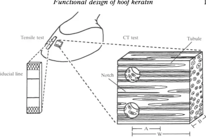
Tensile tests were conducted using an Instron Model 1122 tensile testing machine with a 500-kg load cell and an extension rate of 5 mmmin"1. These tests used strips of hoof-wall with dimensions of approximately 0-6 mm (thickness) X 4 mm (width) X 2-5 cm (length). The test samples were taken from the same region of the hoof-wall as the fracture samples and were oriented with the tubule axes parallel to the tension load (Fig. 2). The specimens were allowed to equilibrate at the four hydration levels and were tested immediately after removal from equilibration chambers. The samples were clamped above and below using pneumatic sample grips. To eliminate end-effects caused by sample gripping, strain was measured by following the movement of surface fiducial marks placed away from the sample grips, using a video dimension analyser (Model 303, Instruments for Physiology & Medicine, La Jolla, CA, USA). Initial Young's modulus (E) was determined from the maximum slope (linear region) of the tensile stress (force/area) vs strain (loaded/original length) records.
Functional design of hoof keratin
123
Tensile test
Fiducial line
[image:5.451.42.390.49.281.2]Tubule
Fig. 2. Location and orientation of longitudinal tension and compact tension (CT) test specimens. Cross-hatched region on tensile specimen indicates pneumatic grip location. Compact tension: A, notch length; W, specimen width; B, specimen thickness. Note: expanded drawings of tensile and compact tension specimens are not to the same scale.
determined by the J-integral method of Rice (1968), as adapted for this material (Bertram & Gosline, 1986).
Toughness is a measure of the resistance to the growth of flaws. The growth of flaws (in the form of microscopic cracks), or fracture when the condition reaches a critical state, is dependent on the formation of new surfaces within the material. The formation of new surfaces requires energy. A thorough analysis of the circumstances in which a material fractures requires a measure of the energy necessary for crack growth. The J-integral is a measure of the instantaneous change in energy with an incremental change in crack length at the critical point of failure. For a given specimen the critical J-integral value (Jcnt) was evaluated by plotting the energy (U)
against the apparent crack length (a) (this value is derived from the mechanical test record using the calibration curve - see description in Results) to specimen width (W) ratio for each interval of extension (all data together produced a series of incremental curves). The derivative of this relationship was taken at the critical failure point (critical failure was defined as the point on the force/deflection curve that intersected a line from the origin of the test having a slope which was 5 % less than the maximum slope). Thus, the J-integral is defined by Broek (1978) as:
J = ( - l / B ) x ( 5 U / 3 a )q,
RESULTS
Hydration properties
Hoof-wall keratin samples equlibrated with a 100% RH environment were found to have a 40-2% ±2-7 (S.D.) (A/ = 24) water content by mass. Relative humidity levels of 75 % and 53 % produced water content values of 18-2 % ±0-3 (N = 12) and 11-7% ±2-7 (N=18), respectively. Periods of 2-3 weeks in 0% RH produced specimens which had a consistent apparent water content of 5-5 % ±1-8 (N = 24) by mass, as indicated by oven drying. It is expected that a period of this length in 0% RH would remove all water from this material. The loss of mass observed probably represents a loss of very tightly bound water or, less likely, of some other volatile component of the tissue. The hydration values presented in this study are given as measured in order to allow direct comparison with previous studies employing the same techniques for determining in vivo hydration. The relationship between environmental humidity and hoof keratin water content, the absorption isotherm, is given in Fig. 3.
Tensile tests
The tensile properties of hoof-wall are profoundly influenced by the hydration conditions of the test specimens (Table 1). The mean Young's modulus value was found to be 410 MPa for longitudinal samples tested at 100% RH. The stiffness increased as the hydration level was decreased. The mean modulus became 2-6 GPa
10
Relative humidity (%)
Functional design of hoof keratin
Table 1. Longitudinal tensile modulus (E) and yield stress in horse hoof-wall keratin
at four relative humidity levels (RH)
RH E
(GPa) S.E.
Yield stress
(MPa) S.E. A"
100 75 S3 0
0-410 2-63 3-36 14-6
0032 0-362 0-629 0071
9 1 8 38-9
0-42
4-70 1
.1 2
S.E., standard error; A', sample number. Cross-head displacement rate, Smmmin
75%
4
-0-05 0-15 0-25
Strain
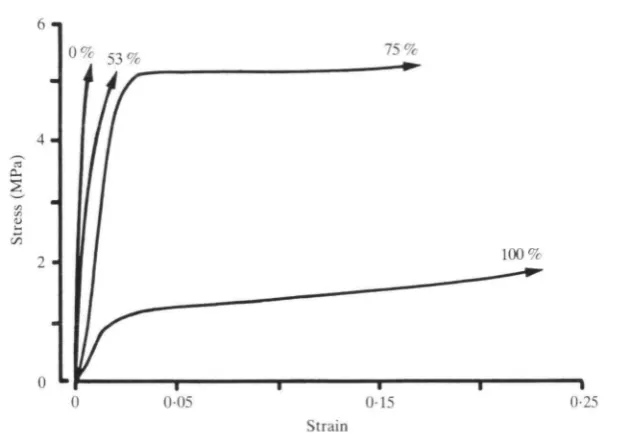
Fig. 4. Representative tensile tests stressed parallel to the tubule axis at four hydration levels. Arrowheads indicate test specimens failed at sample grips and absolute strength was not measured.
at 75% RH and 3-4GPa at 53% RH. The greatest mean modulus, 14-6GPa, was reached at 0% RH.
Table 2. Compact tension-fracture test results according to hydration level
Relative humidity J ^
(%) (kjm'2) S.E. N
100 75 S3 0
11-93 22-82 5-63 8-73
0-90 0-31 0-54 0-97
30 16 28 16
Jcm, mean critical J integral.
greater than the yield stress of the fully hydrated material. At 75 % RH, high stress levels were achieved prior to yield, while strains of up to 0-15 were also seen. The combination of high yield stress and high strain indicates that a great deal of energy can be absorbed by this material at 75 % RH. This observation was confirmed by the fracture mechanics tests which follow.
Compact tension fracture tests Compliance calibration
The resistance provided by a compact tension specimen against a load which tends to open the notch will depend, all other factors being equal, on the length of the notch. Standardized specimen size and shape allows quantification of the influence of notch length on the force required to deform the specimen (which is known as the mechanical compliance). A calibration curve can then be produced relating these two factors. Using the compliance calibration curve, the mechanically apparent length of the crack in a fracturing specimen can be determined from the fracture test record (in which applied load and the resulting deformations are measured). This technique is employed in the study of materials which display complex fracture behaviour. A complex fracture path eliminates the possibility of visual measurement of crack length.
The compliance value determined for a given notch length and specimen size will depend on the stiffness of the material. Since hoof-wall stiffness is dependent on hydration level, four compliance calibration relationships were determined to fit the four hydration levels used in this study. At 100% RH, the raw data were best described by a third-order polynomial regression over the range of notch lengths used in this study (P< 0-001; r2 = 0-88). Less extensive compliance calibration curves were used for the lower hydration levels, since the range of notch lengths employed was also less extensive. These relationships were best described by linear regressions over this range (Fig. 5). All regressions were significant (75% RH, P < 0 - 0 2 5 ; 53% RH, P<0-025; 0% RH, P<0-01).
Fracture toughness
The mean value of the J-integral at the critical failure point (Jcrit) reached a
Functional design of hoof keratin
129
0-8
0-6
0-4
0-2
0 % 100%
10 20 30
Fig. 5. Compliance calibration curves used to determine apparent crack length (a) for the four hydration levels tested. Initial compliance value (Co) on the abscissa and notch length (A) to specimen width (W) ratio ordinate.
100% RH: A/W = 0-211%119+3-4139645xl06Co-6-6S2877xl0l2Co2 + 1018Co3 (P<0-001).
75% RH: A/W = 0-23706+7-18884xl06Co (P<0-025). 53% RH: A/W = 0-257563 +l-20914xl07Co(P<0-025).
0% RH: A/W = 0-282584+l-32607xl07Co ( P < 0-010).
different from that at 53% RH (5-6kJm~2) but could not be distinguished statistically from the 0% RH value (8-7 kj m"2). The mean values at 53% and 0% RH also could not be distinguished from each other statistically.
DISCUSSION
Stiffness
A comparison of stiffness properties of hoof-wall with other keratinous materials indicates that the tensile response of hoof keratin to dehydration is generally similar to that of other keratins (Table 3). However, the level of water content has a more dramatic effect on the tensile properties of hoof-wall keratin. To understand how this can occur it is necessary to view the material at the level of its molecular composition and organization.
5-6 6-8 14-6 2-6(70%) 2-3 (70%) 0-19(70%) 5-1 (60%) 3-36(53%) 2-6(75%) 1-8 1-5 0-13 2-0 2-4 0-41 1 1 1 2 3 Table 3. Longitudinal elastic moduli (stiffness) of keratinized structures and the
effect ofhydration level (in GPa) (after Fraser & MacRae, 1980)
Relative humidity Intermediate
0% (RH in parentheses) 100% Reference Human nail hair stratum corneum Wool Horsehair Hoof wall
1. Baden, Goldsmith & Lee (1974). 2. Meredith (1956). 3. Bendit (1976).
keratin. Again, in this case the microfibrils possess both ar-helical and non-helical domains. However, unlike that of soft keratins, the non-helical microfibril domain of hard keratins is rich in cysteinyl residues. The matrix of hard keratins possesses cysteine-rich protein similar to that found in soft keratin, and in addition contains a protein rich in glycine and tyrosine. The <*-helical portions of the microfibrils are crystalline and can be considered to be quite stable, especially considering that the microfibrils of keratin are composed of several ar-helical chains formed into coiled-coil rope-like structures, reducing the sites available for intermolecular reactivity (McLachlan, 1978). In hard keratins, cysteine—cysteine disulphide cross-links stabilize the non-helical domains of the microfibrils and the cysteine-rich proteins of the matrix. The remainder of the matrix, however, has few covalent cross-links and, therefore, its stabilization depends on secondary cross-linking mechanisms which may be hydration-dependent (e.g. hydrogen bonding).
Functional design of hoof keratin 131
dehydration affects the properties of the matrix to a far greater degree than it does the microfibrils (Fraser & MacRae, 1980). Extensive hydrogen bonding between polymers of the matrix phase in the absence of water has been described as the cause of these changes (Fraser & MacRae, 1980). These may well occur associated with the glycine/tyrosine-rich proteins found in the hard keratin matrix. Such secondary cross-linking would decrease the mobility of the matrix polymers and, in the fully dehydrated state, the matrix would become a 'rigid polymeric glass' and the stiffness would approach that of the crystalline microfibrils.
Alternatively, when keratin is highly hydrated, the matrix lacks much of the secondary bonding it possesses in the dry state. In this case, longer distances occur between cross-link positions giving the matrix polymers greater freedom of move-ment. This translates into lower stiffness, since the secondary cross-links are not available to carry load, and greater extensibility, due to the ability of the polymers to rearrange under load.
Fracture
Rigid structural materials, man-made or natural, generally fail at load levels far below their ultimate theoretical strength because localized stress levels around flaws or cracks can be much greater than the general stress and can lead to crack growth and fracture. For this reason, fracture behaviour is important in determining the functional limitations of a material such as hoof-wall keratin. Over the complete range of hydrations, hoof-wall keratin proved to be a remarkably fracture-resistant material. The lowest mean work of fracture, 5-6 kj m~2 at 53 % RH (measured using the J-integral method), is roughly twice or more the toughness value measured for fresh bone (1 -0-3-0 kj m~2 measured as strain energy release rate, Wright & Hayes, 1977). Under the optimum conditions (i.e. 75% RH), the toughness of hoof-wall keratin (22-0 kjm~2) is an order of magnitude greater than that of bone and even exceeds that of wood (10-0kJm~2), an extremely tough biomaterial (Jeronimidis,
1976).
An indication of the significance of fracture toughness in a material such as hoof-wall can be seen by estimating the critical flaw size (af) or its 'notch sensitivity'. Critical flaw size is a concept developed by Griffith (1921) and emerged from an understanding of the energy balance within a fracturing material. Fracture in a stressed material will be spontaneous when the energy released from the material surrounding a growing crack is equal to the energy required to form new surfaces (i.e. the crack surfaces). For the critical failure stress, the maximum stress a material is capable of withstanding before fracture occurs, a minimum critical flaw size exists in which spontaneous fracture will begin. The fracture criterion for a linearly elastic material is given by:
_
a
f ~~
where E is Young's modulus of elasticity, ac is critical failure stress, y3 is surface
through several processes such as heat dissipation, plastic deformation and fibre pull-out. For most materials yf is several orders of magnitude greater than y3 so af can be
estimated by:
As an example of a notch-sensitive material we can look at a very familiar structural biomaterial, bone. Young's modulus (E) for fresh bone is approximately 20GPa (Wainwright, Biggs, Currey & Gosline, 1982), yield stress (oy) is 200 MPa (Reilly &
Burstein, 1975) and strain energy release rate (GIc) is 20kJm~2 (Wright & Hayes,
1977). It has been determined that 2yf = G,c (ASTM, 1965), where GIc is the strain
energy release rate at failure in the opening mode. The critical flaw size is, therefore, in the range of 300 fim. This figure indicates that any flaw greater than 300/xm in a bone will cause failure when load is applied. Currey (1962) has discussed the design features of bone that help avoid stress concentrations due to such features as the lacunae around osteocytes and the canals for the penetration of blood vessels. Hoof-wall keratin, however, is a very notch-insensitive material. For linear elastic conditions, Jcnt = Gic (Parker, 1981). Critical flaw size can then be estimated by:
_ Jcnt
a — 2 i
~2
where oy is yield stress. At 100% RH, E = 4-1 MPa, Jcril = 8-7 kj m~2 and
oy= 10 MPa. Therefore, under these conditions the critical flaw size for a
cata-strophic rupture would be about l'Sm, a value which is more than an order of magnitude larger than the entire hoof. These, admittedly rough, calculations indicate that, even with a large margin for error, no critically sized crack can exist in the hoof-wall, and it is virtually impossible for catastrophic fracture to occur. The high toughness level (i.e. extreme notch insensitivity) at 100% RH arises from the considerable plasticity that the material displays. By deforming plastically around a stress concentration, the stress is distributed to material farther from the crack tip and greater energy is absorbed. Flaws, such as the test notch introduced into these specimens, are not critical stress concentrators at very high hydration levels because the interconnections between the phases of the composite do not carry stress well. The result, however, is a material with a relatively low yield stress, and therefore it is not capable of supporting loads as large as it could if it were dryer.
Functional design of hoof keratin 133
itself, at cell boundaries within the tissue, and at the level of the tubular and intertubular components (Bertram & Gosline, 1986).
Conditions are appropriate within most of the thickness of the wall to provide the structural integrity the supporting wall requires. A reasonably high degree of rigidity is required of the hoof-wall material, but this rigidity must be balanced with the ability to resist fracture. Hoof-wall keratin at 75 % RH exhibited reasonable stiffness combined with both high yield stress and extensibility. This situation resulted in the maximum energy absorption, and hence fracture resistance, as determined through the J-integral analysis. These results are most probably the result of an intermediate degree of secondary cross-linking that increases the stiffness of the matrix phase while still allowing some extensibility. Thus, an intermediate degree of secondary cross-linking increases the fracture resistance of this material. Leach (1980) found that the major portion of the wall midway between the internal and external surfaces possessed water contents of between 17 and 24% by mass. This is in the same range determined for the 75 % RH tested in this study [18-2 % ±0-3 S.D. (AT = 12); Fig. 3]. Thus, hoof-wall keratin appears to function in vivo at the hydration levels that closely match the optimum condition for fracture toughness. In spite of the notch insensitivity displayed by this material, horse hooves do occasionally crack, probably because of a combination of decreased hydration near the external surface and long-term use causing the fracture toughness to decrease. A decline in fracture toughness by approximately 50% has been reported for the most distal, and consequently the oldest, portions of the hoof-wall (Bertram & Gosline, 1986).
Hoof wall design
Given that hoof-wall keratin functions under hydration conditions which maxi-mize fracture resistance, the question arises: is this an aspect of the mechanical design of this particular biomaterial, or simply a fortuitous result of the hydration conditions which exist in this circumstance? The answer can be suggested by determining whether or not hoof-wall keratin differs from other hard keratins, and if so, whether the differences reflect an adaptation of this biomaterial to the physical circumstances of the functioning hoof.
more than 15-fold, and probably considerably less because of the complex organiz-ation. The 30-fold increase in overall stiffness and the variation in relative stiffness with hydration when compared to wool strongly suggest that the matrix proteins in hoof-wall keratin have an increased potential for secondary bonding relative to other hard keratins such as wool. Just the proper amount, in fact, to provide the maximum fracture resistance within the hydration conditions that normally exist in the hoof. This effect could be derived either from an adjustment in the concentration and organization of amino acid residues within the matrix proteins or from an adjustment in the proportion of the two matrix protein types, if one is indeed more sensitive to hydration effects than the other. Whether or not horse hoof keratin represents a case of molecular adaptation which provides for the complex suite of mechanical properties displayed by this tissue will depend on the results of comparative analysis of matrix protein composition data from various types of hard keratins, including hoof-wall.
The effect of hydration on the mechanical behaviour of hoof keratin and the hydration gradient found in vivo in the hoof-wall provide a mechanism through which the mechanical properties of different areas of the hoof-wall can be adjusted to the requirements of the hoof, in spite of the fact that this material is not alive. This can occur because both the mechanical requirements of the hoof organ and the hydration level are closely associated with specific locations within the wall. It has been shown that the stratum medium, which comprises the majority of the wall, has properties that are capable of providing both support and fracture resistance under the hydration conditions found in that region. The properties measured at other hydration levels also match the requirements that can be expected to occur within the regions of the wall where those hydration conditions are found.
The hoof keratin located on the interior surface, next to the basal membrane and living cell layer, is highly hydrated. Under these conditions it would be far less stiff than in drier areas and also capable of extensive plastic deformation, as is seen in the tensile tests performed at 100% hydration (Fig. 4). The living layer of epidermal cells at the basal membrane is able to transfer the impact load from the hoof-wall to the collagenous connections of the dermis during ground contact simply because three-dimensional interdigitations at the dermal/epidermal boundary increase the area of contact and distribute the load, reducing stress levels (Stump, 1967). Extensibility of the fully keratinized tissue in this area, provided for by the properties of hoof-wall keratin at this hydration level, would also ensure that the stresses were low and distributed evenly across the basal membrane and the vulnerable living cell layer. The fibre organization also differs slightly in this region, which is likely to add to this effect.
Functional design of hoof keratin 135
The hydration environment of the individual cells also changes during the course of their function. Cells produced at the proximal margin of the wall migrate distally as new cells are produced next to the epidermal basal membrane. Even though they are fully keratinized soon after being produced, the high hydration level makes the cells next to the germinative layer quite pliable, again protecting the vulnerable underlying tissue such as that on the attachment surface of the internal wall. These same cells eventually become the cells of the main portion of the wall and finally become contact surface material before being worn away (Fig. 1). As the cells move distally, their hydration state and their properties change.
Intracellular deposition of keratin and the subsequent death of the cells necessarily restricts the manner in which this tissue can be modified during its use. The complex structure of the hoof wall, however, requires that a material be used which has variable mechanical properties. It appears the solution to this problem has been found in the linking of hydration-dependent mechanical properties to the pattern of hydration which exists within the hoof wall.
REFERENCES
ASTM (1965). Fracture toughness testing and its applications. STP 381. Philadelphia.
BADEN, H. P., GOLDSMITH, L. A. & LEE, L. (1974). The importance of understanding the comparative properties of hair and other keratinized tissues in studying disorders of hair. In The
First Human Hair Symposium (ed. A. C. Brown), pp. 388-398. New York: Medicom.
BENDIT, E. G. (1976). Longitudinal and transverse mechanical properties of keratin in compression. In Proceedings of the Fifth International Wool Textile Research Conference, vol. 2 (ed. K. Ziegler), pp. 351-360. Aachen: Deutches Wolforchungsinstitut.
BERTRAM, J. E. A. & GOSLINE, J. M. (1986). Fracture toughness design in horse hoof keratin.
J. exp. Biol. 125, 29-47.
BROEK, D. (1978). Elementary Engineering Fracture Mechanics. The Netherlands: Sitjthoff & Noorhoff International Publishers.
COOK, J. & GORDON, J. E. (1964). A mechanism for the control of cracks in brittle systems. Proc.
R. Soc. Ser. A 282, 508-520.
CURREY, J. D. (1962). Stress concentrations in bone. Q.Jlmicrosc. Sci. 103, 111-133.
DALE, B. A. (1977). Purification and characterization of a basic protein from the stratum corneum of mammalian epidermis. Biochim. biophys. Ada 491, 193-204.
FEUGHELMAN, M. (1959). A two-phase structure for keratin fibres. Text. Res.jf. 29, 223-228. FRASER, R. D. B. & MACRAE, T. P. (1980). Molecular structure and mechanical properties of
keratins. In The Mechanical Properties of Biological Materials. Symp. Soc. exp. Biol. XXXTV (ed. J. F. V. Vincent & J. D. Currey), pp. 211-246.
GRIFFITH, A. A. (1921). The phenomena of rupture and flow in solids. Phil. Trans. R. Soc. Ser. A 221, 163-198.
JERONIMIDIS, G. (1976). The fracture of wood in relation to its structure. In Wood Structure in
Biological and Technological Research (ed. P. Baas, A. J. Bolton & D. M. Catling), Leiden
Botanical Series no. 3, pp. 253-265. Leiden: The University Press.
JONES, L. N. (1980). Protein composition of mammalian stratum corneum. In Fibrous Proteins:
Scientific, Industrial and Medical Aspects, vol. 2 (ed. D. A. D. Parry & L. K. Creamer), pp.
167—175. New York: Academic Press.
LEACH, D. H. (1980). The structure and function of equine hoof wall. Ph.D. thesis, Department of Veterinary Anatomy, University of Saskatchewan, Saskatoon, Saskatchewan, Canada. MCLACHLAN, A. D. (1978). Coiled-coil formation and sequence regulations in the helical regions
of cr-keratin. J. molec. Biol. 124, 297-304.
MEREDITH, R. (1956). The Mechanical Properties of Textile Fibres. Amsterdam: North-Holland. MEITES, L. (1963). Handbook of Analytical Chemistry. Toronto: McGraw-Hill.
NICKEL, R. (1938). Uber den Bau der Hufrohrchen und seine Bedeutung fur den Mechanismus des Pferdehufes. Dt.-ost. tierarztl. Wschr. 46, 449-552.
PARKER, A. P. (1981). The Mechanics of Fracture and Fatigue. An Introduction. London: E. & F. N. Spon Ltd.
REILLY, D. T . & BURSTEIN, A. H. (1975). The elastic and ultimate properties of compact bone tissue. J . Biomech. 8, 393-405.
RICE, J. R. (1968). A path independent integral and the approximate analysis of strain concentration by notches and cracks. J. appl. Mech. 35, 379-384.
STUMP, J. E. (1967). Anatomy of the normal equine foot, including microscopic features of the laminar region. J . Am. vet. med. Ass. 151, 1588-1598.
T U K E Y , J . W. (1951). Quick and Dirty Methods in Statistics, part I I , Simple Analyses for Standard Designs. Proc. 5th Annual Convention, Am. Soc. for Quality Control, pp. 189-197.
WAINWRIGHT, S. A., BIGGS, W. D . , CURREY, J. D. & GOSLINE, J. M. (1982). Mechanical Design in Organisms. Princeton, NJ: Princeton University Press.
WlLKENS, H. (1964). Zur makroskopischen und microskopischen Morphologie der Rinderklaue mit einem Vergleich der Architektur von Klauen-und Hufrohrchen. Zentbl. Vet. Med., Ser. A 11, 163-234.