tVith 1 plate and 7 text-figures Printed in Great Britain
VENTILATORY MUSCLE ACTIVITY
IN INTACT PREPARATIONS OF AESHNID
DRAGONFLY LARVAE
BY R. S. PICKARD AND P. J. MILL
Department of Zoology, University of Leeds
{Received'] October 1971)
INTRODUCTION
Various aspects of rhythmic activity in arthropods have recently been studied in some detail. Interest has centred in particular on: heart beat in decapods (Maynard, 1955; Bullock & Terzuolo, 1957); swimmeret movements in the Astacura (e.g. Hughes & Wiersma, i960; Wiersma & Ikeda, 1964; Davis, 1969); locomotion (e.g. Wilson & Weis-Fogh, 1962; Wilson, 1964; Evoy, Barnes & Spirito, 1971); and insect ventila-tion (e.g. Miller, i960, 1962, 1971a, b; Hughes & Mill, 1966; Farley & Case, 1968; Mill, 1970).
The pacemaker varies in its position in different systems and preparations. How-ever, in the case of swimmeret movements (Hughes & Wiersma, i960; Ikeda & Wiersma, 1964), ventilation in the dragonfly larva (Mill & Hughes, 1966; Mill, 1970), and Limulus gill-plate movements (Fourtner, Drewes & Pax, 1971), it has been shown to lie in one of the posterior ganglia. In these cases activity spreads anteriorly to successive segments.
In this study, intact preparations have been used because they have the obvious advantage that the animal is not subjected to any dissection which may alter or disrupt the activity to be studied. Previous papers on the coordination of ventilation in dragonfly larvae using dissected preparations (Mill & Hughes, 1966; Mill, 1970) have shown that rhythmic bursts of activity emanate from the central nervous system. Expiratory bursts have been recorded from the second segmental nerves from the fourth to the eighth abdominal ganglion and in the corresponding nerve innervating the ninth abdominal segment. These bursts initiate contraction of the respiratory (expiratory) dorso-ventral muscles. Alternating with this activity, inspiratory bursts occur in the median nerves which innervate the two main inspiratory muscles; the 8ubintestinal muscle and the diaphragm. Certain other nerves associated with the intrinsic musculature of the branchial chamber and vestibule show expiratory and inspiratory activity.
528 R. S. PlCKARD AND P . J. MlLL
similar patterns in intact animals. Furthermore, the technique of using intact prepara-tions has made it possible to record simultaneously from several expiratory muscles while obtaining mechanical records of the ventilatory behaviour itself.
MATERIALS AND METHODS
In all experiments late instar larvae of the Emperor dragonfly Anax imperator were used.
The Perspex bath used for these experiments was divided into two compartments by a thin wax partition (Text-fig. 1). In the centre of this partition an inverted triangular hole was cut. The larger compartment was filled with wax to the level of this hole and a depression was made to accommodate a late instar larva. The animal
Water
T.S. light be;im
Electrodes
Inlet
Outlet
Clamps
Wax
Partition
Larva
Text-fig. 1. Side elevation of experimental chamber with larva in recording position.
was positioned ventral surface uppermost, with its anal appendages protruding through the partition hole into the smaller compartment. The body and legs were restrained with clamps. The walls of the hole helped to stop lateral bending of the abdomen. A steady flow of aerated water was passed through the small compartment. Leakage from the smaller compartment to the larger could be almost entirely prevented by using a small amount of vacuum grease to seal the joint between the animal and the hole in the partition.
Muscle recordings. Electrical recordings were made by implanting electrodes
Mechanical recordings. Sternal movement was monitored by passing a narrow light
beam across the fifth abdominal sternum to an OCP 71 photo-transistor.
In some experiments an isotonic SRI strain gauge was used to measure forces being exerted on the pleura by contractions of respiratory dorso-ventral muscles. The follow-ing procedure was used to determine the force exerted by an individual muscle. Two holes of about 0*07 mm diameter were drilled in the pleural cuticle of one segment. They were 1 mm apart and on the posterior margin of the respiratory dorso-ventral attachment. 48 s.w.G. wire was threaded into the animal through one hole and out again through the other. The ends were twisted and attached vertically to the strain gauge.
Records of pleural strain on one side of segment six and activity in the corre-sponding respiratory dorso-ventral muscle were taken simultaneously. The area of cutical containing the respiratory dorso-ventral insertion and the strain gauge attach-ment was then isolated from the entire animal by drilling round it. Simultaneous records at this point were then solely concerned with one respiratory dorso-ventral muscle and the strain it was exerting on the cuticle to which it was attached.
In experiments involving muscle ablation, or muscle isolation, 50 % of the specimens completely recovered after experimentation. In other experiments almost all specimens recovered fully, some even completing their metamorphosis months later.
RESULTS
Normal ventilation (Vn). Of the three pairs of dorso-ventral muscles in each of the
posterior abdominal segments, only the respiratory dorso-ventrals were found to be active during Vn. Muscle potentials from the respiratory dorso-ventral muscle on the
right side of abdominal segment five are shown in Text-fig. 2. It can be seen that these expiratory bursts show exactly the same characteristics observed in dissected preparations by Mill & Hughes (1966) and Mill (1970). There is an increase in fre-quency during the first part of the burst and this is associated with facilitation of the potentials. The upper trace in this figure shows movements of the hind abdominal sterna, upwards indicating lifting of the sterna. It was not surprising to observe that the initial low frequency in the expiratory bursts is associated with a rather gentle lifting of the sterna. As the frequency increases so the strength of the movement in-creases. However, what was not expected was the cessation of firing some 100 msec before the sterna had reached their fully raised position. This was so for all segments from five to eight at least.
Activity on opposite sides of the same segment is very closely synchronized (Text-fig. 3) and there is often a 1:1 relationship between the potentials on the two sides. The closeness to a 1:1 relationship varies somewhat between preparations and within a given preparation. Records (c) and (d) in Text-fig. 3 are single expiratory bursts from the same preparation. In the former there are the same number of potentials (40) on each side; whereas in (d) there are two more on the left side (43) than on the right (41). One of the extra ones on the left side can be accounted for by the single potential at the end of the burst, the other appears in the middle of the burst. It is of interest to note that, when there is a 1:1 relationship, one side does not always lead consistently throughout the burst; nor is there a constant delay between one side and
530 R. S. PlCKARD AND P . J. MlLL
the other. Thus it is almost certain that two expiratory motor neurones are present (one for each side), in spite of the fact that there is some structural evidence that some motor cells send branches into both second roots (Mill, 1964). Perhaps these latter cells are associated with the jet-propulsion mechanism.
] sec
Text-fig. 2. Expiratory bursts (lower trace) in the right respiratory dorso-ventral muscle (rdv.) of segment 5 during normal ventilation (F»). The upper trace monitors sternal movement of segments 4-6. (Upward deflexion indicates lifting of the sterna.) (a) and (6) Same preparation.
^J»i
' 300 msec
_l LllliiiiiliiiliililliiiliililiiSiltbl! 300 msec
Text-fig. 3. Expiratory bursts in right (r) and left (/) rdv. muscles, (a) Segment 5; (b)-{d) segment 6 from a different preparation, cf. pulses in (c) 1: i, with those in (d), not 1:1. Top trace (J): movement of sterna of segments 4-6. (Upward deflexion indicates lifting of the sterna.)
ganglion will start to fire out of sequence. This has been noticed particularly in the case of the sixth ganglion, which in some preparations produces bursts before the seventh, but always after the eighth (PI. 1, fig. 2). However, when this happens the initial frequency of the potentials in the bursts from the sixth ganglion is particularly low. The expiratory bursts all end simultaneously about 100 msec before the sterna are fully raised.
Text-fig. 4. Expiratory bursts in the right rdv. muscles of different abdominal segments, showing serial staggering of burst initiation in adjacent segments (5-8). (a) and (b) are from the same preparation. (1) sternal movement of segments 4-6. (Upward deflexion indicates lifting of the sterna.)
In order to determine the relationship between the premature termination of the expiratory bursts and the lifting of the sterna, preparations were made in which the respiratory dorso-ventral muscles were ablated from segments on either side of the segment from which recordings were being made. Ablations were performed by drilling out the small muscle-attachment points and sealing the holes with Eastman 910. Records from such preparations had reduced amplitude of sternal movement but still showed a delay of about 100 msec between cessation of electrical activity and peak sternal movement (Text-fig. 5). This helped to demonstrate that continued peak sternal movement was not due simply to adjacent segments pulling up the recording segment.
Text-fig. 5. Sternal movement in segments 5 and 6 (upper trace; upward deflexion indicates lifting of sterna) and expiratory bursts in the right rdv. muscle of segment 6. Left and right
rdvs of segments 5 and 7 have been ablated. Note reduced amplitude of sternal movement,
532 R. S. PlCKARD AND P. J. MlLL
Records of the strain developed on a single pleuron in intact preparations (PI. i, fig. i a, b) showed that a delay also existed between the end of electrical activity in the muscle and the development of maximum tension; although in these preparations the delay was less than ioo msec. The tension recorded was in the order of io~* Newtons (about i g wt.). However, the tension exerted by an individual respiratory muscle reached maximum at approximately the same time as the expiratory burst ended (PI. i, fig. ic). Recordings of activity in the subintestinal muscle confirm that this is an inspiratory muscle, and muscle potentials appear in it at or just before peak sternal movement (Text-fig. 6). The subintestinal muscle is innervated by two branches of the same axon from the unpaired median nerve (Zawarzin, 1924; Mill, 1965). The branches enter the muscle on opposite sides of the segment, and this is likely to be the reason for two different impulse sizes appearing in a 1:1 relationship within a single burst recorded from one muscle-attachment point.
Tert-fig. 6. Inapiratory bursts (lower trace) in the subintestinal muscle, and sternal movement (upper trace; upward deflexion indicates lifting of sterna) of segment 5 during Vn.
Other forms of ventilation. Other forms of ventilation have been noted by Tonner
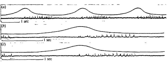
(1936) and by Hughes & Mill (1966) and have generally been described as 'gulping' and 'chewing' ventilation. 'Gulping' has been observed in the current series of ex-periments and the maintained hit of the sterna, characteristic of this form of ventila-tion, is associated with intermittent bursts of activity in the respiratory dorso-ventral muscles. Another type of ventilation frequently observed is shown in Text-fig. 7 and PL 1, fig. 2, and this will now be described.
[image:6.451.54.401.230.360.2]Records from the respiratory dorso-ventral muscles of several segments show that, although those in abdominal segments five to eight at least are active during this form of ventilation, the amount of activity decreases from posterior to anterior. In other words the respiratory dorso-ventral muscles in segment eight are more active than those in segment seven and so on (PI. 1, fig. 26, c).
(h)
J
U)
.'(<•)
J Water removed I sec
Text-fig. 7. Expiratory bursts (lower trace) in the right rdv. muscle of segment 5 during ventilatory activity other than Vn. Sternal movement is shown in the upper trace (upward
deflexion indicates lifting of sterna), (a) and (6) continuous records of spontaneously occurring behaviour; (c) and (d) continuous records of behaviour induced by touching paraprocts with fine needle; (e) behaviour induced by sudden drainage of water from the experimental chamber. Vn continued between (6) and (c) and between (d) and (e).
DISCUSSION
The results obtained using intact preparations confirm the earlier work of Mill & Hughes (1966) and Mill (1970) in showing that ventilation is initiated by segmental expiratory motor neurones which produce bursts of muscle potentials in the respiratory dorso-ventral muscles. Furthermore, the respiratory dorso-ventral muscles at the posterior end of the abdomen are activated first, with successively anterior pairs of muscles becoming involved with a delay of about 100 msec per segment. The bursts in each segment cease simultaneously. There is then a short delay, after which activity can be recorded from the subintestinal muscle. Thus the inspiratory role of the latter is confirmed. Since the diaphragm is active at exactly the same time as the sub-intestinal muscle (Mill & Hughes, 1966) presumably it too has an inspiratory role.
The delay between the end of the expiratory burst and the peak of sternal movement may occur because the role of the respiratory dorso-ventral muscles is to pull the
534 R- S. PICKARD AND P. J. M I L L
sterna past some critical level, after which they 'click' into a stable raised position. Alternatively, it could be due to the inertia of the expiratory flow of water. This, together with the importance of the timing of activity in the inspiratory subintestinal muscle, is discussed elsewhere (Mill & Pickard, 1972).
The fact that the respiratory dorso-ventral muscles of one segment may fire out of turn in some instances, i.e. not sequentially from posterior to anterior, indicates that the model proposed by Mill & Hughes (1966) should be modified to include connexions from the command interneurone(s) to each expiratory centre. It is sug-gested that successively more anterior expiratory motor neurones have higher thres-holds, so that they are normally only activated when the input from the command system is augmented by activity from the next most posterior segment. Future experiments should reveal that more anterior ganglia are capable of acting as pace-makers, as in the crayfish swimmeret system (Ikeda & Wiersma, 1964). There is presumably also a rhythmically firing interneurone between each pair of ganglia, carrying information anteriorly.
Hughes & Mill (1966) described two forms of ventilation in aeshnid larvae other than normal ventilation and jet-propulsion. In one of these, 'gulping' ventilation (Tonner, 1936), the abdominal sterna were raised and held in this position for several seconds. During this time the pressure in the branchial chamber oscillated around 10 cm H2O. In the other type, which was observed less frequently, the abdominal
sterna were extended downwards below their rest position in a series of small steps. The overall downward movement was matched by a corresponding increase in pressure in the branchial chamber to about 4 cm H2O and the steps coincided with
brief increases in pressure of about 2 cm HaO. The latter were reminiscent of normal
respiratory pressure changes, but with the negative phase obscured by the overall pressure increase. The frequency of their occurrence was about twice that of normal ventilation. After reaching a maximum the sterna remained extended, while the pressure started to fall slowly. This form of ventilation was terminated by the occur-rence of a normal ventilatory cycle.
The current study has confirmed the existence of 'gulping' as described by Hughes & Mill (1966). The other type of ventilation described in this paper corresponds to 'gulping' in the initial raising of the sterna; and to the step-like form of ventilation described by Hughes & Mill (1966) in the periodic downward movements of the sterna. It is felt that this other ventilation is more closely allied to the 'stepping' behaviour than to 'gulping'.
Hughes & Mill attributed the pumping up of the branchial chamber pressure during this 'stepping' behaviour to the possibility of intrinsic muscular activity. How-ever, results of the present study suggest that this pumping is due in part to con-tractions of the respiratory dorso-ventral muscles. The anal valve is tightly closed during the procedure. Such modification of Vn may well serve to clear residual pockets
of water or debris from the branchial chamber, and rearrange the contained tracheal gills. The behaviour is always followed with an abnormally strong expiratory move-ment before Vn is resumed. It is hoped that further studies, using free
SUMMARY
1. The expiratory role of the segmental, respiratory dorso-ventral muscles, and the in8piratory role of the subintestinal muscle, have been confirmed using intact preparations of aeshnid dragonfly larvae.
2. The strain developed by individual respiratory dorso-ventral muscles has been measured.
3. The respiratory dorso-ventral muscles all cease firing simultaneously, about 100 msec before the sterna are fully raised, and do not have any mechanical effect on the sterna after this time. It is suggested that the delay is caused either because the role of these muscles is to lift the sterna past some critical position, and/or because of the inertia of the expiratory current.
4. Periodically the sterna are raised and then lowered slowly in a series of steps, each pause in the lowering coinciding with activity in the respiratory dorso-ventral muscles. This form of ventilation is compared with others previously described.
5. In normal ventilation, and in other types of ventilation, activity in the respiratory dorso-ventral muscles shows a pronounced tendency to begin in the most posterior segments and to continue for longer periods in those segments.
6. Some aspects of the central neural connexions involved in normal ventilation are discussed.
Thanks are due to Ethicon Ltd. for samples of their non-toxic Eastman 910 adhesive. R.S.P. wishes to thank the Science Research Council for a studentship, during the tenure of which the above work was carried out.
REFERENCES
BULLOCK, T. H. & TERZUOLO, C. A. (1957). Diverse forms of activity in the somata of spontaneous and integrating ganglion cells. J. Pkysiol. 138, 341-64.
DAVIS, W. J. (1969). The neural control of swimmeret beating in the lobster. J. exp. Biol. 50, 99-117. EVOY, W. H., BARNES, W. J. & SPIRITO, C. P. (1970). Interactions between central commands and
reflexes in crab walking legs. Am. Zool. 10, 202.
FARLEY, R. D. & CASE, J. F. (1968). Sensory modulation of ventilative pacemaker output in the cock-roach, Periplaneta americana. J. Inject Phytiol. 13, 591-601.
FOURTNKR, C. R., DREWES, C. D. & PAX, R. A. (1971). Rhythmic motor outputs co-ordinating the respiratory movement of the gill plates of Limultu polyphemux. Comp. Biochem. Phytiol. 38 A, 751-62. HUGHES, G. M. & MILL, P. J. (1966). Patterns of ventilation in dragonfly larvae. J. exp. Biol. 44,
317-33-HUGHES, G. M. & WIERSMA, C. A. G. (i960). The co-ordination of swimmeret movements in the cray-fish, Procambarut clarkh (Girard). J. exp. Biol. 37, 657-70.
IKEDA, K. & WIERSMA, C. A. G. (1964). Autogenic rhythmicity in the abdominal ganglia of the crayfish: the control of swimmeret movements. Comp. Biochem. Phytiol. ia, 107-15.
MAYNARD, D. M. (1955). Activity in a crustacean ganglion. II. Pattern and interaction in burst forma-tion. Biol. Bull. mar. biol. Lab., Woodt Hole 109, 420-36.
MILL, P. J. (1964). The structure of the abdominal nervous system of Aeschnid nymphs. J. comp.
Neurol. iaa, 157^71.
MILL, P. J. (1965). An anatomical study of the abdominal nervous and muscular systems of dragonfly (Aeshnidae) nymphs. Proc. Zool. Soc. 145, 57-73.
MILL, P. J. (1970). Neural patterns associated with ventilatory movements in dragonfly larvae. J. exp.
Biol. 5a, 167-75.
MILL, P. J. & HUGHES, G. M. (1966). The nervous control of ventilation in dragonfly larvae. J. exp.
Biol. 44, 297-316.
MILL, P. J. & PICKARD, R. S. (1972). Anal valve movement and normal ventilation in aeshnid dragonfly larvae. J. exp. Biol. 56,
536 R. S. PlCKARD AND P. J. MlLL
MILLER, P. L. (i960). Respiration in the desert locust. I. The control of ventilation. J. exp. Biol. 37, 324-36.
MILLER, P. L. (1962). Spiracle control in adult dragonflies (Odonata). J. exp. Biol. 39, 513-35. MILLER, P. L. (1971a). Rhythmic activity in the insect nervous system. I. Ventilatory coupling of a
mantid spiracle. J. exp. Biol. 54, 587—97.
MILLER, P. L. (19716). Rhythmic activity in the insect nervous system. II. Sensory and electrical stimulation of ventilation in a mantid. J. exp. Biol. 54, 599-607.
TONNKR, F. (1936). Mechanik und Koordination der Atem- und Schwimmbewegung bei Libellen-larven. Z. wits. Zool. 147, 433-54.
WIERSMA, C. A. G. & IKEDA, K. (1964). Interneurones commanding swimmeret movements in the crayfish, Procambarus clarkU (Girard). Comp. Biochem. Physiol. ia, 509-25.
WILSON, D. M. (1964). Relative refractoriness and patterned discharge of locust flight motor neurons.
J. exp. Biol. 41, 191-205.
WILSON, D. M. & WEIS-FOGH, T. (1962). Patterned activity of co-ordinated motor units, studied in flying locusts. J. exp. Biol. 39, 643-67.
ZAWARZIN, A. (1924). Uber die histologische Beschaffenheit des unpaaren ventralen Nervs der Insekten. (Histologisch Studien Uber Insekten V.) Z. wist. Zool. IM, 97-115.
EXPLANATION OF PLATE
Fig. 1. (a) and (6) Right pleural strain in segment 6 (upper trace) and expiratory bursts (lower trace) in the rdv. muscle attached to the pleuron. (e) Same preparation with cuticle containing rdv. attachment now isolated from the pleuron and upper trace thus monitoring the strain developed by the muscle alone, ( i g w t . = 9-81 x io~* N.)
Fig. 2. Ventilatory activity other than Vn. (a) Activity in the left (0 and right (r) rdvs of segment 6.
Plate 1
l j . S U .
. • ! • • 1 1 _ „ _ . . 1 1 1.1 I I l l 11 1 11 11 1 1 1 1,
I sec
I g wl. —s.
I sec
I sec
Fig. 2
[image:11.451.20.434.288.504.2]