organic papers
o3656
Nget al. C15H10O4 doi:10.1107/S1600536805031697 Acta Cryst.(2005). E61, o3656–o3658
Acta Crystallographica Section E
Structure Reports
Online
ISSN 1600-5368
1,3-Dihydroxy-2-methyl-9,10-anthraquinone
Shea-Lin Ng,aIbrahim Abdul Razak,aHoong-Kun Fun,a* Sompong Boonsri,b Suchada Chantraprommab* and Uma Prawatc
aX-ray Crystallography Unit, School of Physics,
Universiti Sains Malaysia, 11800 USM, Penang, Malaysia,bDepartment of Chemistry, Faculty of Science, Prince of Songkla University, Hat-Yai, Songkhla 90112, Thailand, andcDepartment of Chemistry, Faculty of Science and Technology, Phuket Rajabhat University, Muang, Phuket 83000, Thailand
Correspondence e-mail: [email protected], [email protected], [email protected], [email protected], [email protected], [email protected]
Key indicators
Single-crystal X-ray study
T= 293 K
Mean(C–C) = 0.002 A˚
Rfactor = 0.045
wRfactor = 0.148
Data-to-parameter ratio = 16.0
For details of how these key indicators were automatically derived from the article, see http://journals.iucr.org/e.
#2005 International Union of Crystallography
Printed in Great Britain – all rights reserved
The non-H atoms of the title compound, C15H10O4, which was
isolated from the roots ofPrismatomeris malayanaRidl., are coplanar. Intramolecular O—H O and C—H O hydrogen bonds are observed in the molecular structure. The molecules form centrosymmetric hydrogen-bonded dimers via inter-molecular O—H O hydrogen bonds. The crystal structure is further stabilized by weak–interactions.
Comment
Prismatomeris malayana Ridl (‘Kradook Kai’ in Thai) is a medicinal plant. The extract from the root of this plant has been used as a folk medicine for the treatment of skin diseases (Perry, 1980). The title compound, rubiadin, (I), has been isolated from the roots ofPrismatomeris malayanaRidl. which were collected from Phuket province in the southern part of Thailand. Rubiadin was previously isolated from Rubia cordifolia (Tripathi et al., 1997) and Hedyotis capitellata
(Ahmad et al., 2005). It possesses an anti-oxidant property which is better than that of EDTA, Tris, mannitol, vitamin E and p-benzoquinone (Tripathi et al., 1997). As part of our systematic studies on chemical constituents of Thai medicinal plants (Chantraprommaet al., 2003, 2004, 2005; Boonnaket al., 2005; Funet al., 2005; Nget al., 2005), we have undertaken the X-ray crystal structure analysis of (I) in order to establish its molecular structure and relative stereochemistry.
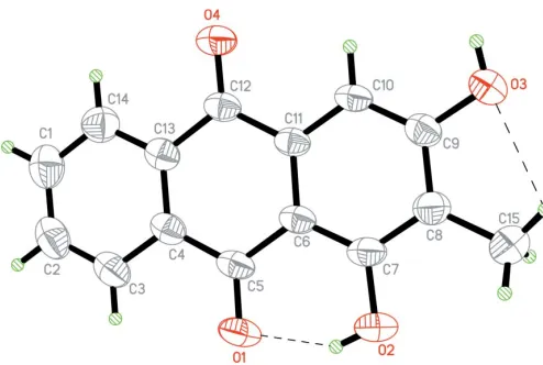
The C—C bond lengths in (I) show normal values (Allenet al., 1987). The C—O and C O bond lengths (Table 1) are comparable to those observed in similar structures (Cotterillet al., 1995; Ohsawa & Ohba, 1993). The non-H atoms of (I) are coplanar (Fig. 1), with a maximum deviation of 0.103 (1) A˚ for atom O4.
All O atoms are involved in hydrogen bonding. Atoms O1, O2 and O3 are involved in intramolecular O2—H2A O1 and C15—H15A O3 hydrogen bonds (Table 2), respectively, while atom O3 forms an intermolecular hydrogen bond with atom O4,viz. O3—H3A O4i[symmetry code: (i)x, 1y,
1 z], leading to the formation of a dimer (Table 2). In addition, the crystal packing is stabilized by weak– inter-actions between the anthraquinone ring system of inversion-related molecules stacked along thebaxis (Fig. 2). The ring system is stacked in such a way that the centroid-centroid distance between the C1–C4/C13/C14 ring at (x,y,z) and the C6–C11 ring at (1x, 1y, 1z) is 3.640 (1) A˚ , while the centroid–centroid separation between the C4–C6/C11–C13 rings at (x,y,z) and (1x, 1y, 1z) is 3.593 (1) A˚ .
Experimental
Air-dried roots of P.malayana were ground and extracted with CH2Cl2at room temperature. The residue obtained after evaporation
of the solvent was repeatedly subjected to column chromatography over silica gel to afford (I). Single crystals of (I) were obtained by recrystallization from a CHCl3–CH3OH (4:1v/v) solvent system (m.p.
576–578 K).
Crystal data
C15H10O4 Mr= 254.23
Monoclinic,P21=c a= 7.3517 (1) A˚
b= 7.3268 (1) A˚
c= 21.1186 (4) A˚ = 97.610 (1)
V= 1127.52 (3) A˚3
Z= 4
Dx= 1.498 Mg m
3
MoKradiation Cell parameters from 4302
reflections = 2.0–28.3
= 0.11 mm1 T= 293 (2) K Plate, light brown 0.780.410.08 mm
Data collection
Bruker SMART APEX2 CCD area-detector diffractometer !scans
Absorption correction: multi-scan (SADABS; Bruker, 2005)
Tmin= 0.828,Tmax= 0.992
11402 measured reflections
2798 independent reflections 2066 reflections withI> 2(I)
Rint= 0.021
max= 28.3 h=9!9
k=9!9
l=26!28
Refinement
Refinement onF2 R[F2> 2(F2)] = 0.046
wR(F2) = 0.148 S= 1.06 2798 reflections 175 parameters
H-atom parameters constrained
w= 1/[2(F
o2) + (0.0833P)2
+ 0.1198P]
whereP= (Fo2+ 2Fc2)/3
(/)max= 0.001
max= 0.24 e A˚
3
min=0.19 e A˚
3
Table 1
Selected geometric parameters (A˚ ,).
O1—C5 1.2408 (14)
O2—C7 1.3501 (14)
O3—C9 1.3566 (15)
O4—C12 1.2255 (14)
O2—C7—C6 121.55 (11)
O2—C7—C8 116.54 (12)
O3—C9—C8 116.73 (12)
[image:2.610.45.292.68.234.2]O3—C9—C10 121.46 (11)
Table 2
Hydrogen-bond geometry (A˚ ,).
D—H A D—H H A D A D—H A
O2—H2A O1 0.82 1.84 2.567 (2) 147
C15—H15A O3 0.96 2.41 2.784 (2) 103
O3—H3A O4i 0.82 1.98 2.796 (1) 171
Symmetry code: (i)x;yþ1;zþ1.
H atoms were placed in calculated positions, with O—H = 0.82 A˚ and C—H = 0.93 or 0.96 A˚ . TheUisovalues were constrained to be
1.5Ueq of the carrier atom for hydroxyl and methyl H atoms and
1.2Ueqfor the remaining H atoms.
Data collection:APEX2(Bruker, 2005); cell refinement:APEX2; data reduction: SAINT (Bruker, 2005); program(s) used to solve structure: SHELXTL (Sheldrick, 1998); program(s) used to refine structure:SHELXTL; molecular graphics:SHELXTL; software used to prepare material for publication:SHELXTLandPLATON(Spek, 2003).
SB thanks Alisa Pathan and Jantana Detpichai for useful information. The authors thank Prince of Songkla University, the Malaysian Government and Universiti Sains Malaysia for the Scientific Advancement Grant Allocation (SAGA) grant No. 304/PFIZIK/635003/A118 and the USM short-term grant No. 304/PFIZIK/635028.
organic papers
Acta Cryst.(2005). E61, o3656–o3658 Nget al. C
15H10O4
o3657
Figure 1The structure of (I), showing 50% probability displacement ellipsoids and the atomic numbering. Hydrogen bonds are shown as dashed lines.
Figure 2
[image:2.610.46.293.281.471.2]References
Ahmad, R., Shaari, K., Lajis, N. H., Hamzah, A. S., Ismail, N. H. & Kitajima, M. (2005).Phytochemistry,66, 1141–1147.
Allen, F. H., Kennard, O., Watson, D. G., Brammer, L., Orpen, A. G. & Taylor, R. (1987).J. Chem. Soc. Perkin Trans. 2, pp. S1–19.
Boonnak, N., Chantrapromma, S., Fun, H.-K., Anjum Ali, S., Rahman, A. A. & Karalai, C. (2005).Acta Cryst.E61, o410–o412.
Bruker (2005).APEX2(Version 1.27),SAINTandSADABS. Bruker AXS Inc., Madison, Wisconsin, USA.
Chantrapromma, K., Saewan, N., Fun, H.-K., Chantrapromma, S. & Rahman, A. A. (2004).Acta Cryst.E60, o312–o314.
Chantrapromma, S., Boonnak, N., Fun, H.-K., Anjum, S. & Rahman, A. A. (2005).Acta Cryst.E61, o2136–o2138.
Chantrapromma, S., Fun, H.-K., Razak, I. A., Laphookhieo, S. & Karalai, C. (2003).Acta Cryst.E59, o1864–o1866.
Cotterill, A. S., Gable, R. W. & Gill, M. (1995).Acta Cryst.C51, 500–502. Fun, H.-K., Razak, I. A., Boonnak, N., Laphookhieo, S. & Chantrapromma, S.
(2005).Acta Cryst.E61, o3086–o3088.
Ng, S. L., Razak, I. A., Fun, H.-K., Boonsri, S., Chantrapromma, S. & Prawat, U. (2005).Acta Cryst.E61, o3234–o3236.
Ohsawa, Y. & Ohba, S. (1993).Acta Cryst.C49, 2149–2151.
Perry, L. M. (1980).Medicinal plants of East and Southeast Asia, p. 620. Cambridge, Mass., USA: The MIT Press.
Sheldrick, G. M. (1998).SHELXTL. Version 5.1. Bruker AXS Inc., Madison, Wisconsin, USA.
Spek, A. L. (2003).J. Appl. Cryst.36, 7–13.
Tripathi, Y. B., Shama, M. & Manickam, M. (1997). Indian J. Biochem. Biophys.34, 302–306.
organic papers
o3658
Nget al. Csupporting information
sup-1 Acta Cryst. (2005). E61, o3656–o3658
supporting information
Acta Cryst. (2005). E61, o3656–o3658 [https://doi.org/10.1107/S1600536805031697]
1,3-Dihydroxy-2-methyl-9,10-anthraquinone
Shea-Lin Ng, Ibrahim Abdul Razak, Hoong-Kun Fun, Sompong Boonsri, Suchada
Chantrapromma and Uma Prawat
1,3-dihydroxy-2-methylanthra-9,10-quinone
Crystal data
C15H10O4 Mr = 254.23
Monoclinic, P21/c Hall symbol: -P 2ybc a = 7.3517 (1) Å b = 7.3268 (1) Å c = 21.1186 (4) Å β = 97.610 (1)° V = 1127.52 (3) Å3 Z = 4
F(000) = 528 Dx = 1.498 Mg m−3
Melting point = 442–443 K Mo Kα radiation, λ = 0.71073 Å Cell parameters from 4302 reflections θ = 2.0–28.3°
µ = 0.11 mm−1 T = 293 K Plate, light_brown 0.78 × 0.41 × 0.08 mm
Data collection
Bruker SMART APEX2 CCD area-detector diffractometer
Radiation source: fine-focus sealed tube Graphite monochromator
Detector resolution: 8.33 pixels mm-1 ω scans
Absorption correction: multi-scan (SADABS; Bruker, 2005) Tmin = 0.828, Tmax = 0.992
11402 measured reflections 2798 independent reflections 2066 reflections with I > 2σ(I) Rint = 0.021
θmax = 28.3°, θmin = 2.0° h = −9→9
k = −9→9 l = −26→28
Refinement
Refinement on F2 Least-squares matrix: full R[F2 > 2σ(F2)] = 0.046 wR(F2) = 0.148 S = 1.06 2798 reflections 175 parameters 0 restraints
Primary atom site location: structure-invariant direct methods
Secondary atom site location: difference Fourier map
Hydrogen site location: inferred from neighbouring sites
H-atom parameters constrained w = 1/[σ2(F
o2) + (0.0833P)2 + 0.1198P] where P = (Fo2 + 2Fc2)/3
supporting information
sup-2 Acta Cryst. (2005). E61, o3656–o3658
Special details
Geometry. All e.s.d.'s (except the e.s.d. in the dihedral angle between two l.s. planes) are estimated using the full covariance matrix. The cell e.s.d.'s are taken into account individually in the estimation of e.s.d.'s in distances, angles and torsion angles; correlations between e.s.d.'s in cell parameters are only used when they are defined by crystal symmetry. An approximate (isotropic) treatment of cell e.s.d.'s is used for estimating e.s.d.'s involving l.s. planes.
Refinement. Refinement of F2 against ALL reflections. The weighted R-factor wR and goodness of fit S are based on F2, conventional R-factors R are based on F, with F set to zero for negative F2. The threshold expression of F2 > σ(F2) is used only for calculating R-factors(gt) etc. and is not relevant to the choice of reflections for refinement. R-factors based on F2 are statistically about twice as large as those based on F, and R- factors based on ALL data will be even larger.
Fractional atomic coordinates and isotropic or equivalent isotropic displacement parameters (Å2)
x y z Uiso*/Ueq
O1 0.76480 (12) 0.16468 (16) 0.43117 (5) 0.0568 (3) O2 0.56385 (13) 0.26507 (17) 0.32832 (5) 0.0603 (3) H2A 0.6560 0.2265 0.3504 0.091* O3 −0.03121 (12) 0.47491 (16) 0.34194 (5) 0.0551 (3) H3A −0.0948 0.5129 0.3682 0.083* O4 0.23316 (13) 0.35636 (15) 0.57119 (5) 0.0533 (3) C1 0.7214 (2) 0.1308 (2) 0.66453 (8) 0.0603 (4) H1 0.7401 0.1132 0.7085 0.072* C2 0.8569 (2) 0.0845 (2) 0.62807 (8) 0.0590 (4) H2 0.9669 0.0355 0.6477 0.071* C3 0.83088 (18) 0.11025 (18) 0.56294 (8) 0.0498 (3) H3 0.9237 0.0800 0.5389 0.060* C4 0.66581 (16) 0.18143 (16) 0.53298 (6) 0.0393 (3) C5 0.63997 (15) 0.20675 (16) 0.46262 (6) 0.0403 (3) C6 0.46581 (15) 0.27964 (16) 0.43235 (6) 0.0368 (3) C7 0.43381 (17) 0.30360 (18) 0.36586 (6) 0.0425 (3) C8 0.26579 (18) 0.37000 (19) 0.33495 (6) 0.0449 (3) C9 0.13032 (16) 0.41185 (17) 0.37266 (6) 0.0411 (3) C10 0.15826 (15) 0.38997 (16) 0.43874 (6) 0.0381 (3) H10 0.0650 0.4188 0.4628 0.046* C11 0.32405 (15) 0.32561 (15) 0.46859 (6) 0.0347 (3) C12 0.35158 (16) 0.30696 (16) 0.53917 (6) 0.0378 (3) C13 0.52759 (16) 0.22863 (16) 0.56968 (6) 0.0392 (3) C14 0.5570 (2) 0.2039 (2) 0.63538 (7) 0.0516 (4) H14 0.4662 0.2364 0.6600 0.062* C15 0.2356 (2) 0.3961 (3) 0.26395 (7) 0.0698 (5) H15A 0.1428 0.4872 0.2531 0.105* H15B 0.1966 0.2829 0.2436 0.105* H15C 0.3480 0.4349 0.2496 0.105*
Atomic displacement parameters (Å2)
U11 U22 U33 U12 U13 U23
supporting information
sup-3 Acta Cryst. (2005). E61, o3656–o3658
O4 0.0429 (5) 0.0687 (7) 0.0517 (5) 0.0092 (4) 0.0184 (4) −0.0012 (5) C1 0.0643 (10) 0.0572 (9) 0.0556 (8) 0.0015 (7) −0.0056 (7) 0.0028 (7) C2 0.0486 (8) 0.0520 (8) 0.0713 (10) 0.0058 (6) −0.0105 (7) −0.0041 (7) C3 0.0357 (6) 0.0442 (7) 0.0682 (9) 0.0021 (5) 0.0021 (6) −0.0084 (6) C4 0.0317 (6) 0.0308 (6) 0.0556 (7) −0.0026 (4) 0.0062 (5) −0.0047 (5) C5 0.0284 (6) 0.0377 (6) 0.0568 (7) −0.0026 (5) 0.0125 (5) −0.0073 (5) C6 0.0290 (6) 0.0350 (6) 0.0480 (7) −0.0025 (4) 0.0115 (5) −0.0049 (5) C7 0.0347 (6) 0.0469 (7) 0.0486 (7) −0.0027 (5) 0.0156 (5) −0.0080 (5) C8 0.0393 (7) 0.0523 (7) 0.0442 (7) −0.0017 (5) 0.0094 (5) −0.0064 (6) C9 0.0315 (6) 0.0428 (7) 0.0492 (7) −0.0008 (5) 0.0057 (5) −0.0042 (5) C10 0.0291 (5) 0.0395 (6) 0.0480 (7) 0.0001 (4) 0.0137 (5) −0.0053 (5) C11 0.0299 (6) 0.0303 (5) 0.0456 (6) −0.0024 (4) 0.0109 (5) −0.0036 (5) C12 0.0325 (6) 0.0338 (6) 0.0492 (7) −0.0023 (4) 0.0131 (5) −0.0025 (5) C13 0.0355 (6) 0.0326 (6) 0.0499 (7) −0.0032 (5) 0.0076 (5) −0.0008 (5) C14 0.0521 (8) 0.0514 (8) 0.0523 (8) 0.0006 (6) 0.0102 (6) 0.0017 (6) C15 0.0573 (9) 0.1075 (14) 0.0452 (8) 0.0100 (9) 0.0093 (7) −0.0063 (8)
Geometric parameters (Å, º)
O1—C5 1.2408 (14) C6—C7 1.4037 (18) O2—C7 1.3501 (14) C6—C11 1.4130 (16) O2—H2A 0.82 C7—C8 1.4053 (19) O3—C9 1.3566 (15) C8—C9 1.3892 (17) O3—H3A 0.82 C8—C15 1.499 (2) O4—C12 1.2255 (14) C9—C10 1.3928 (18) C1—C2 1.380 (2) C10—C11 1.3790 (17) C1—C14 1.389 (2) C10—H10 0.93 C1—H1 0.93 C11—C12 1.4839 (17) C2—C3 1.376 (2) C12—C13 1.4826 (17) C2—H2 0.93 C13—C14 1.3875 (19) C3—C4 1.3934 (18) C14—H14 0.93 C3—H3 0.93 C15—H15A 0.96 C4—C13 1.4005 (18) C15—H15B 0.96 C4—C5 1.4846 (19) C15—H15C 0.96 C5—C6 1.4548 (17)
supporting information
sup-4 Acta Cryst. (2005). E61, o3656–o3658
C3—C4—C5 119.31 (12) C13—C12—C11 117.85 (10) C13—C4—C5 121.13 (11) C14—C13—C4 119.51 (12) O1—C5—C6 121.75 (12) C14—C13—C12 119.78 (12) O1—C5—C4 119.94 (11) C4—C13—C12 120.71 (12) C6—C5—C4 118.31 (11) C13—C14—C1 120.32 (14) C7—C6—C11 118.30 (11) C13—C14—H14 119.8 C7—C6—C5 120.44 (11) C1—C14—H14 119.8 C11—C6—C5 121.26 (11) C8—C15—H15A 109.5 O2—C7—C6 121.55 (11) C8—C15—H15B 109.5 O2—C7—C8 116.54 (12) H15A—C15—H15B 109.5 C6—C7—C8 121.90 (11) C8—C15—H15C 109.5 C9—C8—C7 117.58 (12) H15A—C15—H15C 109.5 C9—C8—C15 121.52 (12) H15B—C15—H15C 109.5
C14—C1—C2—C3 0.1 (2) O3—C9—C10—C11 −179.98 (11) C1—C2—C3—C4 −0.8 (2) C8—C9—C10—C11 0.16 (19) C2—C3—C4—C13 0.8 (2) C9—C10—C11—C6 −0.68 (18) C2—C3—C4—C5 −179.53 (12) C9—C10—C11—C12 178.82 (10) C3—C4—C5—O1 −0.03 (19) C7—C6—C11—C10 0.84 (18) C13—C4—C5—O1 179.67 (11) C5—C6—C11—C10 −178.28 (10) C3—C4—C5—C6 179.42 (11) C7—C6—C11—C12 −178.65 (10) C13—C4—C5—C6 −0.89 (18) C5—C6—C11—C12 2.23 (18) O1—C5—C6—C7 0.35 (19) C10—C11—C12—O4 −3.70 (18) C4—C5—C6—C7 −179.08 (11) C6—C11—C12—O4 175.79 (11) O1—C5—C6—C11 179.45 (11) C10—C11—C12—C13 176.96 (10) C4—C5—C6—C11 0.01 (18) C6—C11—C12—C13 −3.55 (17) C11—C6—C7—O2 179.03 (11) C3—C4—C13—C14 0.02 (19) C5—C6—C7—O2 −1.85 (19) C5—C4—C13—C14 −179.67 (11) C11—C6—C7—C8 −0.50 (19) C3—C4—C13—C12 179.17 (11) C5—C6—C7—C8 178.62 (11) C5—C4—C13—C12 −0.52 (18) O2—C7—C8—C9 −179.55 (12) O4—C12—C13—C14 2.50 (19) C6—C7—C8—C9 0.0 (2) C11—C12—C13—C14 −178.17 (11) O2—C7—C8—C15 −0.1 (2) O4—C12—C13—C4 −176.65 (11) C6—C7—C8—C15 179.41 (14) C11—C12—C13—C4 2.69 (17) C7—C8—C9—O3 −179.69 (11) C4—C13—C14—C1 −0.8 (2) C15—C8—C9—O3 0.9 (2) C12—C13—C14—C1 −179.93 (12) C7—C8—C9—C10 0.2 (2) C2—C1—C14—C13 0.7 (2) C15—C8—C9—C10 −179.22 (13)
Hydrogen-bond geometry (Å, º)
D—H···A D—H H···A D···A D—H···A
O2—H2A···O1 0.82 1.84 2.567 (2) 147 C15—H15A···O3 0.96 2.41 2.784 (2) 103 O3—H3A···O4i 0.82 1.98 2.796 (1) 171