Report
The Human Fetus Preferentially Engages with
Face-like Visual Stimuli
Highlights
d
The third trimester human fetus looks toward three dots
configured like a face
d
The human fetus does not look toward three inverted
configuration dots
d
Postnatal experience of faces is not required for this
predisposition
d
Projecting patterned stimuli through maternal tissue to the
fetus is feasible
Authors
Vincent M. Reid, Kirsty Dunn,
Robert J. Young, Johnson Amu,
Tim Donovan, Nadja Reissland
Correspondence
[email protected]
In Brief
Reid et al. find that the human fetus in the
third trimester prefers to look at face-like
stimuli when contrasted with the same
stimuli in an inverted configuration,
suggesting that this predisposition does
not require postnatal experience and
showing that the delivery of visual stimuli
via maternal tissue to the fetus is
technically feasible.
Reid et al., 2017, Current Biology27, 1–4
The Human Fetus Preferentially Engages
with Face-like Visual Stimuli
Vincent M. Reid,1,6,*Kirsty Dunn,1Robert J. Young,2Johnson Amu,3Tim Donovan,4and Nadja Reissland5
1Department of Psychology, Lancaster University, Lancaster LA1 4YF, UK 2Department of Physics, Lancaster University, Lancaster LA1 4YB, UK
3Department of Obstetrics and Gynaecology, Blackpool NHS Trust, Blackpool FY3 8NR, UK 4Medical and Sports Sciences, University of Cumbria, Lancaster LA1 3JD, UK
5Department of Psychology, Durham University, Durham DH1 3LE, UK 6Lead Contact
*Correspondence:[email protected] http://dx.doi.org/10.1016/j.cub.2017.05.044
SUMMARY
In the third trimester of pregnancy, the human fetus
has the capacity to process perceptual information
[
1–3
]. With advances in 4D ultrasound technology,
detailed assessment of fetal behavior [
4
] is now
possible. Furthermore, modeling of intrauterine
con-ditions has indicated a substantially greater
lumi-nance within the uterus than previously thought [
5
].
Consequently, light conveying perceptual content
could be projected through the uterine wall and
perceived by the fetus, dependent on how light
inter-faces with maternal tissue. We do know that human
infants at birth show a preference to engage with
a top-heavy, face-like stimulus when contrasted
with all other forms of stimuli [
6, 7
]. However, the
viability of performing such an experiment based
on visual stimuli projected through the uterine wall
with fetal participants is not currently known. We
examined fetal head turns to visually presented
up-right and inverted face-like stimuli. Here we show
that the fetus in the third trimester of pregnancy is
more likely to engage with upright configural stimuli
when contrasted to inverted visual stimuli, in a
manner similar to results with newborn participants.
The current study suggests that postnatal
experi-ence is not required for this preferexperi-ence. In addition,
we describe a new method whereby it is possible
to deliver specific visual stimuli to the fetus. This
new technique provides an important new pathway
for the assessment of prenatal visual perceptual
capacities.
RESULTS AND DISCUSSION
In the present study, we examined how the human fetus would respond to upright and inverted face-like stimuli in a paradigm modified from newborn research [6]. Based on a prior computa-tional model of the fetal visual system during the third trimester [8], we propose that the fetus will produce more head turning
to the upright contrasted with the inverted stimuli, in a manner consistent with postnatal studies.
Behavioral responses to stimuli were assessed in 39 fetuses by an ultrasonographer and an experimenter, utilizing 4D ultra-sound. Once comfortable, a set of 2D scans were taken comprising the fetal head position, maternal tissue thickness, fetal biparietal diameter, occipitofrontal diameter, head circum-ference, abdominal circumcircum-ference, femur length, and fetal esti-mated weight. Fetal biometry measurements demonstrated normal fetal growth without fetal anomalies. All participants were then asked not to talk during the study and to remain as still as possible in order to optimize image quality. The initial 2D scan also informed the experimenter of the precise location of the fetal head prior to the presentation of the stimuli.
The stimuli were projected in two orientations (‘‘upright’’ and ‘‘inverted’’) on the maternal abdomen (Figure 1). Both images were presented to the side of the fetal face, such that the stimuli were presented to the fetal retinal visual areas (left, n = 19; right, n = 20). The light was then moved across the maternal abdomen in a horizontal direction away from the fetal central visual loca-tion, for approximately 5 s at an average of 1 cm per second. This is consistent with speeds reported in newborn studies [6] taking into account constraints specific to this population, i.e., the width of maternal abdomen that was accessible in order to present stimuli and the space within the womb available for the fetus to move. Timing was controlled via a stopwatch in view of the experimenter who was delivering the stimuli. This process was repeated a total of five times, with the procedure then imme-diately repeated with the alternate stimulus orientation. The pre-sentation order for upright and inverted orientations of the stimuli was counterbalanced across the sample.
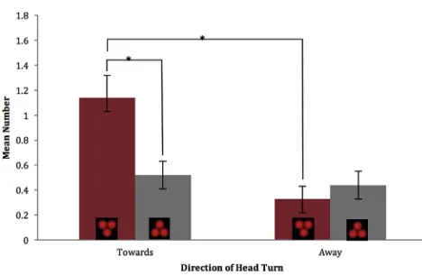
The number of head turns made in response to the stimuli was assessed using condition-blind coding of the 4D scans. All groups presented normally distributed data with similar levels of variation. On average,Figure 2shows that more head turns were made in the direction of the upright (mean [M] = 1.14, SD = 1.09) than in the direction of the inverted (M = 0.52, SD = 0.62) stimuli. There were slightly more head turns in the opposite direction to the inverted (M = 0.44, SD = 0.62) than the upright (M = 0.33, SD = 0.55) stimuli. A Wilcoxon signed-ranks test indi-cated that more head turns were directed toward than away from the upright stimuli (Z = 3.117, p = 0.002). Further, significantly more head turns were directed toward the upright than the
inverted stimuli (Z = 2.380, p = 0.017). No further comparisons were found to be significant.
In addition, a paired-samples t test compared difference scores (looks toward minus looks away) showing a significant difference between upright (M = 0.77, SD = 1.06) and inverted (M = 0.08, SD = 0.13) stimuli, t(38) = 2.924, p = 0.005.
These results indicate that the fetus in the third trimester is more likely to engage with stimuli featuring an upright face-like configuration when contrasted with an inverted configuration. We therefore conclude that postnatal experience is not neces-sary for the emergence of a preferential visual system for face-like stimuli. This finding rules out rapid postnatal learning, such as filial imprinting, as a mechanism for this visual proclivity. These mechanisms may be innate, or, possibly, the perceptual bias is triggered by exposure to patterned light in the womb during prenatal visual experiences.
Prenatal Visual Experience
[image:3.603.58.378.98.388.2]In the third trimester of pregnancy, the human fetus has the capac-ity to process perceptual information [1–3]. Despite this, newborn visual preferences are often attributed to innate mechanisms or to rapid imprinting. Postmortem analysis of the human eye has shown that there is substantial biological development from mid-gestation through to term, with many of the essential compo-nents for visual function present starting around 25 weeks gesta-tional age (GA) [11, 12]. This research also indicated more advanced development in peripheral visual regions. Before post-natal development, peripheral vision is therefore likely to be more sensitive than foveal vision for detecting environmental change. Work on prenatal visual development suggests that visual percep-tual capacities are analogous to newborn functionality well before
Figure 1. A Conceptual Illustration of the Stimuli
(A–D) A conceptual illustration of the stimuli utilized in the current study, depicting upright (A and B) and inverted (C and D) orientations. (A) and (C) illustrate the stimuli prior to contact with maternal tissue. (B) and (D) display the consequence of interaction with 30 mm of maternal tissue based on our equation. To calculate the expected projection size, we used the simple equation for the anisotropy of scatter [9] along with a value for adipose tissue [10] from the corrected version of Figure 8 (expanded view in the Corrigendum, page 2): projected diameter = tan(arccos(g))3thickness of the tissue32. From the figure, g0.98 for adipose, giving a diameter after 30 mm of tissue of12 mm.
term. Evidence also derives from reports of visual function in low-risk pre-term infants. Studies have shown newborns perform fixing and tracking from 32 weeks GA [13–16]. Further, comparing visual evoked potentials in full-term neonates to concep-tion age-matched pre-term infants, no dif-ference was found in neural response to visual stimuli [17]. Postnatal visual experi-ence therefore did not affect the neural correlates of visual processing.
Recent modeling work has indicated a substantially greater luminance within the uterus than previously thought [5]. Animal models have demonstrated not only that light penetrates into the uterus but also that light penetration is critical in mice for preparing the eye and light response pathway for postnatal vision [18]. Together, these studies indicate that visual experi-ence starts prenatally. Prenatal light levels not only are essential for the development of visual pathways but also allow for the innovative methodology used in the current study, with percep-tual content projected through the uterine wall, taking into account how light interfaces with maternal tissue.
From Prenatal to Postnatal Visual Development
Control of the eyes by neonates is relatively advanced when con-trasted with other motor abilities [19]. It is for this reason that vi-sual paradigms are a key aspect of postnatal research. Research on fetal visual perception, however, is limited when compared with our current understanding of fetal abilities in other modal-ities [20]. During avian development, as a consequence of em-bryo orientation in the egg, differential exposure to light for the left or right eye due to the location of the wing results in brain lateralization in chicks [21]. Multiple studies have investigated the response of the human fetal brain to light [22], although none have delivered stimuli that have contained the percept of an image. This absence has been driven by the complexity of delivering visual stimuli to the fetus.
One key, well-replicated finding in newborn research is the preference to engage with a face-like stimulus when contrasted with other forms of stimuli, including the same stimulus pre-sented in an inverted configuration [6, 7]. There has been much debate on how and why this preference is present in the
2 Current Biology27, 1–4, June 19, 2017
emerging visual system [23, 24]. A comprehensive review of two decades of research offers an extension to the original theoret-ical model put forward in explanation of newborn face prefer-ence [25]. The underlying assumptions in much of the newborn visual literature are (1) that no visual experience has taken place prior to birth and (2) that the examination of fetal visual capacities is not possible. The present study illustrates that fetal visual perception can be indexed during the third trimester, given the technical advances in 4D ultrasound that can provide access to fetal fine-grained behavior [26–28]. With appropriate modifica-tions, other aspects of newborn infant perception could also be assessed in the third trimester, including biological motion processing [29]. An exploration of capacities at this stage of development could greatly inform our understanding of visual preferences, as models of development feature different as-sumptions related to the underlying development of visual sys-tems. For example, even though the results of the present study are compatible with superior colliculus activity [8], the same cannot be said for a proposed ‘‘gravity bias’’ for visual stimuli, which has been previously proposed [30].
Even though the results of the current study are analogous to postnatal behaviors, due to the properties of the fetal environ-ment, the paradigm and stimuli are not exactly the same be-tween the current study and postnatal research. For example, only light from the red (or long wave) end of the spectrum pene-trates maternal tissue. Despite this, the results are consistent with a model of fetal visual preferences [8], whereby the largest differential response was for a negative polarity stimulus set with white dots on a black background when contrasted with other stimuli, including black dots on a white background. It should also be noted that the results of the present study do not imply that the fetus can respond to faces presented exter-nally under everyday circumstances. The behavior that has been demonstrated in the current study derives from the specific conditions of the experiment.
The capacity to (1) present visual stimuli through projected light and (2) precisely measure fetal behavior using ultrasound recordings, as demonstrated in the present study, allows for the execution of studies with the human fetus that closely
resemble postnatal methodologies with infant populations. Such an approach will have implications for further understand-ing of the fetus [31] and developmental processes in general. Fetal research can consequently employ similar visual method-ologies and control procedures as those seen in the infancy domain (e.g., [29, 32]). Currently it is unknown how effective these methods would be in terms of producing responses earlier in gestation or whether infant-derived paradigms, such as fixa-tion time measurements, will be as likely to produce meaningful results with the fetus in the third trimester. Such work will un-doubtedly provide more information about the development of the visual system in addition to current animal models [18] and with respect to the transition from fetus to infant.
STAR+METHODS
Detailed methods are provided in the online version of this paper and include the following:
d KEY RESOURCES TABLE
d CONTACT FOR REAGENT AND RESOURCE SHARING
d EXPERIMENTAL MODEL AND SUBJECT DETAILS
d METHOD DETAILS
B Stimuli
B Data Acquisition B Procedure B Data Coding
d QUANTIFICATION AND STATISTICAL ANALYSIS
d DATA AND SOFTWARE AVAILABILITY
AUTHOR CONTRIBUTIONS
Conceptualization, V.M.R.; Methodology, K.D., R.J.Y., N.R., and V.M.R.; Formal Analysis, K.D. and V.M.R.; Resources, J.A. and T.D.; Data Cu-ration, K.D. and V.M.R.; Writing – Original Draft, V.M.R. and K.D.; Writing – Review & Editing, V.M.R., K.D., R.J.Y., J.A., T.D., and N.R.; Visualization, V.M.R., K.D., and R.J.Y.; Supervision, V.M.R.; Project Administration, V.M.R.; Funding Acquisition, V.M.R., N.R., and R.J.Y.
ACKNOWLEDGMENTS
This work was supported by an Economic and Social Research Council Trans-formative Research Grant (grant number ES/L003155/1). V.M.R. is a Professor in the International Centre for Language and Communicative Development (LuCiD) at Lancaster University. The support of the Economic and Social Research Council (ES/L008955/1) is gratefully acknowledged. R.J.Y. acknowl-edges support by the Royal Society through a University Research Fellowship (UF110555). We thank Janette Keit and Linda Walshaw for acting as sonogra-phers; Jane Brooks for research midwife assistance; and Dr. Steve Milan for input throughout the project.
Received: March 3, 2017 Revised: April 20, 2017 Accepted: May 12, 2017 Published: June 8, 2017
REFERENCES
1.DeCasper, A.J., and Fifer, W.P. (1980). Of human bonding: newborns pre-fer their mothers’ voices. Science208, 1174–1176.
[image:4.603.54.288.96.249.2]2.Zoia, S., Blason, L., D’Ottavio, G., Bulgheroni, M., Pezzetta, E., Scabar, A., and Castiello, U. (2007). Evidence of early development of action planning in the human foetus: a kinematic study. Exp. Brain Res.176, 217–226.
Figure 2. The Mean Number of Fetal Head Turns to the Stimuli
3.Witt, M., and Reutter, K. (1996). Embryonic and early fetal development of human taste buds: a transmission electron microscopical study. Anat. Rec.246, 507–523.
4.Lo´pez-Teijo´n, M., Garcı´a-Faura, A´., and Prats-Galino, A. (2015). Fetal facial expression in response to intravaginal music emission. Ultrasound 23, 216–223.
5.Del Giudice, M. (2011). Alone in the dark? Modeling the conditions for vi-sual experience in human fetuses. Dev. Psychobiol.53, 214–219. 6.Johnson, M.H., and Morton, J. (1991). Biology and Cognitive Development:
The Case of Face Recognition (Basil Blackwell).
7.Fantz, R.L. (1963). Pattern vision in newborn infants. Science 140, 296–297.
8.Pitti, A., Kuniyoshi, Y., Quoy, M., and Gaussier, P. (2013). Modeling the minimal newborn’s intersubjective mind: the visuotopic-somatotopic alignment hypothesis in the superior colliculus. PLoS ONE8, e69474. 9.Jacques, S.L. (2013). Optical properties of biological tissues: a review.
Phys. Med. Biol.58, R37–R61.
10.Peters, V.G., Wyman, D.R., Patterson, M.S., and Frank, G.L. (1990). Optical properties of normal and diseased human breast tissues in the visible and near infrared. Phys. Med. Biol.35, 1317–1334.
11.Hendrickson, A., Possin, D., Vajzovic, L., and Toth, C.A. (2012). Histologic development of the human fovea from midgestation to maturity. Am. J. Ophthalmol.154, 767–778.e2.
12.Hendrickson, A., and Drucker, D. (1992). The development of parafoveal and mid-peripheral human retina. Behav. Brain Res.49, 21–31. 13.Dubowitz, L.M.S., Dubowitz, V., and Morante, A. (1980a). Visual function in
the newborn: a study of preterm and full-term infants. Brain Dev.2, 15–29. 14.Dubowitz, L.M.S., Dubowitz, V., Morante, A., and Verghote, M. (1980b). Visual function in the preterm and fullterm newborn infant. Dev. Med. Child Neurol.22, 465–475.
15.Morante, A., Dubowitz, L.M.S., Leven, M., and Dubowitz, V. (1982). The development of visual function in normal and neurologically abnormal pre-term and fullpre-term infants. Dev. Med. Child Neurol.24, 771–784. 16.Romeo, D.M., Ricci, D., Serrao, F., Gallini, F., Olivieri, G., Cota, F.,
Romagnoli, C., and Mercuri, E. (2012). Visual function assessment in late-preterm newborns. Early Hum. Dev.88, 301–305.
17.Baraldi, P., Ferrari, F., Fonda, S., and Penne, A. (1981). Vision in the neonate (full-term and premature): preliminary result of the application of some testing methods. Doc. Ophthalmol.51, 101–112.
18.Rao, S., Chun, C., Fan, J., Kofron, J.M., Yang, M.B., Hegde, R.S., Ferrara, N., Copenhagen, D.R., and Lang, R.A. (2013). A direct and melanopsin-dependent fetal light response regulates mouse eye development. Nature494, 243–246.
19.Stanojevic, M., and Kurjak, A. (2008). Continuity between fetal and neonatal neurobehavior. J. Ultra Obst.Gyne.2, 64–75.
20.DiPietro, J.A., Costigan, K.A., and Voegtline, K.M. (2015). Studies in fetal behavior: revisited, renewed and reimagined. Mono. Soc.r Res. Child Dev.80, 1–94.
21.Rogers, L.J. (1990). Light input and the reversal of functional lateralization in the chicken brain. Behav. Brain Res.38, 211–221.
22.Dunn, K., Reissland, N., and Reid, V.M. (2015). The functional foetal brain: a systematic preview of methodological factors in reporting foetal visual and auditory capacity. Dev. Cogn. Neurosci.13, 43–52.
23.Simion, F., Valenza, E., Umilta`, C., and Dalla Barba, B. (1998). Preferential orienting to faces in newborns: a temporal-nasal asymmetry. J. Exp. Psychol. Hum. Percept. Perform.24, 1399–1405.
24.Wilkinson, N., Paikan, A., Gredeb€ack, G., Rea, F., and Metta, G. (2014). Staring us in the face? An embodied theory of innate face preference. Dev. Sci.17, 809–825.
25.Johnson, M.H., Senju, A., and Tomalski, P. (2015). The two-process theory of face processing: modifications based on two decades of data from in-fants and adults. Neurosci. Biobehav. Rev.50, 169–179.
26.Reissland, N., Francis, B., Mason, J., and Lincoln, K. (2011). Do facial ex-pressions develop before birth? PLoS ONE6, e24081.
27.Myowa-Yamakoshi, M., and Takeshita, H. (2006). Do human fetuses antic-ipate self-directed actions? A study by four-dimensional (4D) ultrasonog-raphy. Infancy10, 289–301.
28.Reissland, N., Francis, B., Aydin, E., Mason, J., and Schaal, B. (2014). The development of anticipation in the fetus: a longitudinal account of human fetal mouth movements in reaction to and anticipation of touch. Dev. Psychobiol.56, 955–963.
29.Simion, F., Regolin, L., and Bulf, H. (2008). A predisposition for biological motion in the newborn baby. Proc. Natl. Acad. Sci. USA105, 809–813. 30.Vallortigara, G., and Regolin, L. (2006). Gravity bias in the interpretation of
biological motion by inexperienced chicks. Curr. Biol.16, R279–R280. 31.Abbott, A. (2015). Neuroscience: the brain, interrupted. Nature518, 24–26. 32.Rochat, P. (2011). What Is It Like to Be a Newborn? Oxford Handbook of
the Self, S. Gallagher, ed. (Oxford University Press).
33.Nijhuis, J.G., Prechtl, H.F.R., Martin, C.B., Jr., and Bots, R.S.G.M. (1982). Are there behavioural states in the human fetus? Early Hum. Dev.6, 177–195.
34.Stockman, A., Jagle, H., Pirzer, M., and Sharpe, L.T. (2008). The depen-€ dence of luminous efficiency on chromatic adaptation. J. Vis.8, 1–26. 35.Johnson, M.H., Dziurawiec, S., Ellis, H., and Morton, J. (1991). Newborns’
preferential tracking of face-like stimuli and its subsequent decline. Cognition40, 1–19.
36.Landis, J.R., and Koch, G.G. (1977). The measurement of observer agree-ment for categorical data. Biometrics33, 159–174.
4 Current Biology27, 1–4, June 19, 2017
STAR
+
METHODS
KEY RESOURCES TABLE
CONTACT FOR REAGENT AND RESOURCE SHARING
Further information and requests for resources and reagents should be directed to and will be fulfilled by the Lead Contact, Vincent Reid ([email protected]).
EXPERIMENTAL MODEL AND SUBJECT DETAILS
The sample size was calculated based on attrition rates for newborn paradigms with the assumption of a 20% larger attrition rate for the current study due to the number of fetal head orientations that would allow for the delivery of stimuli. All pregnant women partici-pating received written information prior to agreeing to take part in the study and gave informed written consent before participation. This study was approved by NHS Health Research Authority National Research Ethics Service, the Lancaster University Research Ethics Committee, the Durham University Psychology Department Ethics Sub-Committee and the Cumbria University Ethics Committee. Behavioral responses to stimuli were assessed in 83 fetuses by an ultrasonographer and an experimenter, utilizing 4d ultrasound. Thirteen were excluded due to poor image resolution. All instances of exclusion in this sample due to fetal head position causing visual stimuli presentation to become unviable, co-occurred with poor image resolution. Twenty-nine were excluded at the coding stage as these fetuses indicated a lack of eye or body movements throughout the scanning period and appeared to be in a deep sleep state, otherwise referred to as behavioral state 1F [33].
The final sample consisted of 39 fetuses, gestational age between 231 and 252 days (M= 240.62 days). Maternal tissue thickness ranged from 13.1 mm to 69.7 mm (M= 27.47, SD = 10.32). Seventeen fetuses were female (44%) with the sample showing an average APGAR score of 9.64 (SD = 1.06), although birth records for 5 participants could not be obtained. Twenty-one scans were performed at Blackpool Victoria Hospital, the remainder at Cumbria University Medical Imaging Unit. All participants had singleton pregnancies with no known complications and a BMI of approximately 30 or lower at the start of pregnancy. A BMI of this level or lower was required in order to ensure a similar quality of presentation of the stimuli through varying amounts of maternal tissue.
METHOD DETAILS
Stimuli
The stimuli were constructed from custom-made semiconductor laser 2 mm dot diodes emitting at 650 nm. Three diodes were ar-ranged in a triangular pattern, with 15 mm distance between each dot. The light source was calibrated to output optical powers of 0.5 mW, 1 mW or 5 mW for maternal tissue thickness (t) below 15 mm, between 15 mm and 30 mm and above 30 mm respectively. This
REAGENT or RESOURCE SOURCE IDENTIFIER
Deposited Data
Data reported in this paper N/A Dataverse Digital Repository: 10.7910/DVN/RLIBYV Software and Algorithms
IBM SPSS statistical package, version 23 https://www.ibm.com/analytics/us/en/ technology/spss/
N/A
Observer XT Behavioral Coding and Analysis software http://www.noldus.com/human-behavior-research/products/the-observer-xt
N/A
Other
GE Healthcare Voluson E8 Expert Advanced 4D HD Live Ultrasound Scanner (location: Blackpool Victoria Hospital)
http://www3.gehealthcare.com/en/ products/categories/ultrasound
BT13
GE Healthcare Voluson 4D Live Ultrasound Scanner (location: Cumbria University)
http://www3.gehealthcare.com/en/ products/categories/ultrasound
iBT07
GE Healthcare 4D Transducer (location: Blackpool Victoria Hospital)
http://www3.gehealthcare.com/en/products/ categories/ultrasound/ultrasound_probes
RM66
GE Healthcare 4D Transducer (location: Cumbria University)
http://www3.gehealthcare.com/en/products/ categories/ultrasound/ultrasound_probes
RAB4-8-RS
ensured that a consistent level of light was delivered to the fetus irrespective of variations in maternal tissue thickness. Intrauterine illuminance (LI) was calculated using the equation:
LI=LE10 :0942+t
:032+:058r
1+r
modified from [5] to remove the clothing factor.LE, the external illuminance was calculated using the output power of the light source,
assuming a projected spot size diameter of 10 mm for a maternal tissue thickness of 30 mm, and correcting for the source wavelength of 650 nm, based on values in [34]. An approximate muscle to fat ratio (r) of 2 was used, as in [5]. Taking three scenarios as examples, with maternal tissue thicknesses of 15 mm, 20 mm and 30 mm, we calculate corresponding intrauterine illuminances of 36, 24 and 16 lx respectively.Figure 1displays the projected input to maternal tissue and the approximate consequence of interaction with maternal tissue for the stimuli. The light levels of the stimuli are all within the range that the fetal visual system is thought to work best, and significantly lower than the illuminances that a fetus may be exposed to on a bright sunny day [5]. The estimated distance between the dots subtended 23 degrees of visual angle. The laser diodes were 15 mm apart and 2 mm in diameter. We estimated an average scatter of 5 mm in either direction, resulting in 5 mm between the dots. This degree of visual angle between the dots is similar to that seen in postnatal studies [e.g.,35].
The dispersion of the input configuration of the light source after traveling through 30 mm of maternal tissue, as illustrated in Fig-ure 1, was estimated using data from [9]. Using a value for the anisotropy of scatter at the source wavelength for adipose taken from [35] results in an expected broadening of a point source to a diameter of 12 mm after transmission through 30 mm of tissue. It should be noted that the minor dispersion, due to amniotic fluid between the fetus’ eye and the uterine wall, has not been taken into account due to uncertainties in quantifying this measure.
Data Acquisition
Maternal tissue was measured from maternal skin to the uterine wall using 2D ultrasound. The 4D live ultrasound technology operated on a bandwidth of 2-8MHz. Fetal behavior was recorded at Blackpool Victoria Hospital using a GE Healthcare Voluson E8 Expert BT13 advanced 4D HD live ultrasound scanner and 4D probe, model RM66. At Cumbria University Medical Imaging Unit, a GE Healthcare Voluson iBT07 4D live ultrasound scanner and 4D probe, model RAB4-8-RS was used. Recordings were saved to DVD for offline coding.
Procedure
The ultrasonographer ensured the ultrasound recording occurred while the experimenter selected the correct strength stimulus, de-pending on maternal tissue depth, and conducted the procedure. Typically the participant was lying on her side. Timings were controlled via a stopwatch and noted on a clipboard, typically by a second experimenter. Once comfortable, an initial set of 2D scans were taken comprising the fetal head position, maternal tissue thickness, fetal biparietal diameter, occipitofrontal diameter, head circumference, abdominal circumference, femur length, fetal weight. Fetal biometry measurements demonstrated normal fetal growth and no fetal anomalies. Note that depth scans were taken in the same maternal position as for when the stimulus was then presented. All participants were then asked not to talk during the study and to remain as still as possible in order to optimize image quality. The 2D scan also informed the experimenter of the precise location of the fetal head prior to the presentation of the stimuli.
The stimuli were delivered in two orientations (‘‘upright’’ and ‘‘inverted’’) to the maternal abdomen (Figure 1) relative to the position of the fetus (breech presentation/head up, n = 6, cephalic presentation/head down, n = 33). Both were presented to the side of the fetal face, such that the stimuli were presented to the fetal peripheral visual areas (left, n = 19, right, n = 20). The experimenter was not blind to the condition that was presented. The ultrasound image, interpreted by the ultrasonographer, informed the initial placement of the light source on the maternal abdomen at the start of each trial. This gave the greatest likelihood that the experimenter could place the light in the peripheral visual field of the fetus. The experimenter utilized the ultrasound image for initial placement but at-tended to the movement of the stimulus across the maternal abdomen, precluding the viewing of the ultrasound image once the trial had started. The administration of the stimulus was standardized across conditions by ensuring equivalent timing between condi-tions. From the 39 scans, the mean distance of the fetal eye to the uterine wall was 12 mm, SD 7.5 mm, with range 2 mm to 30 mm. The stimulus was then moved across the maternal abdomen in a horizontal direction away from the fetal central visual location, for 5 s at an average speed of 1 cm per second. This is consistent with speeds reported in newborn studies [6] taking into account con-straints specific to this population, i.e., the width of maternal tissue that was accessible in order to present stimuli and the space avail-able for the fetus to move. Movement and light offset was simultaneous and timing was controlled via a stopwatch in view of the experimenter who was delivering the stimuli. This process was repeated a total of 5 times, with the procedure then immediately repeated with the alternate stimulus orientation. The presentation order for upright and inverted orientations of the stimuli were coun-terbalanced across the sample. Participants were blind to condition.
Data Coding
During the scan, the position of the light in relation to the recorded image of the fetus was noted. The ultrasound image was rotated by the sonographer such that the fetal face appeared upright. This was done so that, at a later stage, coding consisted of simply ‘‘left’’ and ‘‘right’’ head movements in the coronal plane. Coding of recordings was conducted offline using Observer XT 12.5 software
e2 Current Biology27, 1–4.e1–e3, June 19, 2017
(Tracksys Ltd). The duration of head movements away from the individual fetus’ initial resting head position were measured, along with the direction of the movement for each trial. At the start of each trial, the resting position of the head on the ultrasound image was recorded and coded as ‘‘center.’’ At the onset of a head movement, a code denoted the start time and direction of movement. This code then remained active until a second code was given to indicate a return to the initial resting head position, a head movement in the opposite direction (an opposite head movement was then coded) or the end of a trial. The onset of any head movement was not constrained to the onset of the stimulus but no movements were coded following stimulus offset.
Following blind coding, ‘left’ and ‘right’ head movements were transformed to indicate whether movements were made toward or away from the stimuli. An orientation toward the stimuli was a horizontal head movement in accordance with the position of the light recording during the scan. An orientation away from the stimuli was a horizontal head movement performed in the opposite direction to the recorded position of the light source.
Cohen’s kappa was performed to determine interrater agreement on fetal head movements. There was a substantial agreement between two coders’ judgments,k= 0.797 (95% CI, 0.503 1.00), p = <0.001. Behavioral measurement was thus deemed to be suit-able for use in the hypothesis tests in the present study [36].
QUANTIFICATION AND STATISTICAL ANALYSIS
All groups presented normally distributed data with similar levels of variation. Using SPSS statistical data package, version 23, a com-parison of the number of head turns across conditions was made using the Wilcoxon rank signed test. A comcom-parison of difference scores was also made using a paired-samples t test.
DATA AND SOFTWARE AVAILABILITY