STUDY OF THE ACTIVITY, ENERGY WINDOW AND COLLIMATOR EFFECTS ON SPECT IMAGES BY
USING IMAGES SIMULATED BY SIMIND SOFTWARE
Hamed Hasani, *Alireza
Mohaddeseh Saber
Department of Physics, University of Guilan, Rasht, Iran
ARTICLE INFO ABSTRACT
Radiation dose reduction and improved factor in nuclear medicine imaging. effect of the amount of the injected
the image quality of renal SPECT. We used SIMIND software to simulate SPECT system equipped with LEHR and LEGP collimators. The images of kidneys were obtained by use of the NCAT phantom with activities of 2mCi(75 M
energy windows of 10 and 20%. Then, the images were compared with the reference image by MATLAB software.
obtained by use of the
LEHR collimator, energy window of 20% could be produced images with higher quality in administered activity
width of 10%. The window width of 10% responds better in LEGP collimator and this collimator works well in higher activities with lower window width energy.
Copyright © 2015 Hamed Hasani et al. This is an open access article distributed distribution, and reproduction in any medium, provided the original work is properly cited.
INTRODUCTION
In nuclear medicine, a radioactive tracer is administrated to the specific organ of patient body, through various methods such as perfusion, mouth, respiration, Foley catheter, Spinal cannula and so on. Then, gamma camera provides 2
accumulation of tracer in the organ. Finally, a 3
distribution is performed by use of the reconstruction methods. The quantitative information of organs functions such as renal function, cardiac output and blood flow of the brain can be achieved by means of scintigraphy. Despite the development of anatomical imaging techniques, renal scintigraphy has found its place in the assessment of renal function. Nuclear medicine renal scan can be very helpful in evaluation of renal function, hypertension, renal failure, and even kidney transplant et al., 1994). Renal scintigraphy includes the study of renal blood flow, the parenchyma, and secretion of organs. The most commonly used radioisotope in single photon imaging is Tc99m, which has half-life of 6.1 hours is long enough to get medical information (Zolle, 2007).
*Corresponding author:AlirezaSadremomtaz,
Department of Physics, University of Guilan, Rasht, Iran
ISSN: 0975-833X
Article History:
Received 21st April, 2015
Received in revised form 10th May, 2015
Accepted 30th June, 2015
Published online 31st July,2015
Key words:
Image quality, Kidney scintigraphy, Nuclear medicine imaging.
Citation: Hamed Hasani, Alireza Sadremomtaz, Peyvand
energy window and collimator effects on SPECT images by using images simulated by SIMIND software
Research, 7, (7), 18374-18378.
RESEARCH ARTICLE
STUDY OF THE ACTIVITY, ENERGY WINDOW AND COLLIMATOR EFFECTS ON SPECT IMAGES BY
USING IMAGES SIMULATED BY SIMIND SOFTWARE
*Alireza Sadremomtaz, Peyvand Taherparvar and
Mohaddeseh Saber Zidehsaraiee
Department of Physics, University of Guilan, Rasht, Iran
ABSTRACT
Radiation dose reduction and improved image quality in nuclear medicine scan is most important factor in nuclear medicine imaging. In this paper, we simulated kidney scintigraphy to examine the effect of the amount of the injected radiopharmaceutical,energy window, and type of collimator on the image quality of renal SPECT. We used SIMIND software to simulate SPECT system equipped with LEHR and LEGP collimators. The images of kidneys were obtained by use of the NCAT phantom with activities of 2mCi(75 MBq), 5mCi (185MBq) and 10mCi (370MBq) by use of the energy windows of 10 and 20%. Then, the images were compared with the reference image by MATLAB software. Assessment of comparison between results show that the best result could be obtained by use of the narrow window width energy and injection of the high activity. By use of the LEHR collimator, energy window of 20% could be produced images with higher quality in administered activity, while in high activity, the best result could be obtained
width of 10%. The window width of 10% responds better in LEGP collimator and this collimator works well in higher activities with lower window width energy.
is an open access article distributed under the Creative Commons Attribution License, which distribution, and reproduction in any medium, provided the original work is properly cited.
In nuclear medicine, a radioactive tracer is administrated to the specific organ of patient body, through various methods such as perfusion, mouth, respiration, Foley catheter, Spinal cannula and so on. Then, gamma camera provides 2-D views of of tracer in the organ. Finally, a 3-D image of distribution is performed by use of the reconstruction methods. The quantitative information of organs functions such as renal function, cardiac output and blood flow of the brain can be Despite the development of anatomical imaging techniques, renal scintigraphy has found its place in the assessment of renal function. Nuclear medicine evaluation of renal function, kidney transplant (Weber Renal scintigraphy includes the study of renal blood flow, the parenchyma, and secretion of organs. The most commonly used radioisotope in single photon imaging is is long enough to get
Department of Physics, University of Guilan, Rasht, Iran.
Renal scintigraphy involves the intravenous injection of a Tc99m-labeled radiopharmaceutical (mostly DTPA). Then, imaging were performed under a protocol approved by the National Institutes of Health.
posterior and anterior views are taken in normal and transplanted kidney respectively (
In nuclear medicine, selection of theproper collimators and energy window are considered as one of the most important physical parameters for enhancement of image quality and scatter events suppression. The setting of energy window width allows defining rejection rate of scatter photons from all logging photons in gamma camera.
width rejects more scatter photons and reduces the counts rate from the accumulated activity and vice versa. It seems possible to achieve more signal to noise ratio and better image sensitivity simultaneously if the energy window widt
is considered carefully. On the other hand, spatial resolution and sensitivity of the gamma camera
type of collimator. Different type of collimators are used in the scintigraphy, which their function depends on the particul geometries of holes and septals. Selection of the proper collimator could be improved the signal to noise ratio and
Available online at http://www.journalcra.com
International Journal of Current Research
Vol. 7, Issue, 07, pp.18374-18378, July, 2015
INTERNATIONAL
Sadremomtaz, Peyvand Taherparvar and Mohaddeseh Saber Zidehsaraiee energy window and collimator effects on SPECT images by using images simulated by SIMIND software”,
z
STUDY OF THE ACTIVITY, ENERGY WINDOW AND COLLIMATOR EFFECTS ON SPECT IMAGES BY
USING IMAGES SIMULATED BY SIMIND SOFTWARE
Taherparvar and
in nuclear medicine scan is most important In this paper, we simulated kidney scintigraphy to examine the energy window, and type of collimator on the image quality of renal SPECT. We used SIMIND software to simulate SPECT system equipped with LEHR and LEGP collimators. The images of kidneys were obtained by use of the NCAT Bq), 5mCi (185MBq) and 10mCi (370MBq) by use of the energy windows of 10 and 20%. Then, the images were compared with the reference image by Assessment of comparison between results show that the best result could be narrow window width energy and injection of the high activity. By use of the LEHR collimator, energy window of 20% could be produced images with higher quality in lower , while in high activity, the best result could be obtained by use of the window width of 10%. The window width of 10% responds better in LEGP collimator and this collimator
ribution License, which permits unrestricted use,
Renal scintigraphy involves the intravenous injection of a labeled radiopharmaceutical (mostly DTPA). Then, imaging were performed under a protocol approved by the Usually, in renal scintigraphy, posterior and anterior views are taken in normal and
vely (Wujanto et al., 1987).
In nuclear medicine, selection of theproper collimators and are considered as one of the most important physical parameters for enhancement of image quality and scatter events suppression. The setting of energy window width allows defining rejection rate of scatter photons from all logging photons in gamma camera. Thus, narrower window width rejects more scatter photons and reduces the counts rate from the accumulated activity and vice versa. It seems possible to achieve more signal to noise ratio and better image sensitivity simultaneously if the energy window width setting On the other hand, spatial resolution and sensitivity of the gamma camera depends heavily on the Different type of collimators are used in the scintigraphy, which their function depends on the particular geometries of holes and septals. Selection of the proper collimator could be improved the signal to noise ratio and
INTERNATIONAL JOURNAL OF CURRENT RESEARCH
Zidehsaraiee, 2015. “Study of the activity,
image quality in the particular scan with specific protocols. That is depending on the injected radionuclide and its gamma energy. Low energy, mean energy, and high-energy are three main categories of collimators.
In common clinical condition, low energy collimators are used because technetium with 140 keV energy is mostly used. The common low-energy collimators put into three categories consist of low-energy high-resolution (LEHR), low-energy high-sensitivity (LEHS) and low-energy general-purpose (LEGP) collimators (Moore et al., 1992).
The LEHR collimators have more holes that are both deeper and smaller. This geometry restrictthe solid angle accepted by the collimator holes which improve the resolution, but it also decreases sensitivity (Cherry et al., 2003). LEHS collimator has a good ability in high counting rate of photons and can reduce the statistical noise and improve sensitivity of the image but restrict the spatial resolutioncauses to poor spatial resolution and high noise levels in the obtained images power spectrum.
In LEGP collimator, the holes diameter is relatively larger and the acceptance solid angle is wider. Its spatial resolution is degraded in relative to LEHR but its sensitivity is increased. In fact, this collimator is something between LEHR and LEHS. Image quality plays an important role in nuclear medicine imaging because the goal is to create a reliable image of the irradiated organs to diagnose or treat it correctly. The quality of the SPECT images are limited by several factors such as the attenuation and scatter of gamma ray photons inside the patient body, the detection efficiency of system and the spatial resolution of the collimator-detector system. Amount of the injected activity into the patient body, selection of energy window and intrinsic spatial resolution of collimator are major factors, which affect on the image quality. Because of the quantification of physical parameters are difficult or even impossible to calculate by experimental measurements. Many, papers have been focused on the simulation study based on the Monte carol method. Various simulation programs such as SIMIND, SIMSET, GEANT4 and … has been successfully applied in a variety of nuclear medicine procedure. In 1989, Ljungberg and colleagues at Lund University in Sweden introduced SIMIND as a proprietary code of SPECT. SIMIND software is written in Fortran 90 and includes two main parts: The first section defines the parameter (CHANGE) and the second part (SIMIND) performs the actual simulation (Ljungberg, 2009; Ljungberg, 2012).
Hereby, in this study, we examine the effect of the amount of injected activity into the patient body on the image quality of a gamma camera equipped with the LEHR and LEGP collimators. We used SIMIND program along with a digital advanced cardiac torso phantom such as NCAT to generate image of a radiopharmaceutical that is accumulated by the kidneys. The NCAT phantom was developed base on the Surfaces of a non-uniform rational B-spline (NURBS) in detail by P. Segars. The phantom has a 4D beating heart model which was derived from 4D tagged (MRI) data. NCAT phantom was used to generate CT attenuation maps and activity distribution volumes for the lung regions. In addition
to, the effect of energy window on the image quality of renal scan were studied.
Some criteria such as average difference (AD), maximum difference (MD), mean square error (MSE) and peak signal to noise ratio (PSNR) between images and reference images have been used to obtained the optimized protocol and efficient procedure that produce the best results.
MATERIALS AND METHODS
[image:2.595.317.549.323.500.2]The SIMIND Monte Carlo simulation program version 4.9e were used to create the projection images from NCAT phantom to simulate kidneys with activities of 2mCi (75MBq), 5mCi (185MBq) and 10mCi (370MBq) with two different energy window percentages of 10% and 20%. with increasing of window width energy, recorded events in the SPECT head have been increased. But, as shown in Fig.1, these events also include corrupted events produced by Campton interactions of gamma rays with scintillation crystal of SPECT heads, collimators or patient body.
Figure 1. Representation of the relationship between the energy window width 15%, 20% and 25% of energy spectrum of Tc-99m
and the relative number of counts
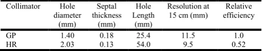
For each cases, we also used the LEHR and LEGP collimators, which their properties were according to the Table 1.
Table 1. Properties of HR and GP collimators (Khalil, 2011)
Collimator Hole
diameter (mm)
Septal thickness
(mm)
Hole Length
(mm)
Resolution at 15 cm (mm)
Relative efficiency
GP 1.40 0.18 25.4 11.5 1.0
HR 2.03 0.13 54.0 9.5 0.52
We set photon energy on 140keV corresponding to Tc, image matrix dimensions on 128×128, pixel size on 0.3cm×0.3cm, 109 disintegration, the time of scan in each
angle on 30 seconds, 64 projections, crystal material of Na I with 1×8×50 cm3 dimensions and thickness of cover of 1mm.
[image:2.595.300.566.619.671.2]activity of 20mCi (750MBq), energy window of10% and LEHR collimator.
Then, all of the simulated images were decoded by XMedCon program and were stored as DICOM (digital reference standard for medical image files). Afterward, the image processing algorithms were proformed by MATLAB software. Individual images were compared with a reference image to find the best results among the simulated images. Comparison criteria, consist of average difference (AD), maximum difference (MD), mean square error (MSE) and peak signal to noise ratio (PSNR), are used to measure the quality of obtained images by different protocols. This criteria compared image matrix of referenced image, K(i,j), with the test images, I(i,j). These objective quality evaluation criteria define as following:
Average Difference is defined from point-to-point comparison of the test image with reference image. A value near zero means that, the test image is more similar to the reference image.
AD = 1
m × n (I(i, j) − K(i, j))
Maximum Difference is the maximum difference between compared points of the test image and reference image.
MD = MAX(MAX(I(i, j) − K(i, j))
Mean Square Erroris the mean square of the difference between the test image and reference image. MSEis defined as follow:
MSE = 1
m × n (I(i, j) − K(i, j))
Peak Signal to Noise Ratio (PSNR) is the maximum possible power of an image and the power of corrupting noise that affects the fidelity of its representation. A higher PSNR value should correlate to a higher quality image (Dewangan and Rathore, 2011).
PSNR = 10 log MAX
MSE
RESULTS
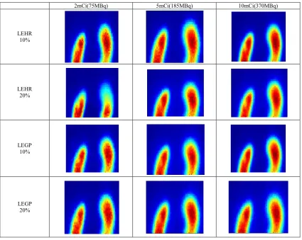
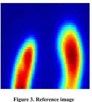
The results of the simulation of kidney scan with different administrated activity are shown in Fig. 2, which are acquired under implementation of different collimators – LEGP and LEHR- with window width energy of 10 and 20%. The test image also cold be seen in Fig. 3. On the other word, the reference image were produced with high-administrated activity to reduce statistical noise of count rate and using of the LEHR collimator and lower energy window to exclude scattered photons. The reference image could be seen in Fig. 2. We compared simulated images (Fig. 2) with reference image by some code written in MATLAB software. The results are shown in Tables 2 and 3, which are related to the LEHR and LEGP collimators, respectively.
2mCi(75MBq) 5mCi(185MBq) 10mCi(370MBq)
LEHR 10%
LEHR 20%
LEGP 10%
[image:3.595.90.516.415.754.2]LEGP 20%
Figure 1. The image of simulated kidney scan with implementation of different factors
Figure 3. Reference image
Table 2. Comparison of image quality parameters in different activities and energy windows by use of the LEHR collimator
LEHR COLLIMATOR
Activity 2mCi(75MBq) 5mCi(185MBq) 10mCi(370MBq)
Energy window
10% 20% 10% 20% 10% 20%
AD 3.52 3.23 3.06 2.85 2.53 3.42
MD 45.88 44.38 40.10 39.28 33.50 45.13
MSE 94.92 79.60 71.24 66.42 48.88 91.52
PSNR 28.30 29.12 29.60 29.90 31.23 28.51
Table 3. Comparison of image quality parameters in different activities and energy windows by use of the LEGP collimator
LEGP collimator
Activity 2mCi(75MBq) 5mCi(185MBq) 10mCi(370MBq)
Energy window
10% 20% 10% 20% 10% 20%
AD 3.60 3.52 2.62 2.61 2.22 3.33
MD 45.37 45.92 36.43 36.66 29.15 44.41
MSE 91.66 94.92 57.83 57.82 38.16 87.30
PSNR 28.42 32.31 30.50 30.50 32.31 28.56
Acquired images by use of the LEHR collimator in the Table 2 show that the values of AD, MD & MSE in administrated activities of 2mCi and 5mCi in kidney and energy window of 20% are less than the energy window of 10%, and the value of PSNR in the energy window of 20% is more than10%. These results show improvement of image quality in the window width of 20%, as could be seen in Fig. 2.
In the third column of Table 2, which are related to theactivity of 10mCi, the results are the oppositeof the activities of 2 and 5mCi. Indeed, better results are related to the energy window of10%. If we just consider window width of 10%, we can see the AD, MD and MSE values decrease, and the values of PSNR increases as activity increases, thus the image quality is improved.
In energy window of 20%, the image quality increases from activities of 2mCi to 5mCibut it decrease inthe administrated activity of 10mCi. According to Table 2, the best image quality for LEHR collimator could be obtained by use of the energy window of 10% in activity of 10mCi.
When we use the LEGP collimator, quality criterions of the image evaluation show that the window width of 10% could be
produced better results in the injected activities of 2mCi and 5mCithan energy window of 20%. The absolute difference between these values in the energy windows of 10 and 20% are sensible in the activities of 2 and10mCi, and negligible in the activity of 5mCi. The best image quality by use of the LEGP collimator could beobtained in the activity of 10mCiand window width of 10%.
The results of the SPECT system equipped with the LEGP collimator, by use of the energy window of 10%, show that the value of the AD, MD and MSE decrease and PSNR increases as activity increases, therefore, the image quality increases. In window width of 20%, image quality increases from 2mCi to 5mCi activities. In administrated activity of 10mCi, the results of image quality assessment are between others. The differences between the data from both tables show that in window width of 10% and activity of 2mCi, the results of the MD, MSE and PSNR, by use of the LEGP collimator, are better than LEHR collimator. Therefore, gamma camera equipped with the LEGP collimator by use of the window width of 10% could be produced better results than gamma camera equipped with the LEHR collimator in administrated activity of 2mCi in the kidney. However, in energy window of 20%, LEHR collimator could be produced better results, because of the having lower AD, MD and MSE values and higher PSNR, than LEGP collimator.
In the injected activity of 5mCi in window width of 10%, LEHR collimator shows better results, whereas, LEGP collimator could be produced better results inactivity of 20%. The better image quality belongs to the images acquired with gamma camera equipped with LEGP collimator in activities of 10mCiand window widths of 10% and 20%.
Setting energy window of 10% could producea higher quality image thanenergy window of 20% by use of the each of the collimators, in activity of 2mCi. In activity of 5mCiand using of the LEHR collimator, image quality in window width of 20% is better than 10%. However, for LEGP collimator and this activity, the result of the image quality evaluation are resemble in energy windows of 10 and 20%. Image quality is better by use of the energy window of 10% than 20% when we set administrated activity in value of 10mCi.
Conclusion
Results show that the image quality of acquired images strongly depend on the selection of the collimator, energy window, and administrated activity. The combination of these parameters could be produced different results. According to the assessments, we should increase the administrated activity to improve the image quality in lower energy window. In this case, reduction of energy window cause noise reduction and improve image quality. When administrated activity in the patient kidney increases, it is better to use lower energy window to suppress noise. However, in lower activities, lower window width leadto a significant decline in the quality of the image.
[image:4.595.37.288.396.475.2]collimator, lower window shows better results in all activity. Moreover, the best image quality could be obtained by higher optimal injected activities. For both LEGP and LEHR collimators, in higher energy window, increasing the activity up to 5mCi increases the image quality and up to 10mCi reduces it. By increasing the activity, the recorded photons in the gamma camera increase, whereas it increases the level of noise output. Then, wider energy window could be used in low activities and lower window could be used in high activity. To reduce the patients’ received dose, it is preferred to reduce the activity, Therefore, in low activities, our results recommend that the better results could be obtained by use of the LEGP collimator with energy window of 10% or LEHR with energy window of 20%. In lower activities with higher window, LEHR is better than the LEGP collimator, and in the higher activities with lower energy window, LEGP collimator is better than the LEHR collimator.
REFERENCES
Cherry S.R., Sorenson J. A., Phelps ME. Physics in Nuclear Medicine, 3rd Edition. Philadelphia, Saunders, 361-417, 2003.
Dewangan D.K., Rathore Y., Image Quality Costing of Compressed Image Using Full Reference Method, J Tech, 1(20), 68-71, 2231-3907, 2011.
Khalil M., Basic Sciences of Nuclear Medicine, springer, Medicine and Health Science, 2011.
Ljungberg M., Strand S.E., King A., Monte Carlo Calculations in Nuclear Medicine.2nd Edition, Applications in Diagnostic Imaging, 2012.
Ljungberg M., The SIMIND Monte Carlo program, Lund, Sweden, 2009. Available from:URL:http://www2.msf.lu. se/simind/Downloads.html.
Moore S.C., Kouris K., Cullum I., Collimator design for single photon emission tomography, Eur J Nucl Med., 19(2), 138– 150, 1992.
Weber D.A. et al, Pinhole SPECT: an approach to Research, in vivo high resolution SPECT imaging in small laboratory animals, J Nucl Med., 35,342–348, 1994.
Wujanto M.B., Lawson R.S., Prescott M.C., Testa H.J., The importance of using anterior and posterior views in the calculation of differential renal function using 99mTc-DMSA, Br J Radio, 160, 869–872, 1987.
Zolle I, Technetium-99m pharmaceuticals. Springer, Berlin, 2007.