Redetermination of the borax structure
from laboratory X-ray data at 145 K
Graeme J. Gainsford,* Tim Kemmitt and Caleb Higham
Industrial Research Limited, PO Box 31-310, Lower Hutt, New Zealand Correspondence e-mail: [email protected]
Received 26 March 2008; accepted 15 April 2008
Key indicators: single-crystal X-ray study;T= 145 K; mean(O–B) = 0.001 A˚; Rfactor = 0.025;wRfactor = 0.077; data-to-parameter ratio = 15.5.
The title compound, sodium tetraborate decahydrate (mineral name: borax), Na2[B4O5(OH)4]8H2O, has been studied previously using X-ray [Morimoto (1956).Miner. J. 2, 1–18] and neutron [Levy & Lisensky (1978).Acta Cryst.B34, 3502– 3510] diffraction data. The structure contains tetraborate anions [B4O5(OH)4]2with twofold rotation symmetry, which form hydrogen-bonded chains, and [Na(H2O)6] octahedra that form zigzag chains [Na(H2O)4/2(H2O)2/1]. The O—H bond distances obtained from the present redetermination at 145 K are shorter than those in the neutron study by an average of 0.127 (19) A˚ .
Related literature
For previous studies of the borax structure, see: Morimoto (1956); Levy & Lisenky (1978). For other structures listed in the Cambridge Structural Database (Allen, 2002) that contain the [B4O5(OH)4]
2
anion, see: Wanget al.(2004); Pan et al. (2007). For related structures, see: Yi et al. (2005). For comparative studies of hydrogen bonds obtained from X-ray and neutron data, see: Allen (1986); Smrcˇoket al.(2006).
Experimental
Crystal data
Na2[B4O5(OH)4]8H2O
Mr= 381.38
Monoclinic,C2=c a= 11.8843 (5) A˚
b= 10.6026 (4) A˚
c= 12.2111 (5) A˚
= 106.790 (2)
V= 1473.06 (10) A˚3
Z= 4
MoKradiation
= 0.22 mm1
T= 145 (2) K 0.650.360.26 mm
Data collection
Bruker–Nonius APEX2 CCD area-detector diffractometer Absorption correction: multi-scan
(SADABS; Bruker, 2006)
Tmin= 0.813,Tmax= 0.94
8429 measured reflections 2275 independent reflections 2137 reflections withI> 2(I)
Rint= 0.018
Refinement
R[F2> 2(F2)] = 0.025
wR(F2) = 0.076
S= 1.08 2275 reflections
147 parameters
All H-atom parameters refined max= 0.37 e A˚
3
min=0.22 e A˚
3
Table 1
Selected bond lengths (A˚ ).
Na1—O8i
2.3815 (6) Na1—O6ii
2.3979 (5) Na1—O7 2.4121 (6) Na2—O7iii
2.4041 (6) Na2—O9 2.4214 (6) Na2—O6 2.4441 (6) B1—O4 1.4451 (8)
B1—O1 1.4657 (7) B1—O2 1.4902 (8) B1—O3 1.5075 (8) B2—O2 1.3655 (8) B2—O3i
1.3757 (8) B2—O5 1.3784 (8)
Symmetry codes: (i)x;y;zþ1
2; (ii)x;y1;zþ 1
2; (iii)x;yþ1;zþ 1 2.
Table 2
Hydrogen-bond geometry (A˚ ,).
D—H A D—H H A D A D—H A
O5—H5iv O3iv
0.836 (15) 1.895 (15) 2.7300 (7) 176.3 (15) O4v
—H4 O9vi
0.828 (14) 2.049 (14) 2.8658 (8) 168.4 (12) O6—H6Avii
O5viii
0.868 (16) 1.978 (16) 2.8323 (8) 167.9 (14) O6ix—H6B O4v 0.846 (16) 2.040 (15) 2.8624 (8) 163.9 (16) O7vii
—H7A O2 0.827 (16) 1.989 (16) 2.8135 (8) 174.1 (12) O7—H7B O4 0.816 (15) 2.135 (15) 2.9233 (8) 162.3 (14) O8v—H8A O1x 0.866 (13) 1.936 (13) 2.7865 (6) 167.0 (14) O8v
—H8B O5xi
0.855 (15) 2.341 (14) 3.1320 (8) 154.2 (12) O9—H9Avii
O3 0.843 (16) 2.253 (16) 3.0894 (8) 171.7 (15) O9—H9Bxii
O8xiii
0.849 (17) 2.069 (16) 2.9034 (8) 167.4 (15)
Symmetry codes: (iv)x;yþ1;zþ1
2; (v)xþ1;y;zþ 1 2; (vi)xþ
1 2;y
1 2;z; (vii)
xþ1 2;yþ
1 2;zþ
1
2; (viii) xþ 1 2;yþ
3
2;zþ1; (ix) xþ1;yþ1;zþ1; (x)
xþ1;y;z; (xi)xþ1
2;yþ12;z12; (xii)x12;yþ12;z; (xiii)x;yþ1;z12.
Data collection:APEX2(Bruker, 2006); cell refinement:APEX2; data reduction: APEX2; program(s) used to solve structure: SHELXS97(Sheldrick, 2008); program(s) used to refine structure:
SHELXL97 (Sheldrick, 2008); molecular graphics: ORTEP-3
(Farrugia, 1997) andMercury(Macraeet al., 2006); software used to prepare material for publication:SHELXL97andPLATON(Spek, 2003).
We thank Dr J. Wikaira of the University of Canterbury, New Zealand, for her assistance with the data collection.
Supplementary data and figures for this paper are available from the IUCr electronic archives (Reference: WM2174).
References
Allen, F. H. (1986).Acta Cryst.B42, 515–522. Allen, F. H. (2002).Acta Cryst.B58, 380–388.
Bruker (2006).APEX2andSADABS. Bruker AXS Inc., Madison, Wisconsin, USA.
Farrugia, L. J. (1997).J. Appl. Cryst.30, 565.
Levy, H. A. & Lisensky, G. C. (1978).Acta Cryst.B34, 3502–3510.
Macrae, C. F., Edgington, P. R., McCabe, P., Pidcock, E., Shields, G. P., Taylor, R., Towler, M. & van de Streek, J. (2006).J. Appl. Cryst.39, 453–457. Morimoto, N. (1956).Miner. J.2, 1—18.
Pan, C.-Y., Wang, G.-M., Zheng, S.-T. & Yang, G.-Y. (2007).Acta Cryst.E63, o1207–o1209.
Sheldrick, G. M. (2008).Acta Cryst.A64, 112–122.
inorganic compounds
i24
Gainsfordet al. doi:10.1107/S1600536808010441 Acta Cryst.(2008). E64, i24–i25Acta Crystallographica Section E
Structure Reports
Online
Acta Cryst.B62, 912–918.
Spek, A. L. (2003).J. Appl. Cryst.36, 7–13.
4654.
supporting information
sup-1
Acta Cryst. (2008). E64, i24–i25
supporting information
Acta Cryst. (2008). E64, i24–i25 [doi:10.1107/S1600536808010441]
Redetermination of the borax structure from laboratory X-ray data at 145 K
Graeme J. Gainsford, Tim Kemmitt and Caleb Higham
S1. Comment
The crystal structure of the title compound was previously studied by Morimoto (1956) using X-ray and later by Levy &
Lisensky (1978, hereafter LL) using neutron diffraction data.
There are 8 other compounds with free tetraborate anions [B4O5(OH)4]2- reported in the Cambridge Structural Database
[C.S.D., version 5.29 with November 2007 updates (Allen, 2002)] with most containing protonated amine-based cations,
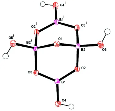
e.g. DALQEN (Wang et al., 2004) and SIBDIR (Pan et al., 2007). The tetraborate anion in borax has 2-fold symmetry
with the axis passing through O1 (Fig. 1) as is observed in five of the related structures. Both Na1 and Na2 cations are on
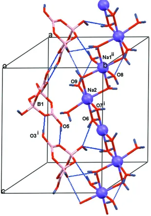
special positions (centre of symmetry and 2-fold axis, respectively) so that they elegantly bind via shared water molecules
in a typical zigzag cationic chain [Na(H2O)4/2(H2O)2/1] parallel to the c axis (e.g. DARNOA, Yi et al., 2005), as shown in
Figure 2. As is found through a C.S.D. search of similar Na+/H
2O cation chains, the Na–O distances to the bridging water
molecules are longer than those to non-bridging water molecules, where the trans related Na–O distances belong to
non-bridging water molecules.
The results of the present study and the LL model are essentially superimposable, but do reflect expected differences
associated with the H atom positions: The systematic pairwise study (Allen, 1986) gave a difference for O–H (X-ray
versus neutron) of -0.155 (10) Å, while a more recent study of levoglucosan (Smrčok et al., 2006) averaged at -0.016 (6)
Å. The mean O—H distance here (0.843 (17) Å) is significantly shorter than for the neutron set (0.97 (1) Å). As the O···O
distances involved in the hydrogen bonding are very similar for both studies (Table 1), the observed H···O distances are
correspondingly longer here than in the LL model. We also note that average Na–O distances are marginally longer
(0.006 (6) Å) and the B–O distances marginally shorter (-0.005 (2) Å) in the LL model, e.g. Na–O6, B1–O2 are 2.458 (3),
1.500 (2) Å compared with 2.4441 (6), 1.5075 (8) Å, respectively, in the present study. These latter differences are barely
significant given that the neutron data set was collected at 296.5 K.
Cell cohesion is provided by strong O—H···O hydrogen bonds of two types: (1) tetraborate anions "head to tail" link via
the O5–H and O2 atoms (entry 1, Table 1) to form anionic chains as also seen in DALQEN (Wang et al., 2004); (2) the
anionic and cationic chains crosslink through the water & tetraborate strong O–H···O hydrogen bond interactions (entries
2–10; see also Fig. 2 and diagrams in the LL study).
S2. Experimental
To a tetrahydrofuran (thf) solution (90 ml) of sodium tetrahydridoborate (0.31 g, 8.4 mmol) was added 0.5 g (4.2 mmol)
of diaminomethane dihydrochloride. After 24 h, the solvent was removed and the remaining product dissolved in water.
Methanol was added and the solution was left in a refrigerator. A small clump of colourless crystals of the title compound
A total of 13 reflections (below 50°/2θ) were not collected. In the present re-determination the same atomic labels and
atomic coordinates have been used as in the previous studies (Morimoto, 1956; Levy & Lisensky, 1978). The positions of
[image:4.610.121.496.133.495.2]the H atoms were fully refined with isotropic thermal parameters for each H atom.
Figure 1
Molecular structure of the tetraborate anion shown with displacement ellipsoids at the 50% probability level. [Symmetry
supporting information
sup-3
[image:5.610.151.461.74.518.2]Acta Cryst. (2008). E64, i24–i25 Figure 2
Part of the crystal structure showing the zigzag [Na(H2O)4/2(H2O)2/1] chain, the hydrogen bonded tetraborate chain and
some interlinking hydrogen bonds, shown as dashed lines. For clarity, only selected atoms and one of each chain is
shown. [Symmetry codes: i) -x, y 1/2 - z; ii) x, 1 + y, z.]
sodium tetraborate decahydrate
Crystal data
Na2[B4O5(OH)4]·8H2O Mr = 381.38
Monoclinic, C2/c Hall symbol: -C 2yc a = 11.8843 (5) Å b = 10.6026 (4) Å c = 12.2111 (5) Å
β = 106.790 (2)° V = 1473.06 (10) Å3 Z = 4
F(000) = 792 Dx = 1.720 Mg m−3
µ = 0.22 mm−1 T = 145 K
0.65 × 0.36 × 0.26 mm
Data collection
Bruker–Nonius APEX2 CCD area-detector diffractometer
Radiation source: fine-focus sealed tube Graphite monochromator
Detector resolution: 8.192 pixels mm-1 φ and ω scans
Absorption correction: multi-scan (SADABS; Bruker, 2006) Tmin = 0.813, Tmax = 0.94
8429 measured reflections 2275 independent reflections 2137 reflections with I > 2σ(I) Rint = 0.018
θmax = 33.2°, θmin = 2.6° h = −17→16
k = −15→15 l = −16→17
Refinement
Refinement on F2
Least-squares matrix: full R[F2 > 2σ(F2)] = 0.025 wR(F2) = 0.076 S = 1.08 2275 reflections 147 parameters 0 restraints
Primary atom site location: structure-invariant direct methods
Secondary atom site location: difference Fourier map
Hydrogen site location: inferred from neighbouring sites
All H-atom parameters refined w = 1/[σ2(F
o2) + (0.0482P)2 + 0.3901P]
where P = (Fo2 + 2Fc2)/3
(Δ/σ)max = 0.001
Δρmax = 0.37 e Å−3
Δρmin = −0.22 e Å−3
Special details
Geometry. All esds (except the esd in the dihedral angle between two l.s. planes) are estimated using the full covariance
matrix. The cell esds are taken into account individually in the estimation of esds in distances, angles and torsion angles; correlations between esds in cell parameters are only used when they are defined by crystal symmetry. An approximate (isotropic) treatment of cell esds is used for estimating esds involving l.s. planes.
Refinement. Refinement of F2 against ALL reflections. The weighted R-factor wR and goodness of fit S are based on F2,
conventional R-factors R are based on F, with F set to zero for negative F2. The threshold expression of F2 > σ(F2) is used
only for calculating R-factors(gt) etc. and is not relevant to the choice of reflections for refinement. R-factors based on F2
are statistically about twice as large as those based on F, and R- factors based on ALL data will be even larger.
Fractional atomic coordinates and isotropic or equivalent isotropic displacement parameters (Å2)
x y z Uiso*/Ueq
supporting information
sup-5
Acta Cryst. (2008). E64, i24–i25
H4 0.7717 (12) 0.2622 (11) 0.2876 (12) 0.032 (3)* H5 0.1187 (13) 0.4667 (13) 0.0305 (12) 0.040 (3)* H6A 0.3089 (13) 0.3828 (14) 0.0413 (12) 0.042 (4)* H6B 0.8662 (14) 0.2018 (16) 0.4941 (13) 0.051 (4)* H7A 0.3098 (13) 0.4817 (11) 0.3051 (12) 0.030 (3)* H7B 0.1304 (12) 0.0776 (14) 0.2014 (12) 0.039 (3)* H8A 0.9099 (12) 0.1906 (13) 0.1075 (11) 0.036 (3)* H8B 0.8131 (12) 0.1365 (12) 0.0352 (11) 0.034 (3)* H9A 0.4018 (13) 0.1300 (15) 0.3385 (12) 0.046 (4)* H9B 0.6140 (15) 0.2331 (15) 0.1058 (14) 0.053 (4)*
Atomic displacement parameters (Å2)
U11 U22 U33 U12 U13 U23
Na1 0.0165 (2) 0.01812 (19) 0.0158 (2) 0.00057 (13) 0.00485 (15) 0.00047 (13) Na2 0.0179 (2) 0.0201 (2) 0.0176 (2) 0.000 0.00773 (16) 0.000
B1 0.0100 (3) 0.0121 (3) 0.0096 (3) 0.0012 (2) 0.0035 (2) −0.00028 (19) B2 0.0108 (3) 0.0133 (3) 0.0111 (3) −0.0014 (2) 0.0040 (2) −0.0014 (2) O1 0.0117 (3) 0.0104 (3) 0.0111 (3) 0.000 0.0037 (2) 0.000
O2 0.0103 (2) 0.0168 (2) 0.0122 (2) −0.00223 (15) 0.00493 (16) −0.00376 (15) O3 0.0103 (2) 0.0180 (2) 0.0130 (2) 0.00309 (15) 0.00519 (16) 0.00559 (15) O4 0.0119 (2) 0.0209 (2) 0.0142 (2) 0.00489 (17) 0.00365 (17) −0.00434 (16) O5 0.0132 (2) 0.0275 (3) 0.0150 (2) −0.00595 (18) 0.00593 (19) −0.00949 (18) O6 0.0146 (2) 0.0219 (2) 0.0186 (2) −0.00109 (18) 0.00414 (19) 0.00229 (18) O7 0.0151 (3) 0.0160 (2) 0.0232 (3) 0.00012 (17) 0.0061 (2) 0.00003 (17) O8 0.0185 (3) 0.0242 (3) 0.0184 (3) 0.00116 (19) 0.0039 (2) 0.00608 (19) O9 0.0217 (3) 0.0196 (2) 0.0229 (3) −0.00047 (19) 0.0041 (2) −0.00352 (19)
Geometric parameters (Å, º)
Na1—O8i 2.3815 (6) Na2—O6ii 2.4441 (6)
Na1—O8ii 2.3815 (6) Na2—O6 2.4441 (6)
Na1—O6iii 2.3979 (5) B1—O4 1.4451 (8)
Na1—O6iv 2.3979 (5) B1—O1 1.4657 (7)
Na1—O7v 2.4121 (6) B1—O2 1.4902 (8)
Na1—O7 2.4121 (6) B1—O3 1.5075 (8) Na2—O7vi 2.4041 (6) B2—O2 1.3655 (8)
Na2—O7vii 2.4041 (6) B2—O3ii 1.3757 (8)
Na2—O9 2.4214 (6) B2—O5 1.3784 (8) Na2—O9ii 2.4214 (6)
O8i—Na1—O8ii 180.00 (2) O9—Na2—O6ii 81.696 (19)
O8i—Na1—O6iii 90.45 (2) O9ii—Na2—O6ii 97.72 (2)
O8ii—Na1—O6iii 89.55 (2) O7vi—Na2—O6 88.29 (2)
O8i—Na1—O6iv 89.55 (2) O7vii—Na2—O6 92.34 (2)
O8ii—Na1—O6iv 90.45 (2) O9—Na2—O6 97.72 (2)
O6iii—Na1—O6iv 180.00 (3) O9ii—Na2—O6 81.70 (2)
O6iii—Na1—O7v 89.177 (19) O4—B1—O2 110.91 (5)
O6iv—Na1—O7v 90.823 (19) O1—B1—O2 109.42 (4)
O8i—Na1—O7 88.283 (19) O4—B1—O3 107.71 (5)
O8ii—Na1—O7 91.717 (19) O1—B1—O3 108.56 (5)
O6iii—Na1—O7 90.823 (19) O2—B1—O3 108.43 (5)
O6iv—Na1—O7 89.177 (19) O2—B2—O3ii 122.44 (6)
O7v—Na1—O7 180.000 (16) O2—B2—O5 117.78 (6)
O7vi—Na2—O7vii 94.98 (3) O3ii—B2—O5 119.78 (6)
O7vi—Na2—O9 172.90 (2) B1ii—O1—B1 110.90 (6)
O7vii—Na2—O9 81.05 (2) B2—O2—B1 116.59 (5)
O7vi—Na2—O9ii 81.05 (2) B2ii—O3—B1 120.25 (5)
O7vii—Na2—O9ii 172.90 (2) Na1vi—O6—Na2 90.952 (19)
O9—Na2—O9ii 103.49 (3) Na2viii—O7—Na1 91.581 (19)
O7vi—Na2—O6ii 92.34 (2) Na2viii—O7—H7B 134.8 (10)
O7vii—Na2—O6ii 88.29 (2) Na1—O7—H7B 96.4 (10)
O4—B1—O1—B1ii −172.74 (6) O7vi—Na2—O6—Na1vi −0.355 (19)
O2—B1—O1—B1ii 64.05 (4) O7vii—Na2—O6—Na1vi 94.56 (2)
O3—B1—O1—B1ii −54.12 (3) O9—Na2—O6—Na1vi 175.84 (2)
O3ii—B2—O2—B1 −4.91 (9) O9ii—Na2—O6—Na1vi −81.56 (2)
O5—B2—O2—B1 174.23 (5) O6ii—Na2—O6—Na1vi −132.911 (15)
O4—B1—O2—B2 −156.88 (5) O8i—Na1—O7—Na2viii −89.93 (2)
O1—B1—O2—B2 −33.19 (7) O8ii—Na1—O7—Na2viii 90.07 (2)
O3—B1—O2—B2 85.06 (6) O6iii—Na1—O7—Na2viii 179.640 (19)
O4—B1—O3—B2ii 136.95 (6) O6iv—Na1—O7—Na2viii −0.360 (19)
O1—B1—O3—B2ii 15.82 (7) O7v—Na1—O7—Na2viii −122 (44)
O2—B1—O3—B2ii −102.97 (6)
Symmetry codes: (i) x, −y, z−1/2; (ii) −x, y, −z+1/2; (iii) x, −y+1, z−1/2; (iv) −x, y−1, −z+1/2; (v) −x, −y, −z; (vi) −x, y+1, −z+1/2; (vii) x, y+1, z; (viii) x,
y−1, z.
Hydrogen-bond geometry (Å, º)
D—H···A D—H H···A D···A D—H···A
O5—H5ix···O3ix 0.836 (15) 1.895 (15) 2.7300 (7) 176.3 (15)
O4x—H4···O9xi 0.828 (14) 2.049 (14) 2.8658 (8) 168.4 (12)
O6—H6Axii···O5xiii 0.868 (16) 1.978 (16) 2.8323 (8) 167.9 (14)
O6xiv—H6B···O4x 0.846 (16) 2.040 (15) 2.8624 (8) 163.9 (16)
O7xii—H7A···O2 0.827 (16) 1.989 (16) 2.8135 (8) 174.1 (12)
O7—H7B···O4 0.816 (15) 2.135 (15) 2.9233 (8) 162.3 (14) O8x—H8A···O1xv 0.866 (13) 1.936 (13) 2.7865 (6) 167.0 (14)
O8x—H8B···O5xvi 0.855 (15) 2.341 (14) 3.1320 (8) 154.2 (12)
O9—H9Axii···O3 0.843 (16) 2.253 (16) 3.0894 (8) 171.7 (15)
O9—H9Bxvii···O8iii 0.849 (17) 2.069 (16) 2.9034 (8) 167.4 (15)