34 Int J Res Med. 2013; 2(4);34-38 e ISSN:2320-2742 p ISSN: 2320-2734
A study to compare various visual field defects between glaucoma and non-glaucomatous diseases.
Hitesh Bhalara1*, Jayesh Sadhu2, S. S. Ganvit3, N. N. Pandya4
1,2,3,4
Resident doctor, co-resident, associate professor, professor and head of the department at S.S.G hospital and govt. Medical college, vadodara
INTRODUCTION
Perimetry, the evaluation of the visual field, is an important diagnostic test, particularly in glaucoma3but also for diagnosing and monitoring the progression of many other eye diseases13,14,15. In routine perimetry, computer supported static perimetry, as it was introduced in 1975 by Fankhauser, proved to be more practical compared to the
*Corresponding Author
Dr. Hitesh Bhalara
Resident doctor, of the department, S.S.G hospital and govt.
Medical college, vadodara
traditional manual Goldmann method. However in special situations such as in neuro-ophthalmology, it is important to have information about the peripheral visual field, and so, in end stage glaucoma and in cases with patients who have difficulty collaborating with an automatic test, computer-assisted (semi-automatic) kinetic testing continues to play an important role in testing the visual field.
Examining visual fields is an integral part of a full ophthalmic evaluation. Several methods for assessing visual field loss are available, and the choice of which to use depends on the patient's age, health, visual acuity, ability to concentrate, and
socio-ORIGINAL ARTICLE
ABSTRACT
BACKGROUND: To compare various visual field defects between glaucoma and non-glaucomatous diseases and to study the progression of disease on visual field defects in glaucoma and non-glaucomatous diseases.
MATERIALS AND METHODS: S.S.G Hospital and Govt. Medical college, Vadodara. Prospective study.50 eyes of glaucomatous patients and 50 eyes of patients of non-glaucomatous diseases between 18 to 65 years of age were taken for study and perimetry done on medmont automated perimeter(M700) using fast threshold strategy on central test. RESULTS: After 1 year of study, glaucomatous visual field defect is more common superiorly(54%) while non-glaucomatous visual field defect is equally common in both superior(44%) and inferior part(42%) of visual field and according to severity, mild visual field defects(62%) are most frequently seen in glaucoma while moderate visual field defects(60%) are most frequently seen in non-glaucomatous diseases. There was no significant progression noted in visual field of glaucoma and non-glaucomatous disease as patients are on anti-glaucoma treatment or in non-glaucomatous diseases, according to diseases patients were on treatment. CONCLUSION: In the study it was concluded that - glaucoma is more common in older age group(90% in >40 years), while non-glaucomatous diseases is equally distributed in younger(42% in < 40 years) and older age group(58% in >40 years). Peripheral depression(18%) is most common visual field defect followed by arcuatescotoma(16%), enlargement of blind spot(14%), nasal step(12%) in glaucoma patients while in non-glaucomatous group peripheral depression(26%) was followed by generalized depression(22%), focal defect(12%), para-central scotoma(12%), arcuatescotoma(10%), hemifield defect(8%). Glaucomatous visual field defect is more common superiorly(56%) while non-glaucomatous visual field defect is equally common in both superior(44%) and inferior part(42%) of visual field. According to severity, mild visual field defects(62%) are most frequently seen in glaucoma while moderate visual field defects(60%) are most frequently seen in non-glaucomatous diseases. There was no significant progression noted in visual field of glaucoma and non-glaucomatous disease as patients are on anti-glaucoma treatment or in non-glaucomatous diseases, according to diseases patients were on treatment.
35 Int J Res Med. 2013; 2(4);34-38 e ISSN:2320-2742 p ISSN: 2320-2734
economic status. Available techniques can test the full field (including confrontation, tangent screen, Goldmannperimetry and automated perimetry), or assess just the
central field of vision, such as the Amsler Grid.
Perimetry, the evaluation of the visual field, is an important diagnostic test in glaucoma3,9,11,12 but also for diagnosing and
monitoring the progression of many other eye diseases8,10,18. Although the visual field examination is mostly used in conjunction withother clinical findings, such as intraocular pressure, and the assessment of structural changes at the optic nerve head and the retina, perimetry remains an indispensable test documenting visual function. After all, patients are not concerned about pressure or appearance of their discs but they are worried about maintaining vision.
The recent past has brought new developments in the field of perimetry, and it has opened up areas for the development of new testing and analysis programs. A larger empiric database for patients with glaucoma and non-glaucomatous diseases will improve the accuracy and detectability of glaucomatous and non-glaucomatous visual field defects. Other psychophysical methods for testing the visual field for damage are now being explored. These methods include contrast sensitivity, acuity perimetry, and color perimetry. Additional studies are needed to determine the role of these and other modalities in the future. MATERIALS AND METHODS
The present prospective analytical study was conducted on 50 eyes of glaucomatous patients and 50 eyes of patients of non-glaucomatous diseases who presented at the ophthalmology department, S.S.G hospital, Vadodara during December, 2012 to November, 2013 after taking permission from The Scientific and Ethical Review Committee, Baroda Medical Collage & SSG Hospital, Vadodara.
Inclusion criteria:-Diagnosed cases of glaucoma. - Diagnosed cases of various non-glaucomatous diseases( patients having
ophthalmology, not only for managing
neurological disease with optic disc changes and retinal problems). - All patients having
visual field defects between 18 to 65 years of age
Exclusion criteria: - Patients below the age of 18 years of age. - Patients above the age of 65 years of age. - Patients having ocular hypertension without visual field defects. - Cases that absconded or lost on follow up. Preliminary data of age, sex, residential address and registration number was recorded. Elaborative data was noted about the duration of complaints, eye to be affected, associated systemic complaints, past or present history of glaucoma and non-glaucomatous disease, or positive family history of glaucoma and non-glaucomatous disease.Visual acuity was determined in both eyes with or without glasses at the time of presentation on standardsnellen visual acuity chart. Intraocular pressure was taken by schiotz tonometer.Fundus examination was done by direct/indirect and 78 D slit lamp fundoscopy.
After full correction automated perimetry using medmont automated perimeter(M700) was done in all patients using central field. Strategy of program used was fast threshold. Visual fields with highpercentage of unreliability (fixation losses ˃ 20% and false negative ˃33% and false positive errors ˃ 33%) were eliminated. Perimetry was repeated every 3 months or 6 months to see any progression was noted if patient comes for follow up.
RESULTS
Data from 50 eyes of glaucomatous patients and 50 eyes of patients of non-glaucomatous diseases were studied for visual field defects over a period of 1 year between 18 to 65 years.
TABLE 1: Age wise distribution of cases of glaucoma and non-glaucomatous diseases.
36 Int J Res Med. 2013; 2(4);34-38 e ISSN:2320-2742 p ISSN: 2320-2734 (Glaucoma) (non-glaucoma) (Glaucoma) (non-glaucoma)
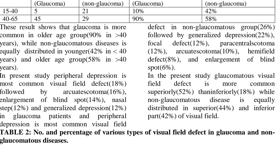
15-40 5 21 10% 42%
40-65 45 29 90% 58%
These result shows that glaucoma is more common in older age group(90% in >40 years), while non-glaucomatous diseases is equally distributed in younger(42% in < 40 years) and older age group(58% in >40 years).
In present study peripheral depression is most common visual field defect(18%) followed by arcuatescotoma(16%), enlargement of blind spot(14%), nasal step(12%) and generalized depression(12%) in glaucoma patients and peripheral depression is most common visual field
defect in non-glaucomatous group(26%) followed by generalized depression(22%), focal defect(12%), paracentralscotoma (12%), arcuatescotoma(10%), hemifield defect(8%), and enlargement of blind spot(6%).
In the present study glaucomatous visual field defect is more common superiorly(52%) thaninferiorly(18%) while non-glaucomatous disease is equally distributed in superior(44%) and inferior part(42%) of visual field.
TABLE 2: No. and percentage of various types of visual field defect in glaucoma and non-glaucomatous diseases.
TABLE : 3 Distribution of visual field defects in glaucoma and non-glaucomatous diseases.
Distribution of
Visual Field Defect
Glaucoma cases
Non-glaucoma cases Percentage (glaucoma)
Percentage (non-glaucoma)
Superiorly 28 22 56% 44%
Inferiorly 9 21 18% 42%
Generalized 13 7 26% 14%
TABLE 4:Severity of visual field defects in glaucoma and non-glaucomatous diseases.
Severity of Visual Field Defect
Glaucoma Non-laucoma Percentage
(glaucoma)
Percentage (non-glaucoma)
Mild 31 17 62% 34%
Moderate 15 30 30% 60%
Severe 4 3 8% 16%
In the present study mild variety of visual field defect is most common type(62%), followed by moderate(30%) and severe(8%) in glaucoma while moderate variety of visual field defect is most common(60%) in non-glaucomatous group followed by
mild(34%) and severe(6%). Data shows that in present study visual field defect is non-progressive in glaucoma and non-glaucomatous diseases.
TABLE 5:Progression of visual field defect in glaucoma and non-glaucomatous diseases.
Progression of Visual Field Defect
Glaucoma Non-glaucoma Percentage(glaucoma) Percentage
(non-glaucoma)
Type of Visual Field Defect Glaucoma Percentage Non-glaucomatous disease Percentage
Nasal step 6 12% 0 0%
Enlargement of blind spot 7 14% 3 6%
Central scotoma 0 0% 1 2%
Paracentralscotoma 6 12% 6 12%
Arcuatescotoma 8 16% 5 10%
Ring scotoma 4 8% 1 2%
Peripheral depression 9 18% 13 26%
Generalized depression 6 12% 11 22%
Hemi field defects 2 4% 4 8%
37 Int J Res Med. 2013; 2(4);34-38 e ISSN:2320-2742 p ISSN: 2320-2734
Yes 4 2 13% 11%
No 30 17 87% 89%
DISCUSSION:
This study was conducted in the department of ophthalmology, S.S.G hospital, Vadodara between December, 2012 to November, 2013.
In this prospective comparative analytical study, visual field defects of 50 eyes of glaucoma patients compared with visual filed defects of 50 eyes of non-glaucoma patients.
Glaucoma is more common in older age group, while non-glaucomatous diseases is equally distributed in younger and older age group.
- Peripheral depression is most common visual field defect followed by arcuatescotoma, enlargement of blind spot, nasal step and generalized depression in glaucoma patients while in non-glaucomatous group peripheral depression was followed by generalized depression, focal defect, para-central scotoma, arcuatescotoma, hemifield defect, and enlargement of blind spot.
In our study nasal steps seen in 12% cases and arcuatescotoma seen in 16% cases of glaucoma which was comparable to the study done to identify the patterns of visual field defects in an older population ( the blue mountains eyes study) at department of ophthalmology, University of Sydney, Sydney, Australia16 in which nasal step seen in 31% cases while arcuatescotoma seen in 23% cases as they include only few types of visual field defects..
- Glaucomatous visual field defect is more common superiorly while non-glaucomatous visual field defect is equally common in both superior and inferior part of visual field In our study superior visual field defects(56%) were more common than inferior visual field defects(18%) in glaucoma which is also the finding in a study which was done to identify characteristics of visual field defects in
primary angle closure glaucoma done at Kunhua Affiliated Hospital of Kunming Medical College, Kunming, China(17). In that study visual field scores with inferior hemifield in primary open angle glaucoma were lower than chronic angle closure glaucoma. The same conclusion also came
in study done at
Univ.-AugenklinikWürzburg that the visual field defects more frequent in the upper than in the lower field in POAG.
- According to severity, mild visual field defects are most frequently seen in glaucoma while moderate visual field defects are most frequently seen in non-glaucomatous diseases.
In our study severe visual field defect found in 8% cases which is comparable to severe visual field defect was found in 15.4% cases in the blue mountain study done at department of ophthalmology, university of Sydney, Sydney, Australia.
- There was no significant progression noted in visual field of glaucoma and non-glaucomatous disease as patients are on anti-glaucoma treatment or in non-anti-glaucomatous diseases, according to diseases patients were on treatment.
Our study which does not show any progression in visual field defect in glaucoma(18) and non-glaucomatous diseases which was also conclusion of study done to Estimate progression of visual field loss in glaucoma done at Department of International Health, Johns Hopkins School of Hygiene and Public Health, Baltimore, Maryland 21205-210. In this study it was concluded that Less than one in three eyes of patients with glaucoma had any progressive field loss and Average changes in threshold sensitivities of less than 1 dB/year could not be detected with seven fields done over 6 years.
38 Int J Res Med. 2013; 2(4);34-38 e ISSN:2320-2742 p ISSN: 2320-2734
older age group, superiorly, and mild type of severity while non-glaucomatous visual field defects are younger age group andequally distributed in younger and older age group and superior and inferior part of visual field with moderate type of severity of visual field defect.
REFERENCES
1. LeBlanc EP, Becker B. Peripheral nasal
field defects. Am J
Ophthalmol.1971;72(2):415–419. 2. Werner EB, Beraskow J. Peripheral
nasal field defects in glaucoma. Ophthalmology.1979;86(10):1875– 1878.
3. Armaly MF. Visual field defects in early open angle glaucoma. Trans AmOphthalmol Soc.1971;69:147–162 4. Schulzer M, Mikelberg FS, Drance SM.
A study of the value of the centraland peripheral isoptores in assessing visual field progression in the presence of paracentralscotoma measurements.Br J Ophthalmol. 1987;71(6):422–427. 5. Ballon BJ, Echelman DA, Shields MB,
et al. Peripheral visual field testing in glaucoma by automated kinetic perimetry with the Humphrey FieldAnalyzer. Arch Ophthalmol. 1992;110(12):1730–1732.
6. Harrington DO. The BjerrumScotoma. Am J Ophthalmol.1965;59:646–656. 7. Gramer E, Gerlach R, Krieglstein GK,
et al. Topography of early glauco- matous visual field defects in computerized perimetry [in German]. KlinMonatsbl Augenheilkd. 1982;180 (6):515–523.
8. Mikelberg FS, Drance SM. The mode of progression of visual field defects in glaucoma. Am J Ophthalmol. 1984;98(4):443–445.
9. Drance SM. The glaucomatous visual field. Br J Ophthalmol.1972;56(3):186– 200.
10. Mikelberg FS, Schulzer M, Drance SM, et al. The rate of progression
ofscotomas in glaucoma.Am J Ophthalmol. 1986;101(1):1–6.
11. Kitazawa Y, Yamamoto T. Glaucomatous visual field defects: their characteristics and how to detect them. ClinNeurosci. 1997;4(5):279–283. 12. Lau LI, Liu CJ, Chou JC, Hsu WM, Liu
JHK. Patterns of visual field defects in chronic angle-closure glaucoma with
different disease
severity.Ophthalmology.
2003;110:1890–1894. [PubMed]
13. Grover S, Fishman GA, Brown J Jr. Patterns of visual field progression in patients with retinitis pigmentosa. Ophthalmology. 1998;105(6):1069-1075.
14. Neuro-Ophthalmology. Basic and Clinical Science Course, Section 5. American Academy of Ophthalmology; 2010:159-165. ISBN 978-1-61525-133-9
15. Borchert MS, Lessell S, Hoyt WF. Hemifield slide diplopia from altitudinal visual field defects. J Neuroophthalmol. 1996 Jun;16(2):107-9. PMID 8797166.
16. Patterns of glaucomatous visual field defects in an older population: the Blue Mountains Eye Study byLee AJ1, Wang JJ, Rochtchina E, Healey P, Chia EM, Mitchell P.Clin Experiment Ophthalmol. 2003 Aug;31(4):331-5. 17. Characteristics of visual field defects in
primary angle-closure glaucoma by Han F1, Yuan YS published in Zhonghua Yan KeZaZhi. 2009 Jan;45(1):14-20. 18. Estimating progression of visual field