Immune Comp
.
lex Reactions in Systemic Lupus
Erythematosus*
**
JOHN ST AIGE DAVIS,
IV,
M.D.
Professor of Internal Medicine, Chief of the Division of
Rheumatology, University of Virginia School of Medicine,
Charlottesville
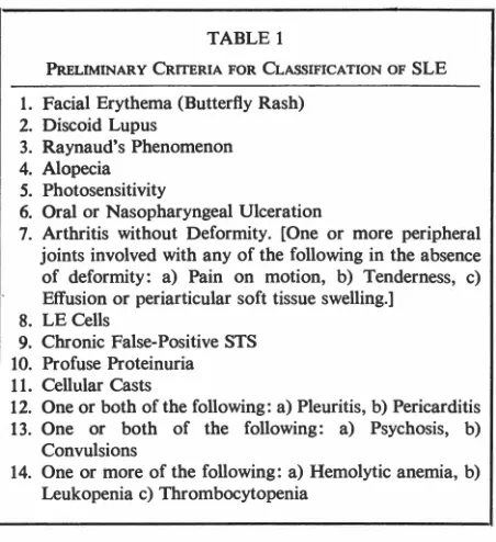
We should first try to define systemic lupus erythematosus (SLE). Table 1 lists criteria proposed by a committee of the American Rheumatism Asso-ciation ( 1). If four of these 14 major criteria are
positive, then there should be about 98% specificity
for SLE. There are clearly many facets to this dis-order. Let us consider mechanisms that might
ac-count for a multisystem disease such as this. A good model is that of experimental serum
sickness. Though Germuth (2) provided much of
the renewed impetus to study serum sickness in the
'SO's, it is Dixon (3) who has fully explored the variables of immune complexes and disease.
When a rabbit is given a single intravenous injection of bovine serum albumin (BSA) labelled with 1811, one notes disappearance of the BSA in serum over a period of about 13 days. First there is a rapid fall, then an equilibrium dev~lops; and
then suddenly there is total disappearance of the injected BSA. Coincident with the rapid disappear-ance of BSA, one can measure antigen-antibody
complexes. Finally, free antibody to BSA is detected. During the period when these immune complexes are
found, the rabbits develop heart disease, arthritis,
nephritis, and other manifestations. It is a
self-limited disease, however, and when free antibody
is found the disease clears up. Hemolytic
comple-ment (C) has been measured also; with the
appear-ance of immune complexes, the C level falls, later
* Presented by Dr. Davis at the 45th Annual McGuire Lecture Series, November 8, 1973, at the Medical College of Virginia, Richmond.
**Supported in part by the USPHS Grant No. 5R01-AM11766 and a grant from the John A. Hartford Founda-tion, Inc.
MCV QUARTERLY 10(2): 65-70, 1974
returning to normal. This then is the model of experimental serum sickness in the rabbit; what
about immune complexes in man?
We are postulating that many manifestations of SLE probably result from deposition of circulating
immune complexes. Numerous data are now avail-able which would support this concept. What can we now do to find these eomplexes and characterize them? If we can show what is in these complexes, we should know something more about pathogenesis. Table 2 lists some of the methods that are
available today. These methods are mostly limited to research laboratories. I have listed these methods
as either analytical or preparative. Cryoprecipitation is both analytical and preparative; furthermore, cryoprecipitates are easily measured since all one needs is a refrigerator and a centrifuge. Recent studies by Mcintosh et al. ( 4) suggest that cryo-precipitates correlate well with immune complex formation in rabbits; evidence in man is not as
convincing since it is difficult to recover a specific pathogenetic antigen in SLE cryoprecipitates.
Clq precipitation was first described by Agnello
et al. (5); here one utilizes purified Clq to pre-cipitate with serum factors in double diffusion systems. Rheumatoid factor (RF) can be used in the same way. Efforts in our lab are now being directed toward an analysis of the characteristics of Clq reactive factors. It is of interest that Clq and RF seem to detect different substances in serum. Passive
agglutination of RF-coated cells is another method which detects complexes, as is complement fixation (which is a way of interpreting "anticomplementary
serum"). Recent work in England has shown com-plement fixation and Clq precipitins not only in
TABLE 1
PRELIMINARY CRITERIA FOR CLASSIFICATION OF SLE
!. Facial Erythema (Butterfly Rash) 2. Discoid Lupus
3. Raynaud's Phenomenon 4. Alopecia
5. Photosensitivity
6. Oral or Nasopharyngeal Ulceration
7. Arthritis without Deformity. [One or more peripheral joints involved with any of the following in the absence of deformity: a) Pain on motion, b) Tenderness, c) Effusion or periarticular soft tissue swelling.]
8. LE Cells
9. Chronic False-Positive STS 10. Profuse Proteinuria 11. Cellular Casts
12. One or both of the following: a) Pleuritis, b) Pericarditis
13. One or both of the following: a) Psychosis, b) Convulsions
14. One or more of the following: a) Hemolytic anemia, b) Leukopenia c) Thrombocytopenia
SLE serum but in sera from patients with dermatitis herpetiformis, ulcerative colitis, regional enteritis,
and coeliac disease ( 6, 7). Lymphocyte inhibition is another technique recently reported: B cells,
which kill sensitized tumor cells, are inhibited by serum containing aggregates or immune complexes ( 8). Platelets can also be agglutinated by com-plexes.
Cryoprecipitation is, at the same time, a pre-parative method. Likewise Clq can be linked to Sepharose® columns through cyanogen bromide to facilitate removal of Clq precipitins from serum for analysis (9). Hopefully, the immune complexes and/
or aggregates will stick and everything else will wash through; then the bound material can be eluted off
TABLE2
DETECTION OF CIRCULATING IMMUNE COMPLEXES AND/OR AGGREGATES
Analytical
Cryoprecipitation
Clq detection in agarose gel RF detection in agarose gel
or reverse hemagglutina
-tion
Complement fixation
Lymphocyte inhibition Platelet agglutination
Preparative
Cryoprecipitation Cl q precipitation Adsorption to columns of
insoluble Clq
Isolation by column chromatography
with salt and the residue analyzed. Serum can also be run directly through agarose columns with ex-clusion of large molecular weight materials. In sum-mary, the main point here is that many methods are being developed to find and characterize aggregates and complexes; the simplest method is probably cryoprecipitation.
Figure 1 shows various factors which we mea-sured longitudinally in one SLE patient several years ago. The relationship between serum cryoglobulins and C levels is particularly noteworthy (our normal range for serum hemolytic complement is 34-48 CH50 units/ml) and one can clearly see the re-ciprocal relationship between C and cryoglobulin during the time span. This negative correlation is not always seen but usually is.
Figure 2 shows findings in another patient.
Here the C level is first found in the normal range at a time when no cryoglobulins are seen. As the cryoglobulins appear, C levels begin to fall. It should also be noted that the antinuclear factor (ANF) remained fairly constant during this time period, though no titers were done. It has been our experi-ence that when we use a 1 :4 dilution of serum for the ANF test, little change in ANF can be noted between acute exacerbations and remissions of SLE. With prolonged remission, however, the staining may become less intense, though the ANF rarely dis-appears entirely in SLE patients. RF titers (usually low titers, when found) also tend to remain quite constant in SLE patients, though RF was not found in either of the patients shown in these figures.
c
Lupus Patient Ca
ANF [
RF
4+ 4+ 4+ 4+ 4+ 3+ 4+ 4+ 4+ 4+ 4+
so ···~ CRYOGLOBULIN
0---0 COMPLEMENT
40 4+ li!l PROTEINURIA
30 3+
Cryo
20 2+
10 1+
12 1 2 3 4
1968
s 6 7 8 9
1969
Months
0 0 0 0 0
6······6····4
Proteinurio
2 3 4
1970
I
-- Ir
DAVIS: IMMUNE COMPLEX REACTIONS
ANF [
"
so
40 4+
30 3+
lupus Patient Wa
2+ 3+ 3+ 3+ 4+ 3+ 4+ 4+ 0 0
···O. CRYOGLOSULIN o - - o COMPLEMENT
lftA PROTE1NURIA
c Cryo Proteinuria
20 2+ 4+
3+
JO I+ 2+
I+
12 I 2 3 4 5 6 7 8 9 10 11 12 I 2 3 4 S 6
1968 1969 1970
Months
Fig. 2-Laboratory findings in another patient with SLE. Very little proteinuria is noted even when the C level is low and large amounts of cryoglobulin are found.
Clq precipitins have been found primarily in
SLE patients with low serum C levels according tb
one report (10). In a group of 30 SLE patients with
low C levels, 23 were positive for Clq precipitins; of those SLE patients who had normal C levels none
were positive for Clq precipitins. A few
p~tients
with miscellaneous diseases were positive ( 6/150).
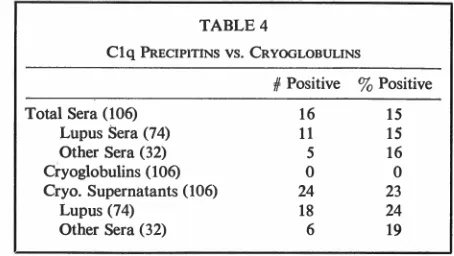
Table 3 shows our results: Of 908 total sera tested for Cl q precipitins, 19 % were positive. (These sera came from patients who had a variety of diseases and were mainly suspected of having a connective
tissue disease or vasculitis.) Of the lupus sera, 15 %
were positive and of the lupus patients, 43 % were
positive on at least one occasion. Sera from patients
with other diseases were positive 27 % of the time,
the 28 % positivity of nonlupus patients reflecting
the fact that many of these patients were only
studied by this technique on one occasion. Of
par-ticular note is the finding of Clq precipitins in other diseases besides SLE-a finding supported by other
investigators ( 6, 7, 10).
TABLE 3
Clq PRECIPITATION
Total #Positive 3 Positive
Total Sera 908 174 19
Lupus Sera 601 90 15
Lupus patients 95 41 43
Other Sera 307 84 27
Other patients 216 61 28
67
TABLE 4
Clq PRECIPITINS VS. CRYOGLOBULINS
#Positive 3 Positive
Total Sera (106) 16 15
Lupus Sera (74) 11 15
Other Sera (32) 5 16
Cryoglobulins (106) 0 0
Cryo. Supernatants (106) 24 23
Lupus (74) 18 24
Other Sera (32) 6 19
Results of the Clq precipitin test as it relates to sera with cryoglobulins ate shown in Table 4. One notes that of the 160 sera tested here, 16 were positive for Clq precipitins; aild of these 16 sera, 11 were SLE seta and 5 were other disease sera.
Only some of these sera had cryoglobulins, but we
treated them all as if they did-that is, they were all centrifuged after 48 hours in the cold and the super-natant transferred to another tube for testing against Clq. The cryoprecipitates, whether visible or not, were resolubilized at a ten-fold concentration and
also retested against Clq. No cryoprecipitates showed
a precipitin line, suggesting that 1 ) there was not enough material, 2) the material did not react with Clq, or 3) that the cryoprecipitate was already
satu-rated with bound Clq. Of more importance,
how-ever, was the finding that of the 106 supernatants,
24 (instead of 16) were now positive.
Ali
of the16 sera which were positive originally remained pos-itive when the supernatant was checked and eight sera which were originally thought to be negative were now positive after removal of any cryoprecipi-tates. These studies have suggested to us that cryo-globulins have properties, at least in part, distinctive from Clq precipitins. Possibly the cooling and cen-trifugation of certain sera lead to aggregation of small
complexes that do not spin down as cryoglobulins,
but do subsequently react with Clq, even at room
temperature!
For several years, we have been interested in how RF might modify IgG aggregates or immune complexes in human disease ( 11, 12). We have wondered whether RF might affect the deposition or metabolism of complexes by changing their size or their physicochemical properties in some other
way. Small, soluble complexes might be made
circum-4,000
3,000
..
:§
·;:;_
.
.,
~
0-2,000
·=
::;:
"-u
1.000
Precipitation of 3H-DNA by SLE Serum with RF-lgM and Controls
- R f.lgM
<>--o C·lgM
•····• Buffer
0.05 ml SLE serum
(F.W. 8/12/ 70)
1.25 2.5 5.0 10 20
micrograms DNA
50 100
Fig. 3-Effect of lgM rheumatoid factor on bNA-anti-DNA complexes. Constant amounts of IgM [either control (C) or containing rheumatoid factor (RF)] are added to constant amounts of anti-DNA (SLE) serum and increasing
amounts of. DNA. Much more radioactive DNA is found in the precipitate when RF is added than in appropriate
control systems.
stances ( 13). Figure 3 shows that RF has the capability of precipitating partially soluble
DNA-anti-DNA complexes form!!d in vitro when one adds
increasing amounts of ~abelled DNA to an SLE
serum. It can be seen that much more precipitate is formed (based on counts of radioactive DNA in the precipitate) in the presence of RF than whert
the complexes are formed in the presence of control-lgM or buffer alone. We interpret these findings to mean that RF can interact with and precipitate
DNA-anti-DNA complexes, thus converting these
partially soluble complexes into insoluble complexes. As mentioned above, such a function for RF in vivo could be either protective or damagihg.
Figure 4 shows effects of RF on cryoprecipitates. It would appear that RF has the potential for either
increasing or decreasing cryoprecipitation, probably depending on characteristics of the cryoprecipitate. Conceivably one might be dealing with a system analagous to an antigen-antibody complex which
would precipitate at equivalence but would resohi-bilize in both antigen and antibody excess.
Table 5 shows the sort of data being
accumu-lated currently on patients seen at the University qf Virginia Lupus Clinic. In the serum of this patient before treatment, a Clq precipitin was found, along with 3+ cryoglobulins, strong anti~DNA activity, a
low C level, and strong ANF staining: After institu-tion of steroid treatment, the Clq precipitin
dis-appeared, but then we found material in the serum reacting with RF! (This often-seen reciprocal rela-tion is of great interest to us, but we don't under-stand it.) Cryoglobulins disappeared (as did the
anti-DNA antibodies) and at the same time the
serum C level returned to normal. Since starting treatment, the patient has done well, and we have continued to be on the lookout for. the reappearance of those factors associated with disease activity as we slowly lower the steroid dose.
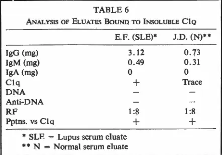
Table 6 is an analysis of serum material bound
to Clq on a cyanogen bromide Sepharose® column.
1.2
1.05
0.9
mg. of o.6
Cryoprotein
0.3
0.15
Cryoglobulin in Lupus Sero before ond after addition of RF
(lml RF added to 2ml Lupus Sera)
Cryo Cryo
+
RF
Wa
(1/8/70)
Cryo Cryo
+
RF
Ca (1/8/70)
Cryo Cryo
+
RF
Fig. 4-Effect of lgM rheumatoid factor on three SLE sera containing cryoglobulins. After addition of RF to fresh sera
Sn and Wa, less cryoglobulin was detected after 48 hours
DAVIS: IMMUNE COMPLEX REACTIONS 69
TABLE 5
S. P. (SLE)
Complexes
Clq RF Cr yo
4/6/72
+
3+Treatment Started
4/11/72
+
+
ND*
4/13/72 ND
4/17/72 ND
4/21/72 1-2+
5/2/72
6/13/72
7 /25/72 11/21/72
* ND = Not Done
One notes the presence in the two eluates of IgG
and IgM but no anti-DNA or DNA. Currently, we are analyzing these eluates by a variety of techniques in-cluding animal immunizations (to elicit antibodies to any infectious agents which might be there).
Charac-tedstics of Clq precipitins are also being compared
with characteristics of cryoglobulins. It should be
noted at this point that normal serum also contains
significant amounts of material which bind to the Clq adsorbent.
In summary, we can draw very few firm
con-clusions about immune complexes in patients with
SLE. SLE patients during episodes of disease activity
often show both cryoglobulins anq Clq precipitins
in their sera, but in our experience, the sera may
show only one of the factors and occasionally neither one is found. When cryoglobulins are removed from sera, Clq precipitins not only remain in the
super-natant but occasionally may be found in the
super-TABLE 6
ANALYSIS OF ELUATES BOUND TO INSOLUBLE CIQ
E.F. (SLE)*
lgG (mg) 3.12
lgM (mg) 0.49
IgA (mg) 0
Clq +
DNA Anti-DNA
RF 1:8
Pptns. vs Clq +
* SLE = Lupus serum eluate
**
N = Normal serum eluateJ.D. (N)**
0.73 0.31 0 Trace
1 :8 +
2+
Anti-DNA
3+
3+ 2-3+ 2-3+ 2+
ANF
4+
ND ND ND
1-2+
ND
3+ 3-4+ 3-4+
CH50
10
13
ND ND ND
40
53
48
45
natant for the first time. This finding suggests that cryoglobulins and Clq precipitins are at least in part distinct. RF may interact with DNA-anti-DNA com-plexes and cryoprecipitates at· least in vitro. Studies
are in progress to furtfler characterize the material
found in, the~e complexes. Hopefully, a particular
antigen or a particular characteristic of SLE
com-plexes will be found which will lead to a better
understanding of the primary cause and intermediate
mechanisms involved in the serious disease kriown
as SLE.
REFERENCES
1. ARA COMMITTEE OF DIAGNOSTIC AND THERAPEUTIC CRITERIA: Preliminary criteria for the classification of systemic lupus erythematosus. Bull Rheum Dis 21:643,
1971.
2. GERMUTH FG JR: A comparative histologic and im-munologic study in rabbits of induced hypersensitivity of the serum sickness type. J Exp Med 97:257, 1953.
3. DIXON FJ: The role of antigen-antibody complexes in diseases. The Harvey Lectures, Series 58:21, 1962-63.
4. McINTOSH RM, GRoss M, LAPLANTE M, KAUFMAN DB,
KuLvINSKAS C: Cryoproteins in immune complex
dis-ease: Role of nephrectomy and implications in recurrent glomerulonephritis in human transplants. Exp Med Surg
29: 108, 1971.
5. AGNELLO V, WINCHESTER RF, KUNKEL HG: Precipitin reactions of the Clq component of complement with aggregated y-globulin and immune complexes in gel
6. MOWBRAY JF, HOLBOROW EJ, HOFFBRAND AV, SEAH PP, FRY L: Circulating immune complexes in dermatitis herpetiformis. Lancet 1: 400, 1973.
7. DOE WF, BOOTH CC, BROWN DL: Evidence for com-plement-binding immune complexes in adult coeliac
disease, Crohn's disease, and ulcerative colitis. Lancet
1:402, l973.
8. JEWELL DP, MACLENNAN ICM: Circulating immune complexes in inflammatory bowel disease. Clin Exp Immunol 14:219, 1973.
9. FATHMAN CG: Characterization of circulating
antigen-antibody complexes in patients with systemic lupus erythematosus. Clin Res 21:577, 1973 (abstract).
10. AGNELLO V, KOFFLER D, EISENBERG JW, WINCHESTER RJ, KUNKEL HG: Clq precipitins in the sera of patients with systemic lupus erythematosus and other hypocom-plementemic states: Characterization of high and low molecular weight types. I Exp Med 134:2285, 1971.
11. DAVIS JS, TORRIGIANI G: Effects of rheumatoid factor on in vivo distribution of aggregated human lgG and antigen-antibody complexes. Proc Soc Exp Biol Med
125:772, 1967.
12. GouGH WW, DAVIS JS: Effects of rheumatoid factor on complement levels in vivo. Arth Rheum 9:555, 1966.
![Fig. 3-Effect constant amounts of lgM rheumatoid factor on bNA-anti-DNA complexes. Constant amounts of IgM [either control (C) or containing rheumatoid factor (RF)] are added to of anti-DNA (SLE) serum and increasing amounts of](https://thumb-us.123doks.com/thumbv2/123dok_us/8793172.1767793/4.541.276.496.312.589/constant-rheumatoid-complexes-constant-control-containing-rheumatoid-increasing.webp)