The Genomic Standards Consortium
Non-contiguous finished genome sequence and
description of
sp. nov.
Jean-Christophe Lagier1, Gregory Gimenez1, Catherine Robert1, Didier Raoult1 and Pierre-Edouard Fournier1*
1 Unité de Recherche sur les Maladies Infectieuses et Tropicales Emergentes, Aix-Marseille Université
*Corresponding author: Pierre-Edouard Fournier ([email protected])
Keywords, genome
strain JC206T sp. nov. is the type strain of H. massiliense sp. nov.,
a new species within the genus
was isolated from the fecal flora of a healthy Senegalese patient. H. massiliense is an aerobic rod. Here we describe the features of this organism, together with the complete genome se-quence and annotation. The 4,186,486 bp long genome (one chromosome but no plasmid) contains 3,847 protein-coding and 54 RNA genes, including 3 rRNA genes.
Introduction
strain JC206T (= CSUR
P159 = DSMZ 25712) is the type strain of H. massiliense sp. nov. This bacterium was isolated from the stool of a healthy Senegalese patient. It is a Gram-negative, aerobic, flagellated, indole-negative bacillus.
The current approach to classification of prokary-otes, generally referred to as polyphasic taxono-my, relies on a combination of phenotypic and genotypic characteristics [1]. However, as more than 3,000 bacterial genomes have been se-quenced [2], we recently proposed that genomic information should be integrated in the descrip-tion of new bacterial species [3,4].
The genuset al. 1986)
was created in 1986 [5,6]. To date, this genus, comprised of nitrogen-fixing, Gram-negative bacil-li, contains 13 species and two subspecies,
includ-ing et al. 2010) [7],
et al. 2011) [8],
and Yokota 2004 [9],et al.
2011) [8], et al. 2004)
[10],et al. 2001) [11],
et al. 2006) [12],
(Leifson 1962) Ding and Yokota
2004 [9],putei (Ding and
Yoko-ta 2004) Dobritsa et al. 2010 [7],
(Valverde et al. 2003) [13],
et al. 2007) [14],
and Edgerton 1930) Baldani et al. 1996 [6],
et al. 1986) [5], and
(Carro et al. 2011) [8]. Members of the genus
environment, in particular from soil, and from plants for which they play the role of growth pro-moters, but have also occasionally been isolated from humans, either as proven pathogens, causing bacteremia in leukemic patients [15,16], as poten-tial pathogens in aortic aneurysms [17], or in res-piratory secretions from cystic fibrosis patients [18,19]. To the best of our knowledge, this is the
first to report the isolation of a
from the normal fecal flora. Here we present a summary classification and a set of features for H. massiliense sp. nov. strain JC206T (= CSUR P159 = DSMZ 25712) together with the description of the complete genomic sequencing and annotation. These characteristics support the circumscription of the species H. massiliense.
Classification and features
the assent procedure were approved by the Na-tional Ethics Committee of Senegal (CNERS) and the Ethics Committee of the Institut Fédératif de Recherche IFR48, Faculty of Medicine, Marseille, France (agreement numbers 09-022 and 11-017). Several other new bacterial species were isolated from this specimen using various culture condi-tions [3,4].
The fecal specimen was preserved at -80°C after collection and sent to Marseille. Strain JC206T (Table 1) was isolated in June 2011 after passive filtration of the stool sample to select motile spe-cies using companion plate, cell culture inserts with 0.4 μm-pore membranes (Becton Dickinson,
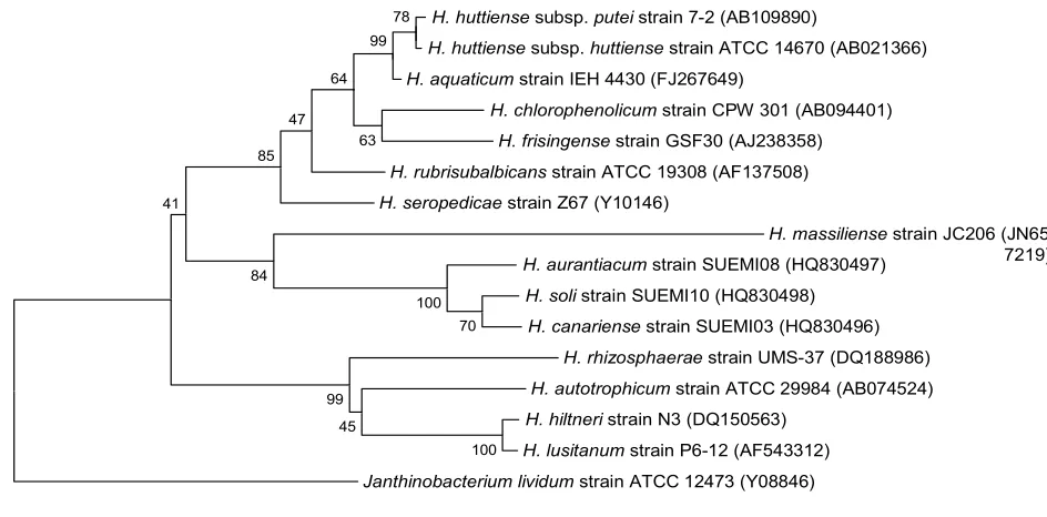
Heildeberg, Germany) and (BioMerieux, Marcy l’Etoile, France). Subsequent-ly, we cultivated strain JC206T on 5% sheep blood agar in an aerobic atmosphere at 37°C. This strain exhibited a 96.7% 16S rDNA nucleotide sequence
similarity withet al. 2012),
the phylogenetically closest validly published
vated from volcanic soil in Canary Islands. This value was lower than the 98.7% 16S rRNA gene sequence threshold recommended by Stackebrandt and Ebers to delineate a new species without carrying out DNA-DNA hybridization [29].
Table 1. Classification and general features o strain JC206T according to the MIGS rec-ommendations [21]
MIGS ID Property Term Evidence codea
Domain TAS [22]
Phylum TAS [23]
Cla TAS [24,25]
Current classification Orde TAS [24,26]
Family Oxalobacteriaceae TAS [24,27]
Genu TAS [5,6]
Species IDA
Type strain JC206T IDA
Gram stain Negative IDA
Cell shape Rod IDA
Motility Motile IDA
Sporulation Nonsporulating IDA
Temperature range Mesophile IDA
Optimum temperature 37°C IDA
MIGS-6.3 Salinity Growth in BHI medium + 5% NaCl IDA
MIGS-22 Oxygen requirement Aerobic IDA
Carbon source Unknown Energy source Unknown
MIGS-6 Habitat Human gut IDA
MIGS-15 Biotic relationship Free living IDA
Pathogenicity Unknown Biosafety level 2
MIGS-14 Isolation Human feces
MIGS-4 Geographic location Senegal IDA
MIGS-5 Sample collection time September 2010 IDA
MIGS-4.1 Latitude 13.7167 IDA
MIGS-4.1 Longitude – 16.4167 IDA
MIGS-4.3 Depth Surface IDA
MIGS-4.4 Altitude 51 m above sea level IDA
H. huttiensesubsp. puteistrain 7-2 (AB109890)
H. huttiensesubsp. huttiensestrain ATCC 14670 (AB021366) H. aquaticumstrain IEH 4430 (FJ267649)
H. chlorophenolicumstrain CPW 301 (AB094401) H. frisingensestrain GSF30 (AJ238358) H. rubrisubalbicansstrain ATCC 19308 (AF137508) H. seropedicaestrain Z67 (Y10146)
H. massiliensestrain JC206 (JN65 7219) H. aurantiacum strain SUEMI08 (HQ830497)
H. solistrain SUEMI10 (HQ830498) H. canariensestrain SUEMI03 (HQ830496)
H. rhizosphaeraestrain UMS-37 (DQ188986) H. autotrophicumstrain ATCC 29984 (AB074524) H. hiltneristrain N3 (DQ150563)
H. lusitanumstrain P6-12 (AF543312) Janthinobacterium lividumstrain ATCC 12473 (Y08846)
100 70 100
45 99 84
41
85 47
63 64
99 78
Figure 1. Phylogenetic tree highlighting the position of strain JC206T relative to other type strains
within the CLUSTALW, and phylogenetic inferences obtained using the maximum-likelihood method within the MEGA software. Numbers at the nodes are bootstrap values obtained by repeating 500 times the analysis to generate a majority consensus tree.
Different growth temperatures (25, 30, 37, 45°C) were tested. No growth occurred at either 25°C or 45°C, growth occurred at either 30 or 37°C. Opti-mal growth was observed at 37°C. Colonies were light brown, opaque and 0.5 mm in diameter on blood-enriched Columbia agar and Brain Heart Infusion (BHI) agar. Growth of the strain was test-ed under anaerobic and microaerophilic condi-tions using GENbag anaer and GENbag microaer systems, respectively (BioMérieux), and in the presence of air, of 5% CO2 and in anaerobic condi-tions. Optimal growth was obtained aerobically, with weak growth being observed under microaerophilic condition and with 5% CO2. No growth occurred under anaerobic conditions. Gram staining showed Gram negative curved rods (Figure 2). A motility test was positive. Cells grown on agar have a mean diameter of 0.44 µm by electron microscopy and have several polar flagella (Figure 3).
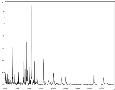
Matrix-assisted laser-desorption/ionization time-of-flight (MALDI-TOF) MS protein analysis was carried out as previously described [30] using a Microflex spectrometer (Bruker Daltonics, Ger-many). Spectra were compared with the Bruker database that contained no spectrum from
obtained with any other taxon. We incremented our database with the spectrum from strain JC206 T (Figure 4).
Strain 206T exhibited catalase and oxidase activi-ties. Using an API 20 NE strip (BioMerieux), ni-trate reduction, indole formation, glucose fermen-tation and urease were negative. Arginine dihydrolase and esculin hydrolysis were positive. H. massiliense is susceptible to ticarcillin, imipenem, trimethoprim/sulfamethoxazole, gen-tamicin, amikacin, and colimycin but resistant to fosfomycin and nitrofurantoin.
Figure 3. Transmission electron microscopy of H. massiliense strain JC206T, using a Morgani 268D (Philips) at an operating voltage of 60kV.The scale bar represents 900 nm.
0.0 0.2 0.4 0.6 0.8 1.0 1.2 4 x10
Inte
ns
. [a
.u.]
2000 4000 6000 8000 10000 12000 14000 16000 18000
Genome sequencing information
Genome project history
The organism was selected for sequencing on the basis of its phylogenetic position and 16S rRNA
simi-larity to other members of the genus
and is part of a “culturomics” study of the human digestive flora aiming at isolating all bacterial spe-cies within human feces. It was the second genome
of H.
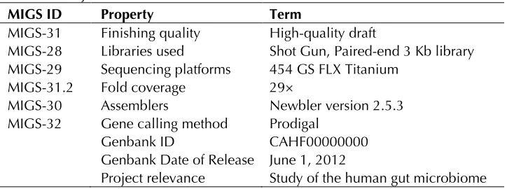
massiliense sp. nov. A summary of the project infor-mation is shown in Table 2. The Genbank accession number of the genome is CAHF00000000 and con-sists of 27 contigs. Table 2 shows the project infor-mation and its association with MIGS version 2.0 compliance.
Table 2. Project information
MIGS ID Property Term
MIGS-31 Finishing quality High-quality draft
MIGS-28 Libraries used Shot Gun, Paired-end 3 Kb library MIGS-29 Sequencing platforms 454 GS FLX Titanium
MIGS-31.2 Fold coverage 29×
MIGS-30 Assemblers Newbler version 2.5.3 MIGS-32 Gene calling method Prodigal
Genbank ID CAHF00000000 Genbank Date of Release June 1, 2012
Project relevance Study of the human gut microbiome
Growth conditions and DNA isolation
H. massiliense sp. nov. strain JC206T (= CSUR P159, = DSM 25712), was grown aerobically on 5% sheep blood-enriched Columbia agar at 37°C. Cell growth from eight petri dishes (“spread plates”) was resuspended in 4×100µl of G2 buffer (EZ1 DNA Tissue kit, Qiagen). A first mechanical lysis was performed by glass powder on the Fastprep-24 device (MP Biomedicals, USA) during 2×20 se-conds. DNA was then incubated with lysozyme for 30 minutes at 37°C and extracted using the EZ 1 Advanced XL BioRobot (Qiagen). DNA was concen-trated and purified using the QiAmp kit (Qiagen). The yield and concentration were measured using the Quant-it Picogreen kit (Invitrogen) and the Genios_Tecan fluorometer at 52.5 ng/µl.
Genome sequencing and assembly
Both a shotgun and a 3-kb paired end sequencing were performed on a 454 GS FLX pyrosequencer. Both projects were loaded on a ¼ and a 1/8 re-gions of a PTP Picotiterplate. The shotgun library was constructed with 500 ng DNA as recommend-ed by the manufacturer (Roche). For pairrecommend-ed end sequencing, five µg of DNA were mechanically fragmented using the Hydroshear device (Digilab, Holliston, MA) with an enrichment size at 3-4kb. The DNA fragmentation was visualized using the BioAnalyzer 2100 on a DNA labchip 7500 (Agilent) with an optimal size of 3.944 kb. The library was
constructed according to the 454 GS FLX Titanium paired end protocol. Circularization and nebuliza-tion were performed and generated a pattern with an optimal at 418 bp. After PCR amplification through 15 cycles followed by double size selection, the single stranded paired end library was then quantified on the Quant-it Ribogreen kit (Invitro-gen) on the Genios Tecan fluorometer at 128 pg/µL. The library concentration equivalence was calculated as 5.62 × 10⁸ molecules/µL. The library was stored at -20°C until further use.
Genome annotation
Open Reading Frames (ORFs) were predicted us-ing Prodigal [31] with default parameters but the predicted ORFs were excluded if they were span-ning a sequencing GAP region. The predicted bac-terial protein sequences were searched against the National Center for Biotechnology Information (NCBI) nonredundant (NR) and the Clusters of Orthologous Groups (COG) databases using BLASTP. The tRNAScanSE tool [32] was used to find tRNA genes, whereas ribosomal RNAs were found by using RNAmmer [33] and BLASTn against the NR database. ORFans were identified if their BLASTP E-value were lower than 1e-03 for alignment length greater than 80 amino acids. If alignment lengths were smaller than 80 amino acids, we used an E-value of 1e-05. Such
parame-ter thresholds have already been used in previous works to define ORFans.
Genome properties
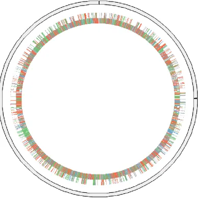
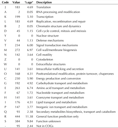
The genome is 4,186,486 bp long (one chromo-some but no plasmid) with a 59.73% GC content (Table 3 and Figure 5). Of the 3,901 predicted genes, 3,847 were protein-coding genes, and 54 were RNAs. A total of 2,924 genes (74.95%) were assigned a putative function. ORFans accounted for 312 (8.0%) of the genes. The remaining genes were annotated as hypothetical proteins. The dis-tribution of genes into COGs functional categories is presented in Table 4. The properties and the statistics of the genome are summarized in Tables 3 and 4.
Table 3. Nucleotide content and gene count levels of the genome
Attribute Value % of totala
Genome size (bp) 4,186,486
DNA coding region (bp) 3,655,584 87.32 DNA G+C content (bp) 2,500,588 59.73
Total genes 3,901 100
RNA genes 54 1.4
Protein-coding genes 3,847 98,61
Genes with function prediction 2,924 74.95
Genes assigned to COGs 3,135 80.36
Genes with peptide signals 378 9.68 Genes with transmembrane helices 955 24.48 aThe total is based on either the size of the genome in base pairs or the total number of protein coding genes in the annotated genome.
Table 4. Number of genes associated with the 25 general COG functional categories Code Value %agea Description
J 183 4.69 Translation
A 2 0.05 RNA processing and modification K 199 5.10 Transcription
L 183 4.69 Replication, recombination and repair B 2 0.05 Chromatin structure and dynamics D 45 1.15 Cell cycle control, mitosis and meiosis Y 0 0 Nuclear structure
V 44 1.13 Defense mechanisms
T 234 6.00 Signal transduction mechanisms M 272 6.97 Cell wall/membrane biogenesis N 142 3.64 Cell motility
Z 0 0 Cytoskeleton
W 0 0 Extracellular structures
U 141 3.61 Intracellular trafficking and secretion
O 168 4.31 Posttranslational modification, protein turnover, chaperones C 230 5.90 Energy production and conversion
G 192 4.92 Carbohydrate transport and metabolism E 263 6.74 Amino acid transport and metabolism F 67 1.72 Nucleotide transport and metabolism H 141 3.61 Coenzyme transport and metabolism
I 176 4.51 Lipid transport and metabolism
P 147 3.77 Inorganic ion transport and metabolism
Q 93 2.38 Secondary metabolites biosynthesis, transport and catabolism R 444 11.38 General function prediction only
S 384 9.84 Function unknown - 95 2.44 Not in COGs
Comparison with
To date, the genome fro
SmR1 is the only genome fr
species that has been sequenced [34]. By
compari-son withH. massiliense exhibited a
smaller genome (4,186,486 bp vs 5,513,887 bp, respectively), a lower G+C content (59.73% vs 63.4%, respectively) and a smaller number of genes (3,901 vs 4,804). In contrast, H. massiliense had higher ratios of genes per Mb (0.93 vs 0.87) and genes with assigned functions (74.9% vs 64.7%).
Conclusion
On the basis of phenotypic, phylogenetic and ge-nomic analyses, we formally propose the creation
o sp. nov. that contains
the strain JC206T. This bacterium has been found in Senegal.
Description of
sp.
nov.
(mas.il.ien’se. L. gen.
neutr. n. massiliense, o
Marseille where strain JC206T was cultivated).
Colonies are 0.5 mm in diameter on blood-enriched Columbia agar and Brain Heart Infusion (BHI) agar. Cells are rod-shaped with a mean di-ameter of 0.44 µm. Motile with tufts of polar flagellae optimal growth occurs under aerobic conditions. Weak growth is observed under microaerophilic conditions and with 5% CO2. No growth is observed under anaerobic conditions. Growth occurs between 30-37°C, with optimal growth observed at 37°C.
Cells stain Gram-negative. Catalase, oxidase and arginine dihydrolase activities, as well as esculin hydrolysis are present. Nitrate reduction and indole production are absent. Cells are susceptible to ticarcillin, imipenem, trimethoprim/sulfa-methoxazole, gentamicin, amikacin, and colimycin. The G+C content of the genome is 59.73%. The 16S rRNA and genome sequences are deposited in Genbank under accession numbers JN657219 and CAHF00000000, respectively. The type strain JC206T (= CSUR P159 = DSMZ 25712) was isolated from the fecal flora of a healthy patient in Senegal.
References
1. Tindall BJ, Rossello-Mora R, Busse HJ, Ludwig W, Kämpfer P. Notes on the characterization of pro-karyote strains for taxonomic purposes. Int J Syst Evol Microbiol 2010; 60:249-266
2. Liolios K, Mavromatis K, Tavernarakis N, Kyrpides NC. The Genomes On Line Database (GOLD) in 2007: status of genomic and metagenomic pro-jects and their associated metadata. Nucleic Acids Res 2008; 36:D475-D479
3. Lagier JC, El Karkouri K, Nguyen TT, Armougom F, Raoult D, Fournier PE. Non-contiguous finished genome sequence and description of
Stand
Ge-nomic Sci 2012; 6:116-125
4. Mishra AK, Lagier JC, Robert C, Raoult D, Four-nier PE. Non-contiguous finished genome se-quence and description of
senegalense sp. nov. Stand Genomic Sci 2012; 6:386-395.
5. Baldani JI, Baldini VL, Seldin L, Döbereiner J.
Characterization of
gen. nov., sp. nov., a root-associated nitrogen-fixing bacterium. Int J Syst Bacteriol 1986; 36
:86-93
6. Baldani JI, Pot B, Kirchhof G, Falsen E, Baldani VL, Olivares FL, Hoste B, Kersters K, Hartmann A, Gillis M, Dobereiner J. Emended description of
rubrisubalbicans, a milk plant pathogen, as
classification of a group of clinical isolates (EF
group 1) asInt J Syst
Bacteriol 1996; 46:802-810
7. Dobritsa AP, Reddy MC, Samadpour M.
Reclassi-fication o
typic synonym o
the description ohuttiense subsp. nov. aputei subsp. nov., comb. nov., and description of
Int J Syst Evol
Microbiol 2010; 60:1418-1426
8. Carro L, Rivas R, Leon-Barrios M, Gonzalez-Tirante M, Velazquez E, Valverde A. 2011.
isolated in Tenerife (Canary Islands). Int J Syst Evol Microbiol 2012; 62:1300-1306
9. Ding L, Yokota A. Proposals of
well water and reclassification o huttiensislanceolata,
delicatum a
autotrophicum a
nov
Int J
Syst Evol Microbiol 2004; 54:2223-2230
10. Im WT, Bae HS, Yokota A, Lee ST
degrading bacterium. Int J Syst Evol Microbiol 2004; 54:851-855
11. Kirchhof G, Eckert B, Stoffels M, Baldani JI, Reis
VM, Hartmann A
nov., a new nitrogen-fixing bacterial species that occurs in C4-fibre plants. Int J Syst Evol Microbiol 2001; 51:157-168
12. Rothballer M, Schmid M, Klein I, Gattinger A, Grundmann S, Hartmann A
wheat roots. Int J Syst Evol Microbiol 2006; 56:1341-1348
13. Valverde A, Velazquez E, Gutierrez C, Cervantes
E, Ventosa A, Igual
sp. nov., a novel nitrogen-fixing bacterium asso-ciated with root nodules of Phaseolus vulgaris. Int J Syst Evol Microbiol 2003; 53:1979-1983
14. Jung SY, Lee MH, Oh TK, Yoon JH.
from rhizosphere soil of Allium victorialis var. platyphyllum. Int J Syst Evol Microbiol 2007; 57:2284-2288
15. Ziga ED, Druley T, Burnham CA species bacteremia in a pediatric oncology pa-tient. J Clin Microbiol 2010; 48:4320-4321
16. Chen J, Su Z, Liu Y, Sandoghchian S, Zheng D,
Wang S, Xu H
pathogenic bacteria isolated from acute
lympho-blastic leukemia patient. Curr Microbiol 2011; 62:331-333
17. Marques da Silva R, Caugant DA, Eribe ER, Aas JA, Lingaas PS, Geiran O, Tronstad L, Olsen I. Bacterial diversity in aortic aneurysms determined by 16S ribosomal RNA gene analysis. J Vasc Surg 2006; 44:1055-1060
18. Spilker T, Uluer AZ, Marty FM, Yeh WW, Levison JH, Vandamme P, Lipuma JJ. Recovery of
fibrosis. J Clin Microbiol 2008; 46:2774-2777
19. Coenye T, Goris J, Spilker T, Vandamme P, Lipuma JJ. Characterization of unusual bacteria isolated from respiratory secretions of cystic fibro-sis patients and description o gen. nov., sp. nov. J Clin Microbiol 2002; 40:2062-2069
20. Trape JF, Tall A, Diagne N, Ndiath O, Ly AB, Faye J, Dieye-Ba F, Roucher C, Bouganali C, Badiane A, et al. Malaria morbidity and
pyrethroid resistance after the introduction of in-secticide-treated bednets and artemisinin-based combination therapies: a longitudinal study. Lan-cet Infect Dis 2011; 11:925-932
21. Field D, Garrity G, Gray T, Morrison N, Selengut J, Sterk P, Tatusova T, Thomson N, Allen MJ, Angiuoli SV, et al. The minimum information about a genome sequence (MIGS) specification. Nat Biotechnol 2008; 26:541-547
22. Woese CR, Kandler O, Wheelis ML. Towards a natural system of organisms: proposal for the do-mains ArchaEukarya. Proc Natl Acad Sci USA 1990; 87:4576-4579
23. Garrity GM, Bell JA, Lilburn T. Phylum XIV.
DJ, Krieg NR, Staley JT (eds), Bergey's Manual of Systematic Bacteriology, Second Edition, Volume 2, Part B, Springer, New York, 2005, p. 1. 24. Validation List No. 107. List of new names and
new combinations previously effectively, but not validly, published. Int J Syst Evol Microbiol 2006;
56:1-6
25. Garrity GM, Bell JA, Lilburn T. Class II.
Brenner DJ, Krieg NR, Staley JT (eds), Bergey's Manual of Systematic Bacteriology, Second Edi-tion, Volume 2, Part C, Springer, New York, 2005, p. 575.
26. Garrity GM, Bell JA, Lilburn T. Order I.
DJ, Krieg NR, Staley JT (eds), Bergey's Manual of Systematic Bacteriology, Second Edition, Volume 2, Part C, Springer, New York, 2005, p. 575. 27. Garrity GM, Bell JA, Lilburn T. Family II.
Brenner DJ, Krieg NR, Staley JT (eds), Bergey's Manual of Systematic Bacteriology, Second Edi-tion, Volume 2, Part C, Springer, New York, 2005, p. 623.
28. Ashburner M, Ball CA, Blake JA, Botstein D, But-ler H, Cherry JM, Davis AP, Drolinski K, Dwight SS, Eppig JT, et al. Gene ontology: tool for the unification of biology. The Gene Ontology Con-sortium. Nat Genet 2000; 25:25-29
29. Stackebrandt E, Ebers J. Taxonomic parameters revisited: tarnished gold standards. Microbiol To-day 2006; 33:152-155.
30. Seng P, Drancourt M, Gouriet F, La Scola B, Fournier PE, Rolain JM, Raoult D. Ongoing revo-lution in bacteriology: routine identification of bacteria by matrix-assisted laser desorption ioni-zation time-of-flight mass spectrometry. Clin In-fect Dis 2009; 49:543-551
31. Prodigal. http://prodigal.ornl.gov
32. Lowe TM, Eddy SR. t-RNAscan-SE: a program for imroved detection of transfer RNA gene in ge-nomic sequence. Nucleic Acids Res 1997; 25:955-964
33. Lagesen K, Hallin P, Rodland EA, Staerfeldt HH, Rognes T, Ussery DW. RNAmmer: consistent and rapid annotation of ribosomal RNA genes. Nucle-ic Acids Res 2007; 35:3100-3108
34. Pedrosa FO, Monteiro RA, Wassem R, Cruz LM, Ayub RA, Colauto NB, Fernandez MA, Fungaro MH, Grisard EC, Hungria M. Genome of
cialized diazotrophic endophyte of tropical grass-es. PLoS Genet 2011; 7:e1002064
![Table 1. Classification and general features of Herbaspirillum massiliense strain JC206T according to the MIGS rec-ommendations [21]](https://thumb-us.123doks.com/thumbv2/123dok_us/694840.2067483/2.612.67.550.280.673/table-classification-general-features-herbaspirillum-massiliense-according-ommendations.webp)