RESEARCH
Palm kernel oil-based polyester
polyurethane composites incorporated
with multi-walled carbon nanotubes
for biomedical application
Nurul Nabilah bt Zulkifli
1, Khairiah bt Hj Badri
2and Khairul Anuar Mat Amin
1,3*Abstract
Background: In this study, polyurethane (PU) films from palm kernel oil-based polyester (PKO-p) incorporated multi-walled carbon nanotubes (MWNTs) were prepared via evaporative casting method. Nanoparticle fluid disper-sions containing 0.01, 0.04 and 0.08 % wt. of MWNTs were added into PKO-p-based resin and mixed by digital probe sonicator for 20 min followed by mixing with isocyanate to produce PU-MWNTs composite films. The mechanical properties, water resistance, water vapor transmission rates (WVTR), biocompatibility, and antibacterial activities of the PU-MWNTs composite films were examined.
Results: Results show that PU containing 0.01 wt. % of MWNTs demonstrated optimum mechanical properties as it possessed high tensile strength, modulus, and good flexibility compared to PU film and other PU-MWNTs com-posite films. There are no significant difference in swelling values as well as water vapor transmission rates for PU film and PU-MWNTs composite films. All the films showed low swelling values (17–23 %) and WVTR values in the range 181–269 g m−2 d−1. Cell studies revealed that PU and PU-MWNTs composite films are non-cytotoxic to human skin
fibroblast cells (CRL2522) and the cell proliferation was increased after incubation of 72 h. The in vitro qualitative anti-bacterial results showed both PU and PU-MWNTs composite films exhibited bactericidal effect against Gram-positive (Staphylococcus aureus and Bacillus cereus) and Gram-negative bacteria (Escherichia coli and Klebsiella pneumonia).
Conclusions: In summary, incorporation of MWNTs improved the mechanical properties of the polyurethane films with no cytotoxic effect against normal human skin fibroblast cells.
Keywords: Polyurethane, Palm kernel oil-based polyester, MWNTs, Biocompatibility, Antibacterial activities, Composites
© 2016 The Author(s). This article is distributed under the terms of the Creative Commons Attribution 4.0 International License (http://creativecommons.org/licenses/by/4.0/), which permits unrestricted use, distribution, and reproduction in any medium, provided you give appropriate credit to the original author(s) and the source, provide a link to the Creative Commons license, and indicate if changes were made.
Background
Polyurethane (PU) has been widely used in many areas such as automotive, medical, and chemical industries due to their wide range of properties. Polyurethanes are highly preferred materials in the constructions of medical applications due to their physiochemical, bio-chemical, and mechanical design criteria (Campos et al. 2011). These criteria’s include, but are not limited to,
blood compatibility, biocompatibility, biodegradability, and high mechanical performances. In biomedical appli-cation, it has been used since 1980 in producing such as the artificial heart, pacemaker leads, heart valves, and catheters (Kütting et al. 2011; Alves et al. 2014). In the recent years, polyurethane has been showing a promis-ing result as a wound dresspromis-ing material and bio-scaffolds for biomedical applications (Yari et al. 2012). Not limited to that, a few studies have reported successfully in pro-ducing the PU materials using the polyol-based polyester from vegetable oils such as oil palm, soybean, and linseed oil (Badri et al. 2001; Petrović2008). These PU materials
Open Access
*Correspondence: [email protected]
1 School of Fundamental Science, Universiti Malaysia Terengganu, 21030 Kuala Terengganu, Terengganu, Malaysia
are expected to minimize the toxicity from synthetic pet-rochemical-based polyester and enhanced the biocom-patibility of the PU products.
Polyurethane nanocomposites using carbon nanotubes and graphite have gained a great attention because they offer a great potential to enhance mechanical proper-ties and increased electrical conductivity due to their large specific surface area. These fillers are considered as an ideal reinforcing agent for high-strength nanocom-posites. Recent studies show that single-walled carbon nanotubes (SWNTs) and multi-walled carbon nano-tubes (MWNTs) are among attracting nanofillers to produce advance polyurethane (PU) materials with high mechanical properties and conducting materials (Kim et al. 2010). These nanocomposites need only a small amount of nanofillers to give a tremendous improvement in mechanical properties than pure PU composites. For example, incorporation of SWNTs into polyurethane [polyester-based (polycaprolactone (PCL)] improved the Young’s modulus compared to pristine PU (Xia and Song 2006). The Young’s modulus of 0.7 wt. % SWNT-grafted-PU increased by ~278 % compared to the pure SWNT-grafted-PU. Other study reported that the incorporation of MWNTs in waterborne polyurethane exhibits a huge increase in the tensile properties to 370 % due to a strong covalent bond-ing in the system (Kuan et al. 2005).
For biocompatibility of polyurethane nanocomposites, some recent studies indicated that carbon nanotubes may have promising potentials to be used at molecular and cellular levels (Smart et al. 2006; Lee et al. 2011). For example, Meng et al. (Meng et al. 2009) reported that polyurethane (polyether-based polyol) contain-ing multi-walled carbon nanotubes (PU-MWNT) films are non-cytotoxic to the mouse fibroblast cell (3T3). In other study, Verdejo et al. reported that the addition of MWNTs (0.01, 0.05, and 0.10 %, wt. %) into PU (pol-yether-based, Bayer Group, UK) did not significantly affect the cytotoxicity of human osteosarcoma cell line (SaOs-2) (Verdejo et al. 2009). The blood compatibility of PU (polyether-based polyol)-MWNT composites also reported as promising than pure PU composites with the MWNT significantly improved the anticoagulant func-tion as well as the platelets activafunc-tion (Meng et al. 2005).
For antibacterial activities, a limited study has been reported on the polyurethane-carbon nanotubes (CNTs). Yadav et al. (Yadav et al. 2012) observed the hyper-branched polyurethane nanocomposites [polyether-based (polytetramethylglycol)] incorporated with 0.04 % wt of multi-walled carbon nanotubes (MWNT) which inhibited the growth of Escherichia coli (E. coli) about 50 % after 21 h. These nanocomposites inhibit the growth of E. coli even at low concentration (0.01 % wt) and the inhibition become stronger as the concentration of
MWNT is increased. This could be due to specific physi-ochemical and structural characteristics such as surface area, functional group, length, and diameter of CNTs.
In our review, there has been limited studies using polyols-based polyester from vegetable oils in produc-ing PU-CNTs film (Ali et al. 2014; Liu et al. 2016). Most of the studies produced PU-CNTs nanocomposites using polyols-based polyether and polyester derived from petroleum based and shows a promising result in mechanical characteristics and electrical conductivity (Cho et al. 2005; Jell et al. 2008; Khan et al. 2010, 2011; Pokharel 2014). Therefore, in this study, we fabricated and characterized the polyurethane films from palm ker-nel oil (PKO)-based polyester incorporating multi-walled carbon nanotubes. The mechanical characteristics, water vapor transmission rates, and swelling of the pol-yurethane multi-walled carbon nanotubes (PU-MWNTs) composite films were carried out. The cell viability and cell proliferation through in vitro study of the PU nano-composites were assessed against human fibroblast skin cell (CRL-2522, American Type Tissue Collection). The antibacterial activities were examined via in vitro quali-tative studies against two bacterial strains, i.e., Gram-positive (Staphylococcus aureus, Bacillus cereus) and Gram-negative (Escherichia coli, Klebsiella pneumoniae) bacteria.
Methods Materials
The palm kernel oil-based monoester polyol (PKO-p) was prepared as reported by (Badri et al. 2001). 2, 4-diphenylmethane diisocyanate (MDI) was obtained from Cosmopolyurethane (M) Sdn. Bhd., Klang, Malay-sia. Acetone and polyethylene glycol (PEG 400) were pur-chased from Merck Sdn. Bhd., Malaysia. A multi-walled carbon nanotube (MWNT, product number 773840 and lot number MK13N5858 V, diameter = 10 ± 1 nm and length = 3–6 µm) was purchased from Sigma Aldrich, Malaysia. Penicillin disks (product number CT0043B, lot number 523987) were obtained from Oxoid, Eng-land. All materials were used as received without further purification.
Preparation of polyurethane (PU) films
5 min. The acetone (35 %) was also added into MDI to accommodate the evaporation process. The mixture and MDI were then stirred at 100 rpm for 5 min at room tem-perature and then casted onto a Teflon plate and dried in humidity chamber (37 °C, 50 % ± 5 relative humidity) for 24 h to remove the solvent. The PU films were then pre-conditioned at the same temperature for next 24 h prior to any characterizations.
Preparation of PU‑MWNT composite films
The nanoparticle fluid dispersions containing PKO-p, PEG400, and acetone (35 %) with 0.01, 0.04, and 0.08 % (w/w) of multi-walled carbon nanotubes (MWNTs) were prepared using a digital sonicator horn (Qsonica-Q500, USA) with a probe diameter of 6 mm, in a pulse mode (0.5 s on/off) with an amplitude set at 40 %. Different intervals of sonication were carried out to determine the optimum sonication time (min). During sonication, the sample vials were placed inside a water bath to keep the dispersion temperature constant. The dispersions were then mixed with MDI (containing 35 % acetone) and stirred for 5 min. The mixtures were casted onto a Teflon plate and dried in a humidity chamber (37 °C, 50 % ± 5 relative humidity) for 24 h to remove the solvent. The PU composite films containing 0.01, 0.04, and 0.08 % (w/w) of MWNTs hereafter known as PU-MWNT01, PU-MWNT04, and PU-MWNT08, respectively. The PU-MWNT composite films were then pre-conditioned at the same temperature for next 24 h prior to any characterizations.
UV–visible spectroscopy
UV–visible absorption spectra of homogenous PU-MWNTs dispersions were obtained with a dual-beam UV–vis spectrophotometer (Shimadzu UV-1800) using quartz cuvettes (path length = 10 mm). All samples were measured after a 10-fold dilution with Triton X-102. The transmittance spectra of PU films and PU-MWNTs com-posite films were measured in the range of 200–700 nm wavelengths light.
Optical microscopy
Dispersions of PU films containing multi-walled carbon nanotubes (PU-MWNTs) were imaged using an optical microscope (Olympus IX73) with cell Sens Dimension software.
Mechanical properties
Stress–strain measurements were obtained using an Instron Universal Testing machine (model 3366) with ±10 kN grips and the cross speed set at 20 mm/min. Film thickness (2.0 × 6.0 cm) was measured by a hand-held micrometer (Mitutoyo). Young’s modulus (E), tensile
strength (TS), and toughness (T) were calculated from the slope of the linear part of the stress–strain curve and maximum stress and through integration of the area under the curve, respectively. Strain-at-break (γ) was also recorded.
Swelling ratio
Water uptake was measured by weighing the dried films (Wdry) prior to immersion into solutions of pH 7 in water bath (37 ± 0.5 °C). The temperature was set at 37 °C to mimic the condition of wound which simi-lar to body temperature. The films were removed after 24 h, wiped gently with a filter paper to expel surface solution, and then weighed (Wwet). Water uptake was then determined from the equilibrium-swelling ratio
Wwet −Wdry
/Wdry
×100.
Water vapor transmission rates
The water vapor transmission rates (WVTR) of the sam-ples were measured by following a modified ASTM E96 standard method (Wu et al. 1995). Each membrane was fixed on the circular opening of a permeation bottle (diameter = 1.5 cm, height = 5.0 cm) with the effective transfer area (A) of 1.33 cm2. The permeation bottle was placed in the temperature humidity chamber at 37 °C and 50 ± 5 % relative humidity. The WVTR was then deter-mined by the rate of change of mass (m) in these water-filled permeation bottles at 24 h exposure time as follows:
where m/Δt is the water gain per unit time of transfer and A is the area exposed to water transfer (m2).
Cell studies
Normal human skin fibroblast cells (CRL2522-ATCC) were cultured in Eagle’s minimal essential medium (EMEM, Sigma Aldrich) supplemented with 10 % (v/v) fetal bovine serum (FBS, ATCC) and 1 % antibiotic (Peni-cillin/Streptomycin, ScienCell) at 37 °C in a humidified atmosphere of 5 % CO2. Cells were sub-cultured every 3 days using established protocols and then were har-vested at 60–80 % confluence.
To investigate the cell viability, PU and PU-MWNTs composite films were placed into a 96-well culture plates and sterilized in a laminar air flow chamber using UV radiation for 20 min. Prior to cell seeding, PU and PU-MWNTs composite films were soaked in medium (EMEM only) for 24 h and the supernatant removed. Suspensions of normal human skin fibroblast cells (5000 cells/well) were seeded into wells containing PU and PU-MWNTs composites and cultured at 37 °C in humidified atmosphere of 5 % CO2 for 24, 48 and 72 h. Tissue cul-ture polystyrene plates (TCPP) used as control contained
the same number of cells. Cell viability of PU and PU-MWNTs composite films were examined through a staining procedure of calcein-AM (Life Technologies, USA) and observed by light microscope (Olympus TH4-200) equipped with fluorescence filter (Olympus U-RFL-T UV with blue light excitation).
Cell proliferations were quantified using a CellTiter 96 aqueous one solution assay (Promega, USA) which con-tained tetrazolium compound [3-(4,5-dimethylthiazol-2-yl)-5-(3-carboxymethoxyphenyl)-2-(4-sulphonyl)-2H tetrazolium, inner salts; MTS(a)] with electron coupling reagent (phenazine ethosulfate). Prior to addition of assay solution (20 µL in each wells), the media in all wells that contained film samples, except for the negative con-trol, were replaced with fresh media and later incubated for 4 h at 37 °C in humidified atmosphere of 5 % CO2. Then, 100 µL of the inoculants was transferred to a new well and the absorbance was then measured at 492 nm with a microplate reader (TECAN, Infinite M200, USA). The absorbance was then related to cell numbers using calibration curves.
Antibacterial studies
Gram-positive (Staphylococcus aureus-S. Aureus, Bacillus cereus-B. Cereus) and Gram-negative (Escherichia coli
-E. coli, Klebsiella pneumonia-K. pneumoniae) microbes were used for antibacterial assays. Mueller–Hinton (MH, Difco, Malaysia) agar was used for the growth of both bacterial types. It was prepared by suspending 38 g of the MH powder in 1 L purified water, heated with frequent agitation, and boiled for 1 min to dissolve the powder. The prepared agar was autoclaved at 121 °C for 15 min with the final pH at 7.3 ± 0.1. (S. Aureus, B. Cereus) and (E. coli, K. Pneumoniae) were grown on Mueller–Hinton (MH) agar and incubated aerobically at 37 °C in an incu-bator. (S. Aureus, B. Cereus) and (E. coli, K. Pneumoniae)
suspensions were evenly spread on the solid MH agar and dried in a laminar air flow chamber. The polyure-thane (PU) and polyurepolyure-thane multi-walled carbon nano-tubes (PU-MWNTs) composite films (diameter ≈ 6 mm) were then placed on the MH agar and incubated at 37 °C for 24 h. The presence of any clear zone around the disks on the MH agar was recorded as an indication of inhibition against (S. Aureus, B. Cereus) and (E. coli, K. Pneumoniae).
Results and discussion
Dispersing multi‑walled carbon nanotubes
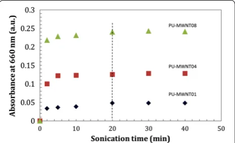
To prepare the PU-MWNTs composite films, the opti-mum sonication time is required to successfully disperse the multi-walled carbon nanotubes (MWNTs) in the PKO-p solution. According to Aldalbahi and co-work-ers, the optimum sonication time was defined as the
minimum amount of time required to effectively disperse the multi-walled carbon nanotubes (MWNTs) in the dis-persion (Aldalbahi 2011), which an excess sonication can shorten or cause defects to the tubes and thereby dimin-ish their properties (O’connell MJ, Bachilo SM, Huffman CB, Moore VC, Strano MS, Haroz EH, Rialon KL, Boul PJ, Noon WH, Kittrell C 2002; Benedict et al. 2005; Vais-man et al. 2006). The optimization process was deter-mined by monitoring the level of the UV–Vis absorption intensity at different sonication times and disappearance of visible aggregates. Figure 1 shows that the UV–Vis absorbance intensity (λ = 660 nm) increases with sonica-tion time, reflecting to an increasing amount of MWNTs [0.01, 0.04, and 0.08 % (w/w)] which became dispersed over time. The absorbance of the dispersions at 660 nm was plotted as a function of sonication time and showed that the sonication becomes independent after 20 min for all dispersions. This particular wavelength was selected as it correlated with the maxima of an absorption band arising from the Van Hove singularities for single-walled carbon nanotubes (SWNTs) (Ryabenko et al. 2004; Gros-siord et al. 2005; Attal et al. 2006).
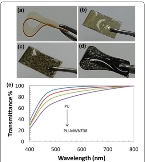
Physical appearance of PU‑MWNTs composites
Polyurethane films and polyurethane multi-walled car-bon nanotubes (PU-MWNTs) composite films were successfully prepared via solution casting (Fig. 2). The polyurethane films exhibit smooth surface, void-free and transparent (Fig. 2a). However, the additions of multi-walled carbon nanotubes (MWNTs) have decreased the transparency of the films (Fig. 2e). The polyurethane films with 0.01 wt. % of multi-walled carbon nano-tubes (PU-MWNT01) were still clear at transmittance (λ = 700 nm), T = 97 %, but became opaque when the amount of MWNTs increased (Fig. 2c, d). The T values
of PU-MWNT04 and PU-MWNT08 films were at 93 and 90 %, respectively.
Stress–strain analysis
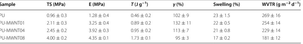
The stress–strain properties of PU-MWNTs compos-ite films are shown in Fig. 3 and summarized in Table 1.
The tensile strength of PU-MWNT01, PU-MWNT04, and PU-MWNT08 composite films were higher than that of PU film, indicating that the modulus (E) as well as the toughness (T) of the composites were improved by the addition of the multi-walled carbon nano-tubes (MWNTs). For example, the T values of the PU-MWNT01 and PU-MWNT04 films were increased about twofold and fourfold, respectively, than PU films. The increment of strain-at-break (γ) of the PU films also can be observed after the inclusion of MWNTs. PU-MWNT01 films recorded highest γ value at 132 ± 11 % followed by PU-MWNT04 films at 113 ± 7 %. The addi-tion of higher amount of MWNTs (0.08 % wt. %) into PU composite films (PU-MWNT08) caused the γ value to decrease 95 ± 3 %, which was below than PU films at 102 ± 9 %. The decreasing value of the γ may be caused by uneven dispersion of multi-walled carbon nanotubes (MWNTs) in polymer matrix (Xiong et al. 2006). Optical microscopy revealed that there were large aggregates pre-sent in MWNT08 composite films followed by PU-MWNT04 composite films (Fig. 3d, e). This aggregation resulted to the uneven dispersion of multi-walled carbon nanotubes (MWNTs) in the matrix, which contributed to the lower value of the strain-at-break of the films (Xiong et al. 2006). Interfacial stress and aspect ratio between the MWNTs and PU as well as orientation of the MWNTs are among important factors to obtain the effective load transfer and could improve the mechanical performances of the composites (Tijing et al. 2013).
Swelling ratio and water vapor transmittance rates (WVTR) Swelling ratio and water vapor transmittance rate (WVTR) values of the PU-MWNTs composite films are Fig. 2 Photograph of the sample; a PU film, b PU-MWNT01 film, c
PU-MWNT04 film, d PU-MWNT08 film, and e decreased transmittance (%) of PU film at higher loading of MWNTs
shown in Table 1. PU films recorded highest swelling ratio at 23 ± 1.5 %, and the values were decreased upon addition of MWNTs. These low swelling ratio values of PU-MWNT composite films were considered as hydro-phobic as they showed less water absorption (Yücedag et al. 2010) and consistent with the previous study (Badri 2012).
The water vapor transmission rate (WVTR) values of PU films and PU-MWNTs composite films are in the range of 181–269 g m−2 d−1 (Table 1). Addition of multi-walled carbon nanotubes (MWNTs) slightly decreased the WVTR values which could be due to dispersion of MWNTs in the polyurethane matrix and blocked the water vapor transmission (Kavoosi et al. 2014). However, these values still remain within the range of WVTR of commercial wound dressings values at 90–2893 g m−2 d−1 (Wu et al. 1995).
Biocompatibility of films
Biocompatibility of PU and PU-MWNTs composite films was determined by examining the cell viability and cell proliferation that involved human skin fibroblast cells (CRL2522-ATCC). The cell morphological attached onto the surface of PU film were changed from rounded shape (24 h incubation) to elongated/spindle-like shape after being incubated for 72 h (Fig. 4a–o). Even though the cell viability on the PU films and PU-MWNTs compos-ite films was low compared to control (TCPP), it showed that the cell growth increased over time on PU film as well as on PU-MWNTs composite films from 24 to 72 h. In addition, it also showed that the PU and MWNTs were unlikely to be cytotoxic to the CRL2522 cell and consist-ent with the prior reports (Shim et al. 2002; Meng et al. 2005; Lee et al. 2011).
To enumerate the cell growth on the PU films and PU-MWNT composite films, cell proliferations were carried out using cellTiter 96 aqueous one solution assay after being cultured for 24, 48 and 72 h (Fig. 4p). The cell proliferation observed was comparable with the cell viability results, in which the cell numbers were increased for PU films and PU-MWNTs composite
films after being incubated for 24 to 72 h. PU films containing MWNTs showed higher cell proliferation than PU films. For example, the cell numbers cultured on PU-MWNT01, PU-MWNT04, and PU-MWNT08 composite films after incubation of 72 h were 2740, 3000, and 3200 cell/well, respectively (Fig. 4p). Com-pared to PU films, the cell number was recorded at 2600 cell/well after being incubated for 72 h. PU-MWNT08 composite films showed the highest cell increment at 45 % from 24 to 72 h of incubation. Higher cell growth enumerated on PU-MWNT08 composite films than the other PU films could be due to the influence of the material’s surface such as roughness, hardness, hydro-phobicity, and hydrophilicity of the substrate (Hu et al.
2011). An addition of high amount of MWNTs in PU
composite films (PU-MWNT08) contributed to the lowest water sorption (17 ± 0.2 %), or in another word improved the hydrophobicity and led to a positive bio-logical response of viable cells on the substrate (Zhou et al. 2007; Tanodekaew et al. 2004; Muzzarelli et al. 2005). Incorporation of nanoparticles also has been reported to increase the surface area-to-volume ratio of nanocomposites for the cell growth, and therefore increased cell proliferation on the substrate (Azad et al. 2004). For that reason, this factor also could explain the higher cell proliferation obtained on the PU-MWNT08 composite films compared to other samples. Not lim-ited to that, incorporation of MWNTs also has been reported to change surface topology of the composites which could affect cell proliferation (Sharma and Elis-seeff 2004).
Antibacterial performances
The antibacterial performance of PU films and PU-MWNTs composite films were studied through quali-tative method (disk diffusion) against Gram-positive, i.e., Staphylococcus aureus (S. Aureus), Bacillus cereus (B. Cereus) and Gram-negative, i.e., Escherichia coli
(E. coli), Klebsiella pneumoniae (K. Pneumoniae). The zone of inhibition was not observed on all films against all bacteria strains, i.e., S. Aureus, B. Cereus, E. coli and Table 1 Summary of the tensile strength (TS), Young’s modulus (E), toughness (T), strain-at-break (γ), swelling and water vapor transmission rates (WVTR) of polyurethane (PU) films, PU film containing 0.01 % wt % MWNT (PU-MWNT01), PU film containing 0.04 % wt % MWNT (PU-MWNT04), and PU film containing 0.08 % wt % MWNT (PU-MWNT08)
Sample TS (MPa) E (MPa) T (J g−1) γ (%) Swelling (%) WVTR (g m−2 d−1)
PU 0.96 ± 0.3 1.28 ± 0.4 0.46 ± 0.2 102 ± 9 23 ± 1.5 269 ± 16
PU-MWNT01 2.11 ± 0.3 3.25 ± 0.4 0.89 ± 0.2 132 ± 11 22 ± 0.5 254 ± 14
PU-MWNT04 2.45 ± 0.2 3.92 ± 0.3 0.95 ± 0.2 113 ± 7 21 ± 0.8 229 ± 14
K. pneumoniae after being incubated for 24 h. Figure 5 shows the representative images of the bacteria strains cultured on penicillin disks and PU-MWNT08 compos-ite films. To examine the bactericidal effect of the sample indirectly, we have removed the sample on the MH agar and further incubated the agar for 24 h to observe if there was any bacteria growth on the open area once occupied by the samples. The results showed that there is no bac-teria growth on the samples’ area after further incubated (24 h) and could support that the PU films containing MWNTs have the antibacterial activities. Our result was in agreement with the study conducted by Yücedag and co-workers on the bactericidal effect of MWNTs samples (Yücedag et al. 2010).
act as a potential barrier against foreign molecules with high molecular weight (Jung et al. 2007).
Conclusions
The polyurethane (PU) films and polyurethane (PU) composite films with 0.01 wt. %, 0.04 wt. %, and 0.08 wt. % of multi-walled carbon nanotubes (PU-MWNT) were successfully prepared using palm kernel oil-based polyester. The effect of multi-walled carbon nanotubes (MWNTs) on the mechanical properties of PU films was investigated. It showed that incorporation of MWNTs gave significant improvement in mechanical proper-ties of PU films. Biocompatibility study revealed that PU-MWNTs composite films exhibited enhancement to both the cell viability and cell proliferation than PU film. Moreover, this study also concluded that both samples, PU films and PU-MWNTs composite films, exhibited bactericidal effect against Gram-positive (Staphylococcus aureus, Bacillus cereus) and Gram-negative (Escherichia coli and Klebsiella pneumoniae) bacteria.
Authors’ contributions
NNZ performed the experiment and analysis, interpreted the results, and helped in preparing the manuscript. KHB helped in synthesis and preparation of palm kernel oil-polyester polyol to be used throughout the experiment. KAMA planned, supervised the whole study, and wrote the manuscript. All authors read and approved the final manuscript.
Author details
1 School of Fundamental Science, Universiti Malaysia Terengganu, 21030 Kuala Terengganu, Terengganu, Malaysia. 2 Polymer Research Centre, Faculty of Sci-ence and Technology, Universiti Kebangsaan Malaysia, 43600 Bangi, Selangor, Malaysia. 3 Institute of Marine Biotechnology, Universiti Malaysia Terengganu, 21030 Kuala Terengganu, Terengganu, Malaysia.
Acknowledgements
The authors wish to thank Ministry of Higher Education, Malaysia for financial assistance under Fundamental Research Grant Scheme (FRGS–vote no. 59294), Central Lab (UMT) and Institute of Marine Biotechnology (UMT) for providing the facilities for undertaking this work.
Competing interests
The authors declare that they have no competing interests.
Received: 29 December 2015 Accepted: 5 May 2016
References
Aldalbahi A (2011) Preparation and characterisation of conducting biopoly-mer-carbon nanotube composite materials. World J Eng 8:39 Ali A, Yusoh K, Hasany S (2014) Synthesis and physicochemical behaviour of
polyurethane-multiwalled carbon nanotubes nanocomposites based on renewable castor oil polyols. J Nanomater 2014:165
Alves P, Cardoso R, Correia T, Antunes B, Correia I, Ferreira P (2014) Surface modification of polyurethane films by plasma and ultraviolet light to improve haemocompatibility for artificial heart valves. Colloids Surf, B 113:25–32
Attal S, Thiruvengadathan R, Regev O (2006) Determination of the concentra-tion of single-walled carbon nanotubes in aqueous dispersions using UV-visible absorption spectroscopy. Anal Chem 78(23):8098–8104 Azad AK, Sermsintham N, Chandrkrachang S, Stevens WF (2004) Chitosan
membrane as a wound-healing dressing: characterization and clinical application. J Biomed Mater Res B Appl Biomater 69B(2):216–222. doi:10.1002/jbm.b.30000
Badri KH (2012) Biobased polyurethane from palm kernel oil-based polyol. In: Zafar F, Sharmin E (eds) Polyurethane. INTECH Open Access Publisher. doi:10.5772/2416
Badri K, Ahmad S, Zakaria S (2001) Production of a high-functionality RBD palm kernel oil-based polyester polyol. J Appl Polym Sci 81(2):384–389 Benedict B, Pehrsson PE, Zhao W (2005) Optically sensing additional sonication
effects on dispersed HiPco nanotubes in aerated water. J Phys Chem B 109(16):7778–7780
Campos E, Cordeiro R, Santos AC, Matos C, Gil M (2011) Design and characteri-zation of bi-soft segmented polyurethane microparticles for biomedical application. Colloids Surf, B 88(1):477–482
Cho JW, Kim JW, Jung YC, Goo NS (2005) Electroactive shape-memory polyu-rethane composites incorporating carbon nanotubes. Macromol Rapid Commun 26(5):412–416
Grossiord N, Regev O, Loos J, Meuldijk J, Koning CE (2005) Time-dependent study of the exfoliation process of carbon nanotubes in aqueous disper-sions by using UV-visible spectroscopy. Anal Chem 77(16):5135–5139 Hu X, Park S-H, Gil ES, Xia X-X, Weiss AS, Kaplan DL (2011) The influence of
elas-ticity and surface roughness on myogenic and osteogenic-differentiation of cells on silk-elastin biomaterials. Biomaterials 32(34):8979–8989 Jell G, Verdejo R, Safinia L, Shaffer MS, Stevens MM, Bismarck A (2008) Carbon
nanotube-enhanced polyurethane scaffolds fabricated by thermally induced phase separation. J Mater Chem 18(16):1865–1872 Jung KH, Huh MW, Meng W, Yuan J, Hyun SH, Bae JS, Hudson SM, Kang IK
(2007) Preparation and antibacterial activity of PET/chitosan nanofi-brous mats using an electrospinning technique. J Appl Polym Sci 105(5):2816–2823
Kavoosi G, Dadfar SMM, Dadfar SMA, Ahmadi F, Niakosari M (2014) Investiga-tion of gelatin/multi-walled carbon nanotube nanocomposite films as packaging materials. Food Sci Nutr 2(1):65–73
Khan U, Blighe FM, Coleman JN (2010) Selective mechanical reinforcement of thermoplastic polyurethane by targeted insertion of functionalized SWCNTs. J Phys Chem C 114(26):11401–11408
Khan U, May P, O’Neill A, Vilatela JJ, Windle AH, Coleman JN (2011) Tuning the mechanical properties of composites from elastomeric to rigid thermoplastic by controlled addition of carbon nanotubes. Small 7(11):1579–1586
Kim H, Miura Y, Macosko CW (2010) Graphene/polyurethane nanocompos-ites for improved gas barrier and electrical conductivity. Chem Mater 22(11):3441–3450. doi:10.1021/cm100477v
Kuan H-C, Ma C-CM, Chang W-P, Yuen S-M, Wu H-H, Lee T-M (2005) Synthesis, thermal, mechanical and rheological properties of multiwall carbon nanotube/waterborne polyurethane nanocomposite. Compos Sci Tech-nol 65(11):1703–1710
Kütting M, Roggenkamp J, Urban U, Schmitz-Rode T, Steinseifer U (2011) Polyurethane heart valves: past, present and future. Expert Rev Med Devices 8(2):227–233
Lee HH, Sang Shin U, Lee JH, Kim HW (2011) Biomedical nanocomposites of poly (lactic acid) and calcium phosphate hybridized with modified carbon nanotubes for hard tissue implants. J Biomed Mater Res B Appl Biomater 98(2):246–254
Liu W, Xu K, Wang C, Qian B, Sun Y, Zhang Y, Xie H, Cheng R (2016) Carbon nanofibers reinforced soy polyol-based polyurethane nanocomposites. J Therm Anal Calorim 123(3):2459–2468
Meng J, Kong H, Xu H, Song L, Wang C, Xie S (2005) Improving the blood compatibility of polyurethane using carbon nanotubes as fillers and its implications to cardiovascular surgery. J Biomed Mater Res, Part A 74(2):208–214
Meng J, Kong H, Han Z, Wang C, Zhu G, Xie S, Xu H (2009) Enhancement of nanofibrous scaffold of multiwalled carbon nanotubes/polyurethane composite to the fibroblasts growth and biosynthesis. J Biomed Mater Res, Part A 88A(1):105–116. doi:10.1002/jbm.a.31862
Muzzarelli RAA, Guerrieri M, Goteri G, Muzzarelli C, Armeni T, Ghiselli R, Cor-nelissen M (2005) The biocompatibility of dibutyryl chitin in the context of wound dressings. Biomaterials 26(29):5844–5854. doi:10.1016/j. biomaterials.2005.03.006
O’connell MJ, Bachilo SM, Huffman CB, Moore VC, Strano MS, Haroz EH, Rialon KL, Boul PJ, Noon WH, Kittrell C (2002) Band gap fluorescence from indi-vidual single-walled carbon nanotubes. Science 297(5581):593–596 Petrović ZS (2008) Polyurethanes from vegetable oils. Polym Rev 48(1):109–155 Pokharel P (2014) High performance polyurethane nanocomposite films
pre-pared from a masterbatch of graphene oxide in polyether polyol. Chem Eng J 253:356–365
Reinthaler F, Posch J, Feierl G, Wüst G, Haas D, Ruckenbauer G, Mascher F, Marth E (2003) Antibiotic resistance of E. coli in sewage and sludge. Water Res 37(8):1685–1690
Ryabenko A, Dorofeeva T, Zvereva G (2004) UV–VIS–NIR spectroscopy study of sensitivity of single-wall carbon nanotubes to chemical processing and
Van-der-Waals SWNT/SWNT interaction. Verification of the SWNT content measurements by absorption spectroscopy. Carbon 42(8):1523–1535 Shahverdi AR, Fakhimi A, Shahverdi HR, Minaian S (2007) Synthesis and effect
of silver nanoparticles on the antibacterial activity of different antibiotics against Staphylococcus aureus and Escherichia coli. Nanomed Nanotech-nol Biol Med 3(2):168–171
Sharma B, Elisseeff JH (2004) Engineering structurally organized cartilage and bone tissues. Ann Biomed Eng 32(1):148–159. doi:10.1023/b:a bme.0000007799.60142.78
Shim M, Shi Kam NW, Chen RJ, Li Y, Dai H (2002) Functionalization of carbon nanotubes for biocompatibility and biomolecular recognition. Nano Lett 2(4):285–288. doi:10.1021/nl015692j
Smart S, Cassady A, Lu G, Martin D (2006) The biocompatibility of carbon nanotubes. Carbon 44(6):1034–1047
Tanodekaew S, Prasitsilp M, Swasdison S, Thavornyutikarn B, Pothsree T (2004) Pateepasen R Preparation of acrylic grafted chitin for wound dressing application. Biomaterials. 25(7–8):1453–1460. doi:10.1016/j. biomaterials.2003.08.020
Tijing LD, Park C-H, Choi WL, Ruelo MTG, Amarjargal A, Pant HR, Im I-T, Kim CS (2013) Characterization and mechanical performance comparison of multiwalled carbon nanotube/polyurethane composites fabricated by electrospinning and solution casting. Compos B Eng 44(1):613–619. doi:10.1016/j.compositesb.2012.02.015
Vaisman L, Wagner HD, Marom G (2006) The role of surfactants in dispersion of carbon nanotubes. Adv Colloid Interface Sci 128:37–46
Verdejo R, Jell G, Safinia L, Bismarck A, Stevens MM, Shaffer MS (2009) Reactive polyurethane carbon nanotube foams and their interactions with osteo-blasts. J Biomed Mater Res, Part A 88(1):65–73
Wu P, Fisher A, Foo P, Queen D, Gaylor J (1995) In vitro assessment of water vapour transmission of synthetic wound dressings. Biomaterials 16(3):171–175
Xia H, Song M (2006) Preparation and characterisation of polyurethane grafted single-walled carbon nanotubes and derived polyurethane nanocom-posites. J Mater Chem 16(19):1843–1851
Xiong J, Zheng Z, Qin X, Li M, Li H, Wang X (2006) The thermal and mechanical properties of a polyurethane/multi-walled carbon nanotube composite. Carbon 44(13):2701–2707
Yadav SK, Mahapatra SS, Cho JW (2012) Synthesis of mechanically robust antimicrobial nanocomposites by click coupling of hyperbranched polyurethane and carbon nanotubes. Polymer 53(10):2023–2031 Yari A, Yeganeh H, Bakhshi H (2012) Synthesis and evaluation of novel
absorp-tive and antibacterial polyurethane membranes as wound dressing. J Mater Sci - Mater Med 23(9):2187–2202
Yücedag F, Atalay-Oral C, Erkal S, Sirkecioglu A, Karasartova D, Sahin F, Tantekin-Ersolmaz SB, Güner FS (2010) Antibacterial oil-based polyurethane films for wound dressing applications. J Appl Polym Sci 115(3):1347–1357 Zhou Y, Yang D, Chen X, Xu Q, Lu F, Nie J (2007) Electrospun water-soluble