Ly m p h N o d e
S o n o g r a p h y
Gary J. Whitman,
MDa,*, Tracy J. Lu
a,b,
Margaret Adejolu,
MRCP, FRCRa,c,
Savitri Krishnamurthy,
MDd, Declan Sheppard,
FRCReThe sonographic appearances of benign (normal and reactive) and malignant (metastatic and lymphomatous) lymph nodes can be explained by an understanding of normal nodal anatomy and nodal pathophysiology. In this article, we review the sonographic features of benign and malignant regional (axillary, infraclavicular, internal mam-mary, and supraclavicular) lymph nodes. As axillary lymph nodes are those most frequently involved in patients with breast cancer, this review focuses mainly on axillary lymph nodes.
NORMAL NODAL ANATOMY
Lymph nodes are vital immunologic organs distrib-uted widely throughout the body and linked by lymphatic vessels. Lymph nodes are usually small and bean-shaped, and range from a few millimeters to 1 to 2 cm in size. B, T, and other immune cells are stored in and circulate through these lymph nodes, which act as filters for foreign particles. Humans have approximately 500 to 600 lymph nodes, with clusters found in the axillae, groin, neck, chest, and abdomen.1
A single lymph node consists of multiple lymphoid lobules surrounded by lymph-filled sinuses and en-closed by a capsule. The smallest lymph nodes may contain only a few lobules whereas large lymph nodes contain many lobules. Lobules within the
same lymph node may have different levels of immunologic stimulation and activity; therefore, the lobules will not necessarily have a uniform appear-ance.2There are 3 parts to each lobule: the cortex,
the paracortex, and the medulla. The cortex and the paracortex are also sometimes referred to as the superficial cortex and the deep cortex, respec-tively. The paracortex consists of deep cortical units (DCUs), and each DCU can in turn be anatomically and functionally divided into a central DCU and a surrounding peripheral DCU. Subcompartmentali-zation of the lobule creates separate areas for T and B cells to interact with their antigen-presenting cells (APCs), and to undergo clonal expansion during times of infection and/or disease.2
A single afferent lymphatic vessel delivers a con-stant stream of lymph to the subcapsular sinus over each lobule. Lymph spreads through the sub-capsular sinus at the top of the lobule and flows down the sides of the lobule through the trans-verse sinuses, and into the medullary sinuses. The medullary sinuses converge at the hilum, and lymph then leaves the lymph node through a single efferent lymphatic vessel.2
The space between the lobules of a lymph node is filled with a reticular meshwork made of a deli-cate, porous, sponge-like tissue. This tissue forms the framework of the lobules and criss-crosses the lumens of the sinuses. The reticular meshwork
a
Departments of Diagnostic Radiology and Radiation Oncology, The University of Texas MD Anderson Cancer Center, Unit 1350, PO Box 301439, Houston TX 77230-1439, USA
b
Harvard College, 86 Brattle Street, Cambridge, MA 02138, USA c
Department of Radiology, King’s College Hospital, Denmark Hill, London SE5 9RS, UK d
Department of Pathology, The University of Texas MD Anderson Cancer Center, 1515 Holcombe Boulevard, Houston, TX 77030, USA
e Department of Radiology, Portiuncula Hospital, Ballinasloe, Galway, Ireland
* Corresponding author. Departments of Diagnostic Radiology and Radiation Oncology, The University of Texas MD Anderson Cancer Center, Unit 1350, PO Box 301439, Houston TX 77230-1439.
E-mail address:[email protected]
KEYWORDS
Benign lymph nodesMalignant lymph nodes
Axillary lymph nodesLymph node biopsy
Ultrasound Clin 6 (2011) 369–380 doi:10.1016/j.cult.2011.05.005
PATHOPHYSIOLOGY
Lymph arrives via the afferent lymphatics, and filters from the subcapsular/marginal sinus through the cortex and paracortex, via the trabecular sinuses, to the hilum. In inflammatory disease, the diffuse nature of the process is more likely to preserve the nodal shape and the echogenic hilum.3,5,6In malignant disease, carcinoma enters
the lymph node via the afferent lymphatics, pene-trates the capsule, and enters the subcapsular sinus.6,7 Metastatic disease is arrested in the
periphery of the nodes, causing cortical enlarge-ment, which may be eccentric. Consequently, a cortical bulge often precedes generalized cortical enlargement and distortion or destruction of the intranodal architecture, with loss of the hilum. However, microscopic tumor deposits may not cause changes in lymph node morphology, and consequently may be invisible sonographically. In addition, some gross morphologic features seen in malignant nodes may be observed in benign hypertrophic inflammatory nodes.
Sonographic features that have been used to characterize lymph nodes as benign or malignant include size, shape, presence or absence of an echogenic hilum, cortical morphology, echo-genicity, nodal border, calcification, cystic change/ necrosis, and vascular patterns. There is, however,
without breast cancer have benign axillary lymph nodes.11,12Therefore, the mere presence of axil-lary lymph nodes does not indicate malignancy.
Size
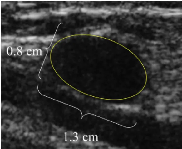
Size cannot be used as the sole criterion in differen-tiating benign (normal and reactive) from malignant (metastatic or lymphomatous) lymph nodes (Fig. 2).3,13–17Microscopic metastatic deposits in
axillary lymph nodes, beyond the resolution of existing imaging technology, occur in 9% of patients11 and therefore it is unlikely that any
imaging technique will have a sensitivity greater than 91%. While larger nodes have a higher inci-dence of malignancy (80% positive predictive value [PPV] if the long axis is >2 cm), reactive nodes can be large and malignant nodes can be small. There is a significant overlap in size between benign (Fig. 3) and malignant lymph nodes, and attempting to differentiate based solely on maximal size is unreli-able. There is less overlap using short axis rather than long axis dimensions (90% PPV if short axis is >1 cm).15,18,19Using a smaller cutoff value can
Fig. 1.Normal axillary lymph node anatomy.
Fig. 2. Small (long axis <2 cm and short axis <1 cm) malignant axillary lymph node.
give very high sensitivities but at the expense of a lower specificity.20
Shape
Malignant lymph nodes, including nodes involved by lymphoma, tend to be round, whereas normal and reactive nodes tend to be oval or elliptical. The degree of roundness is assessed by using the longest-to-shortest axis ratio (L/S). Reported sensitivities for malignancy using an L/S ratio <2 (ie, more rounded) are approximately 85%, with specificities varying from 61% to 85%.3,5,21 The
reported average cancer content by volume in lymph nodes with an L/S ratio >2.0 was 26%, compared with 59.1% in lymph nodes with an L/S ratio <2.0.3,5,22,23 Malignant lymph nodes are,
however, frequently oval (Fig. 4).
Echogenic Hilum
Previously, the presence or absence of a central echogenic hilum had been proposed as a reliable indicator of benignity or malignancy.3,4,21,23,24
While the absence of a hilum is very suggestive of malignancy, the presence of a hilum does not
necessarily imply benignity.3,24,25The hilum may
be absent in 14% of normal lymph nodes,26and
up to 30% of malignant lymph nodes may retain their hili.26As tumor infiltrates the hilum there is
loss of the normal echogenicity, resulting in apparent narrowing (hilar compression), hilar displacement (the hilum, rather than lying in the center of the node, lies to one side of the node in at least one plane), and subsequent disappearance of the central hilum (Fig. 5).5In some benign lymph
nodes, the node is largely replaced by a hypere-choic hilum with no visible cortex demonstrable. The lymph nodes of older patients or patients on chemotherapy may be small and have an isoechoic or a hyperechoic appearance.27
Cortical Morphology
The appearances of the hilum and cortex must be interpreted together. Concurrent changes in the shape of the central echogenic hilum and the peripheral concentric hypoechoic cortex may suggest the presence of nodal disease even in the absence of nodal enlargement.3 The normal
cortical rim measures 1 to 2 mm (Fig. 6).12,13
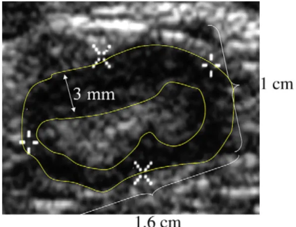
Neoplastic involvement of the cortex may not affect the cortical echogenicity, but may result in concen-tric or eccenconcen-tric cortical widening (Fig. 7).5,15
Malig-nant lymph nodes may demonstrate cortical thickening with or without hilar displacement (Figs. 8 and 9). Eccentric cortical hypertrophy measuring greater than 2 mm has been reported Fig. 3. Large (long axis >2 cm) benign axillary lymph
node.
Fig. 4. Rounded and oval malignant axillary lymph nodes.
Fig. 5. Malignant axillary lymph node. A rounded (L/S ratio <2), hypoechoic (no visible hilum), 1.5-cm (short axis >1 cm) lymph node. This combination of sono-graphic features is highly suggestive of malignancy.
as suggestive of malignancy.13It has also been
re-ported that eccentric cortical widening occurs only in malignancy,3 and that focal doubling of the
cortical rim thickness is specific for malignancy. Eccentric cortical widening may be due to focal nodular areas of intranodal metastatic disease.23
Other studies have used differing cortical thickness cutoff values (3 mm, 4 mm, 5 mm, and 6 mm) with the usual trade-off of decreasing specificity with increasing sensitivity and vice versa. Although a lymph node with a narrow or concentrically wide cortex is generally felt to be benign, there are reports of concentric widening in malignant lymph nodes.5
Echogenicity
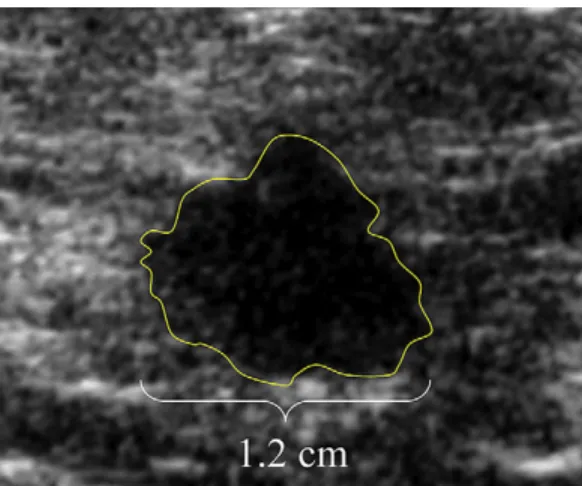
There have been several reports looking at the internal echo patterns in lymph nodes. Benign nodes are typically reported as being homogeneous, whereas malignant lymph nodes are typically heterogeneous and hypoechoic (Fig. 10).11,19,26,28–31 However, malignant nodes
may be homogeneous17,26,28 and benign lymph
nodes may be hypoechoic.19Alterations in
echoge-nicity in benign nodes may be due to infection or hemorrhage, and these findings may be mistaken for metastatic disease.29Hyperechoic nodes are Fig. 6.Benign axillary lymph node. A small oval lymph
node (long axis <2 cm, short axis <1 cm) with a thin cortex (<3 mm) and a central hilum (no compression or displacement). This combination of sonographic features is highly suggestive of benignity.
Fig. 7.Malignant axillary lymph node. A small lymph node (long axis <2 cm, short axis >1 cm) with an asym-metrically thickened (>3 mm) cortex with hilar displacement (although no hilar compression). This combination of sonographic features is suggestive of malignancy.
Fig. 8. Malignant axillary lymph node. A small lymph node (long axis <2 cm, short axis >1 cm) with an asym-metrically thickened (>3 mm) cortex with hilar compression (although no hilar displacement). This combination of sonographic features is suggestive of malignancy.
Fig. 9. Malignant axillary lymph node. A small lymph node (long axis <2 cm, short axis >1 cm) with an asym-metrically thickened (>3 mm) cortex with hilar compression and hilar displacement. This combination of sonographic features is suggestive of malignancy.
typically believed to be inflammatory,31 although
focal hyperechoic areas can be seen in malignant nodes.17Lymphomatous nodes may demonstrate a pseudocystic appearance with hypoechogenicity and posterior enhancement (Fig. 11). True cystic change and/or intranodal necrosis is unusual in lymphoma except after treatment. A micronodular pattern has also been reported in lymphoma. Microcalcifications in lymph nodes have been described in the neck in the setting of papillary thyroid cancer. Calcifications in axillary nodes are less commonly reported, but these findings have been reported in treated lymphoma.
Nodal Borders
Benign nodes often have indistinct borders where-as malignant nodes tend to have well-defined borders,26,28,30,31 due to altered echogenicity in
the replaced node compared with the surrounding tissue. However, with local infiltration, the borders of malignant lymph nodes may become indis-tinct.19,23,25Nodal borders are therefore not reliable
predictors of benignity or malignancy.
Vascular Patterns
Small nodes (benign or malignant) may have no demonstrable blood flow because the vessels are small, but flow is demonstrated in 90% of nodes measuring greater than 5 mm. In metastatic lymph nodes, angiogenesis factors may stimulate the growth of new vessels with thin walls, leading to high systolic and diastolic flow and abnormal vascular shunting, resulting in abnormal flow patterns and angioarchitecture.
Normal and reactive lymph nodes may be avas-cular or have only hilar vasavas-cularity. Mixed hilar and peripheral vascularity is associated with lymphoma, whereas pure peripheral vascularity is reportedly more suggestive of metastatic disease.19,21,32,33Tumor deposits within a lymph
node may compress the intranodal vessels, with resultant increased vascular resistance.21,32,34,35
Yang and colleagues36evaluated the reliability
of unenhanced and echo-enhanced color Doppler ultrasonography in distinguishing benign breast masses and axillary lymph nodes from malignant masses and axillary lymph nodes in patients with known breast cancer. Thirty-two enlarged axillary lymph nodes in 32 patients with invasive cancer underwent power Doppler sonography with and without contrast material. Vascular features and contrast material transit times were recorded. The investigators found that the significant predic-tors of lymph node malignancy were an increase in peripheral vessel number after contrast material administration and duration of enhancement. Yang and colleagues also found that malignant lymph nodes were enhanced more than the corre-sponding primary breast cancers, whereas benign lymph nodes were enhanced less than the primary breast tumors.
PATTERNS
No single sonomorphological feature reliably differentiates benign, reactive, or malignant (meta-static or lymphomatous) lymph nodes. Combina-tions of features either as nodal patterns or as scoring systems better differentiate benign from malignant nodes (Fig. 12).5,19
Mills and colleagues37conducted a retrospective
study of 653 consecutive patients presenting with mixed histologic types of invasive breast cancer. The investigators performed 232 ultrasound-guided axillary lymph node biopsies, resulting in Fig. 10. Malignant axillary lymph node. An
ill-defined, lobulated, hypoechoic (with no visible hilum), 1.2-cm (short axis >1 cm), rounded (L/S ratio <2) lymph node. This combination of sonographic features is suggestive of malignancy.
Fig. 11. Lymphomatous lymph node demonstrating hilar and peripheral vascularity.
a positive diagnosis in 150 cases. The morphologic criteria for metastatic involvement of lymph nodes on ultrasonography were diffuse or focal cortical thickening of more than 2 mm, replacement of the fatty hilum, and abnormal or increased periph-eral blood flow. Mills and colleagues found that axillary ultrasound assessment with selected fine-needle aspiration (FNA) or core fine-needle biopsy had a sensitivity of 59%, a specificity of 100%, a PPV of 100%, a negative predictive value (NPV) of 79%, and an accuracy of 84% in the diagnosis of axillary lymph node metastases.
Bedi and colleagues38performed high-resolution in vitro sonography on 171 lymph nodes from 19 axillae in 18 patients with unknown axillary nodal status who underwent axillary lymph node dissec-tion for early invasive breast cancer. Each lymph node was classified into 1 of 6 types based on the cortical morphologic features. Type 1 lymph nodes were hyperechoic with no visible cortex; type 2 had a thin (<3 mm) hypoechoic cortex; type 3 had a hy-poechoic cortex thicker than 3 mm; type 4 had a generalized lobulated hypoechoic cortex; type 5 had focal, hypoechoic cortical lobulations; and type 6 had a totally hypoechoic lymph node with
no hilum. Types 1 to 4 were considered benign whereas types 5 and 6 were considered metastatic. Interobserver agreement was 77% for classification of nodal morphology (types 1–6) and 88% for char-acterization of a lymph node as benign or malignant. The NPVs of types 1 to 4 were 100%, 100%, 93%, and 89%, respectively. The PPVs of types 5 and 6 were 29% and 58%, respectively. Sensitivity, spec-ificity, PPV, NPV, and overall accuracy for cortical shape in the prediction of metastatic involvement of axillary lymph nodes were 77%, 80%, 36%, 96%, and 80%, respectively.
Cho and colleagues39 prospectively evaluated the role of axillary lymph node classification on sonography in 191 patients. The axillary lymph node that had the thickest cortex was prospec-tively classified on a scale of 1 to 6 according to the cortical thickness and then removed following sonographically guided needle localization and surgical excision. The rates of malignancy, ac-cording to the sonographic classification, were as follows: 2% for grade 1 (cortical thickness <1.5 mm), 6% for grade 2 (cortical thickness >1.5 mm and2.5 mm), 40% for grade 3 (cortical thick-ness >2.5 mm and 3.5 mm), 70% for grade 4 Fig. 12.Lymph node patterns.
(cortical thickness >3.5 mm with an intact fatty hilum), and 90% for grade 5 (cortical thick-ness >3.5 mm with loss of the fatty hilum). When a cutoff point of a cortical thickness of 2.5 mm was used for determining the presence of malig-nancy, the sonographic classification showed a sensitivity of 85% (35/41), a specificity of 78% (117/150), an NPV of 95% (117/123), a PPV of 51% (35/68), and an accuracy of 80% (152/ 191) for the diagnosis of axillary lymph node metastases.
Combining sonographic features such as the presence or absence of a hilum, hilar compression, hilar displacement, smooth or lobulated borders, short axis size, and cortical thickening, it is possible to predict with reasonable accuracy those lymph nodes that are suspicious for malignancy (Fig. 13).
LYMPH NODES AFFECTED BY BREAST CANCER
Axillary, infraclavicular, internal mammary, and supraclavicular lymph nodes are located in close proximity to the breast, and these lymph nodes are the most commonly affected nodes in patients with breast cancer. In addition, intramammary nodes may be involved by breast cancer and by lymphoma.
Metastases from Breast Cancer
When cancer metastasizes, nearby lymph nodes are usually affected earlier than distant lymph nodes. Regarding breast cancer, the malignancy metastasizes first to the nearby axillary lymph nodes then to more distant axillary lymph nodes.40
Thus, metastases to lymph nodes are viewed as indicators of tumor progression. Nodal status is also considered a marker of tumor biology, with node-positive tumors having a worse prognosis than node-negative tumors.40,41Furthermore,
ac-cording to studies by Jatoi and colleagues40and
Nouh and colleagues,41 there is a correlation
between the number of lymph nodes involved and the aggressiveness of the cancer. The total number of lymph nodes involved is more important than the extent to which the disease has spread within the nodes.42
As breast cancers increase in size, the likelihood of axillary lymph node involvement increases. In a study of 3747 mastectomy specimens by Nouh and colleagues,41 71.6% of T1 (2 cm) tumors
metastasized to lymph nodes, along with 75.4% of T2 (2–5 cm) tumors, and 85% of T3 (>5 cm) tumors. Multiple tumors were almost twice as likely as single tumors to result in lymph node metastases (24.1% vs 12.4%, respectively).
Tumor grade is a measure of the amount of differ-entiation in the cancer cells of the tumor, with grade 1 being the most differentiated with a better prog-nosis, and grade 3 being the least differentiated with a worse prognosis. Node positivity showed a marked increase with an increase in tumor grade, as 49.3% of grade 1 tumors were node positive compared with 76.8% of grade 3 tumors.41 A surprising finding in the study by Nouh and colleagues41 was the effect of the laterality of
breast cancer on node positivity. Left-sided breast cancer was less prone to cause metastasis to lymph nodes in comparison with right-sided breast cancer. This conclusion may be explained by the more frequent use of the right arm in the predomi-nantly right-handed population.
LYMPHOMA
Lymphoma is the most common type of blood-related malignancy in the United States. Often the first sign of lymphoma (Fig. 14) is lymphade-nopathy, or swelling of the lymph nodes. The swelling is initially painless and is usually located in the neck, the axillae, or the groin. There are two major types of lymphoma, namely Hodgkin lymphoma and non-Hodgkin lymphoma, and more than 30 subtypes. Hodgkin lymphoma develops from a specific type of abnormal B cell, whereas non-Hodgkin lymphoma may derive from abnormal B or T cells. Risk factors for lymphoma include chronic infection, immunosup-pression, hereditary traits, and autoimmune disease. Autoimmune disease constantly stimu-lates the immune system, and thus can potentially give rise to irregular cloning of autoimmune cells. Fig. 13. Malignant axillary lymph node. A large (long
axis >2 cm, short axis >1 cm) lymph node with an asymmetrically thickened (>3 mm) cortex with hilar compression and hilar displacement. This combination of sonographic features is highly suggestive of malignancy.
The diagnosis of lymphoma is often based on lymph node biopsy.43
The differentiation of metastatic lymph nodes from lymphomatous nodes (Fig. 15) can be diffi-cult. It has been suggested that within the axilla, lymphoma tends to involve all the nodes in a rela-tively uniform fashion, whereas with carcinoma the lymph node morphology may be different, reflect-ing differential nodal involvement. With advanced disease, the lymphomatous nodes often become matted together.
SENTINEL LYMPH NODE BIOPSY
The sentinel lymph node is the first lymph node or group of nodes that are expected to be affected
by breast cancer metastases. Because the spread of cancer usually follows an orderly progression, a negative sentinel lymph node means that it is unlikely that the cancer has spread to any other, more distant nodes. To assess the sentinel lymph nodes, a sentinel lymph node biopsy is performed. Sentinel lymph node biopsy is advantageous, as it decreases the number of axillary lymph node dissections.44Axillary lymph node dissections are more likely to cause postoperative problems such as lymphedema, pain, impaired shoulder mobility, and arm weakness.45Furthermore, by identifying
the nodes most likely to contain metastases, more attention can be paid to the specific nodes, and micrometastases will have a higher likelihood of being detected.44 In sentinel lymph node
mapping, a radioisotope (usually technetium-99m sulfur colloid), a blue dye (isosulfan blue or methy-lene blue), or both, are injected before the biopsy is performed. These mapping agents aid in the detection of the sentinel lymph nodes. Studies have shown that the use of both mapping agents yields higher sentinel lymph node identification rates when compared with the use of a single agent.46During the procedure, the surgeon uses a gamma probe to detect which nodes have taken up the most radioactive material. These nodes, along with the lymph nodes that have taken up the blue dye, are the sentinel lymph nodes. The pathologist then assesses the sentinel lymph nodes to determine the presence of cancer. Most sentinel lymph nodes are located in the inferior aspect of the axillary region.45
There are also shortcomings to sentinel lymph node biopsy. There may be false-negative results. Also, cancers may drain by alternative pathways, to the internal mammary or infraclavicular lymph Fig. 14.Lymphomatous involvement of an internal mammary node. (A) Ultrasonography shows an oval hypoe-choic internal mammary lymph node (arrow). (B) Computed tomography (CT) shows the suspicious internal mammary lymph node (arrow) and additional prevascular and paratracheal lymph nodes.
Fig. 15. Sonography demonstrates a 20-mm lympho-matous axillary node with a cortical thickness of 7 mm (arrow). The appearance is not specific for lymphoma, and these findings could also represent metastatic disease in a patient with breast cancer.
nodes rather than to the axillary lymph nodes. Increasing patient age also affects lymphatic mapping, as sentinel lymph nodes may be more difficult to identify and appear less frequently in older patients.46
ULTRASONOGRAPHY WITH CONTRAST AGENTS
Recent reports have noted that sentinel lymph nodes may be identified and localized with contrast-enhanced sonography after the injection of microbubbles. Contrast-enhanced ultrasono-graphy is a developing technique that adds the injection of a contrast agent to traditional sonography.47Most studies have used gas-filled
microbubbles, and the contrast agent may be administered by intradermal, subareolar, or intra-venous injections. Studies targeting sentinel lymph nodes have been performed with intradermal, peri-tumoral, and subareolar injections.48–50The
micro-bubbles have a high degree of echogenicity compared with normal tissue, creating increased contrast in the resulting sonographic images. Lymph node sonography is an area in which the addition of contrast agents may be beneficial, especially regarding identification of sentinel lymph nodes. Contrast-enhanced sonography has the ability to increase the specificity of ultra-sound. As ultrasonography with contrast agents advances in the future, its advantages and limita-tions will be better understood.
PERCUTANEOUS BIOPSY PROCEDURES
In addition to its role in identifying normal and abnormal lymph nodes, sonography plays a major role in guiding biopsies of suspicious lymph nodes. Nearly all biopsies of the regional (axillary, infracla-vicular, supraclainfracla-vicular, and internal mammary) lymph nodes are performed with sonographic guidance. Ultrasound-guided FNA may be per-formed when adequate cytology support is available. If appropriate cytology support is unavail-able, core needle biopsy is suggested. Percuta-neous axillary lymph node biopsy determines whether the patient proceeds to sentinel lymph node biopsy or to axillary dissection. Patients with negative ultrasonograms and/or negative ultrasound-guided axillary lymph node biopsies proceed to sentinel lymph node biopsy, while those with metastatic disease documented by percuta-neous biopsy will undergo axillary dissection. In addition, patients with proven axillary lymph node involvement will usually be treated with neoadjuvant or adjuvant chemotherapy.
Fine-Needle Aspiration
FNA employs a thin needle (18–25 gauge) to biopsy breast masses and regional lymph nodes. Ultrasonography is used to detect the lymph node in question, and the needle is then inserted into the node and moved in a back-and-forth motion to obtain cellular material under sono-graphic guidance. Once the cells are extracted, they are stained and evaluated by a cytologist.
In a study by Kuenen-Boumeester and
colleagues,51ultrasonography combined with
ultra-sound-guided FNA identified evidence of meta-static disease in 44% (37 of 85) of histologically node-positive patients and in 20% of the patients evaluated in the study. In these cases, cytology identified metastases in the lymph nodes, sparing the patient sentinel lymph node biopsy. In addition, FNA can document extensive metastatic involve-ment, which may be associated with false-negative sentinel lymph node biopsies.
There is a risk of false-negative axillary lymph node FNAs because the sampling size is small, potentially allowing tumor cells to be missed. Also, FNA may sometimes fail to identify any lymph tissue and instead demonstrate only blood, making the diagnosis inconclusive. In cases where no lymph tissue is obtained, the physician should perform additional passes to retrieve lympho-cytes. FNA is particularly useful for sampling deep lymph nodes in the axillary and infraclavicu-lar regions (Fig. 16). FNA is a minimally invasive procedure with a low cost and high specificity. When adequate cytology support is available, FNA is a reliable preoperative staging procedure that can eliminate unnecessary sentinel lymph node biopsies.51
Core Needle Biopsy
Core needle biopsy removes cores of tissue. Core needle biopsy samples are larger than the samples obtained with FNA. The larger samples allow pathol-ogists to evaluate abnormal cells in the context of the surrounding environment. Ultrasound-guided core needle biopsy is considered to be minimally invasive and safe. In a study by Topal and colleagues52 of 39 patients who underwent
ultrasound-guided axillary lymph node biopsy, the sensitivity and the specificity of ultrasound-guided core needle biopsy of axillary lymph nodes were 90% and 100%, respectively. No significant compli-cations were noted in this study other than pain, which responded to analgesics.
Core needle biopsy is considered a good alter-native to FNA when adequate cytology support is lacking. FNA is more dependent on operator expertise than is core needle biopsy. Core needle
biopsy, like FNA, can be used to document meta-static disease and to avoid sentinel lymph node biopsy. Core needle biopsy has a higher reproduc-ible success rate in comparison with FNA.45 SUMMARY
In the future, there will be increased emphasis on sonography of the regional lymph nodes in patients with breast cancer. Gray-scale sonog-raphy is an efficient, reliable tool in classifying regional lymph nodes. In addition, ultrasound-guided FNA and ultrasound-ultrasound-guided core needle biopsy are safe, quick, reliable, low-cost proce-dures that can be used to document metastatic involvement. Contrast-enhanced ultrasonography will likely improve clinicians’ ability to classify regional lymph nodes. In addition, contrast-enhanced ultrasonography has the potential to transform sentinel lymph node biopsy into a less invasive procedure.
ACKNOWLEDGMENTS
The authors thank Barbara Almarez Mahinda for assistance in manuscript preparation.
REFERENCES
1. Warwick R, Williams PL. Angiology. In: Warwick R, Williams PL, editors. Gray’s anatomy. 35th edition. Philadelphia: WB Saunders; 1973. p. 588–785. Chapter 6.
2. Willard-Mack CL. Normal structure, function, and histology of lymph nodes. Toxicol Pathol 2006;34:409–24. 3. Vassallo P, Wernecke K, Roos N, et al. Differentiation of benign from malignant superficial lymphadenop-athy: the role of high-resolution US. Radiology 1992;183:215–20.
4. Rubaltelli L, Proto E, Salmaso R, et al. Sonography of abnormal lymph nodes in vitro: correlation of sono-graphic and histological findings. Am J Roentgenol 1990;155:1241–4.
5. Vassallo P, Edel G, Roos N, et al. In-vitro high-resolution ultrasonography of benign and malignant nodes: a sonographic-pathological correlation. Invest Radiol 1993;28:698–705.
6. Kendall BE, Arthur JF, Patey DH. Lymphangiography in carcinoma of the breast. A comparison of clinical, radiological, and pathological findings in axillary lymph nodes. Cancer 1963;16:1233–42.
7. Fajardo LF. Lymph nodes and cancer: a review. In: Meyer JL, editor. The lymphatic system and cancer. Frontiers of radiation therapy and oncology, vol. 28. Basel (Switzerland): Karger; 1994. p. 1–10. 8. Bruneton JN, Carmella E, Hery M, et al. Axillary lymph
node metastases in breast cancer: preoperative detection with ultrasound. Radiology 1986;158:325–6. 9. De Freitas R Jr, Costa MV, Schneider SV, et al. Accu-racy of ultrasound and clinical examination in the diagnosis of axillary lymph node metastases in breast cancer. Eur J Surg Oncol 1991;17:240–4. 10. Mustonen P, Farin P, Kosunen O. Ultrasonographic
detection of metastatic axillary lymph nodes in breast cancer. Ann Chir Gynaecol 1990;79:15–8.
Fig. 16. Metastatic infraclavicular lymph node. A 55-year-old woman with known right breast cancer presented for staging. (A) In the right infraclavicular region a deep, suspicious, hypoechoic lymph node was noted. Ultrasound-guided right infraclavicular FNA with a 21-gauge needle (small arrow) was used to sample the deep lymph node (large arrow). (B) Cytology revealed a cluster of tumor cells, representing metastatic adenocar-cinoma (Papanicolaou stain, original magnification40).
11. Bonnema J, van Geel AN, van Ooijen B, et al. Ultra-sound-guided aspiration biopsy for detection of non-palpable axillary node metastases in breast cancer patients: new diagnostic method. World J Surg 1997;21:270–4.
12. Yang WT, Ahuja A, Tang A, et al. Ultrasonographic demonstration of normal axillary lymph nodes: a learning curve. J Ultrasound Med 1995;14:821–2. 13. Yang WT, Ahuja A, Tang A, et al. High resolution sono-graphic detection of axillary lymph node metastases in breast cancer. J Ultrasound Med 1996;16:241–6. 14. Chang DB, Yuan A, Yu CJ, et al. Differentiation of
benign and malignant lymph nodes with color Doppler sonography. Am J Roentgenol 1994;162:965–8. 15. Sutton RT, Reading CC, Charboneau JW, et al.
US-guided biopsy of neck masses in postoperative management of patients with thyroid cancer. Radi-ology 1988;168:769–72.
16. March DE, Wechsler RJ, Kurtz AB, et al. CT-Patho-logic correlation of axillary lymph nodes in breast cancer. J Comput Assist Tomogr 1991;15:440–4. 17. Maurer J, Willam C, Steinkamp HJ, et al. Keratinization
and necrosis: morphological aspects of lymphatic metastases in ultrasound. Invest Radiol 1996;31:545–9. 18. van den Brekel MW, Stel HV, Castelijns JA, et al. Cervical lymph node metastasis: Assessment of radiologic criteria. Radiology 1990;177:379–84. 19. Toriyabe Y, Nishimura T, Kita S, et al. Differentiation
between benign and metastatic cervical lymph nodes with ultrasound. Clin Radiol 1997;52:927–32. 20. Van den Brekel MW, Castelijns JA, Stel HV, et al. Occult metastatic neck disease: detection with US and US-guided fine-needle aspiration cytology. Radiology 1991;180:457–61.
21. Na DG, Lim HK, Byun HS, et al. Differential diag-nosis of cervical lymphadenopathy: usefulness of color Doppler sonography. Am J Roentgenol 1997; 168:1311–6.
22. Tohnosu N, Onoda S, Isono K. Ultrasonographic eval-uation of cervical lymph node metastases in esopha-geal cancer with special reference to the relationship between short to long axis ratio (S/L) and the cancer content. J Clin Ultrasound 1989;17:101–6.
23. Tregnaghi A, De Candia A, Calderone M, et al. Ultraso-nographic evaluation of superficial lymph node metas-tases in melanoma. Eur J Radiol 1996;24:216–21. 24. Yang WT, Metreweli C. Color Doppler flow in normal
axillary lymph nodes. Br J Radiol 1998;71:381–3. 25. Evans RM, Ahuja A, Metreweli C. The linear
echogen-ic hilus in cervechogen-ical lymphadenopathy-a sign of benig-nity or malignancy? Clin Radiol 1993;47:262–4. 26. Ahuja A, Ying M, King W, et al. A practical approach
to ultrasound of cervical lymph nodes. J Laryngol Otol 1997;111:245–56.
27. Spaulding K. Ultrasound imaging of the lymph nodes: normal & abnormal appearance. J Veterinary Radiology & Ultrasound 2008;49:277–81.
28. Ahuja A, Ying M, Yang WT, et al. The use of sonog-raphy in differentiating cervical lymphomatous lymph nodes from cervical metastatic lymph nodes. Clin Radiol 1996;51:186–90.
29. Sakai F, Kiyono K, Sone S, et al. Ultrasonic evaluation of cervical metastatic lymphadenopathy. J Ultrasound Med 1988;7:305–10.
30. Noritomi T, Machi J, Feleppa EJ, et al. In vitro inves-tigation of lymph node metastasis of colorectal cancer using ultrasonic spectral parameters. Ultra-sound Med Biol 1998;24:235–43.
31. Hildebrandt U, Feifel G. Endosonography in the diag-nosis of lymph nodes. Endoscopy 1993;25:243–5. 32. Steinkamp HJ, Maurer J, Cornehl M, et al. Recurrent
cervical lymphadenopathy: differential diagnosis with color-duplex sonography. Eur Arch Otorhinolar-yngol 1994;251:404–9.
33. Walsh JS, Dixon JM, Chetty U, et al. Colour Doppler studies of axillary node metastases in breast carci-noma. Clin Radiol 1994;49:189–91.
34. Choi MY, Lee JW, Jang KJ. Distinction between benign and malignant causes of cervical, axillary, and inguinal lymphadenopathy: value of Doppler spectral waveform analysis. Am J Roentgenol 1995; 165:981–4.
35. Mountford RA, Atkinson P. Doppler ultrasound exam-ination of pathologically enlarged lymph nodes. Br J Radiol 1979;52:464–7.
36. Yang WT, Metreweli C, Lam PK, et al. Benign and malignant breast masses and axillary nodes: evalu-ation with echo-enhanced color power Doppler US. Radiology 2001;220:795–802.
37. Mills P, Sever A, Weeks J, et al. Axillary ultrasound assessment in primary breast cancer: an audit of 653 cases. Breast J 2010;16:460–3.
38. Bedi DB, Krishnamurthy R, Krishnamurthy S, et al. Cortical morphologic features of axillary lymph nodes as a predictor of metastases in breast cancer: in vitro sonographic study. AJR Am J Roentgenol 2008;191: 646–52.
39. Cho N, Moon WK, Han W, et al. Preoperative sono-graphic classification of axillary lymph nodes in patients with breast cancer: node-to-node correla-tion with surgical histology and sentinel node biopsy results. AJR Am J Roentgenol 2009;193:1731–7. 40. Jatoi I, Hilsenbeck SG, Clark GM, et al. Significance
of axillary lymph node metastasis in primary breast cancer. J Clin Oncol 1999;17:2334–40.
41. Nouh MA, Ismail H, Ali El-Din NH, et al. Lymph node metastasis in breast carcinoma: clinicopathologic correlations in 3747 patients. J Egypt Natl Canc Inst 2004;16:50–6.
42. Lam WW, Yang WT, Chan YL, et al. Detection of axil-lary lymph node metastases in breast carcinoma by technetium-99m sestamibi breast scintigraphy, ultra-sound and conventional mammography. Eur J Nucl Med 1996;23:498–503.
47. Wilson SR, Greenbaum LD, Goldberg BB. Contrast-enhanced ultrasound: what is the evidence and what are the obstacles? AJR Am J Roentgenol 2009; 193:55–60.
48. Sever AR, Mills P, Jones SE, et al. Preoperative sentinel node identification with ultrasound using
cancer patients. A preoperative staging procedure. Eur J Cancer 2003;39:170–4.
52. Topal U, Punar S, Tasdelen I, et al. Role of ultrasound-guided core needle biopsy of axillary lymph nodes in the initial staging of breast carcinoma. Eur J Radiol 2005;56:382–5.