Eric G. Bend, PhD Yue Si, PhD
David A. Stevenson, MD Pinar Bayrak-Toydemir,
MD, PhD
Tara M. Newcomb, MS Erik M. Jorgensen, PhD Kathryn J. Swoboda, MD
Correspondence to Dr. Swoboda:
[email protected] or Dr. Jorgensen: [email protected]
Supplemental data at Neurology.org
NALCN channelopathies
Distinguishing gain-of-function and loss-of-function mutations
ABSTRACT
Objective:To perform genotype–phenotype analysis in an infant with congenital arthrogryposis due to a de novo missense mutation in the NALCN ion channel and explore the mechanism of pathogenicity using aCaenorhabditis elegansmodel.
Methods:We performed whole-exome sequencing in a preterm neonate with congenital arthrog-ryposis and a severe life-threatening clinical course. We examined the mechanism of pathogenic-ity of the associatedNALCNmutation by engineering the orthologous mutation into the nematode
C elegansusing CRISPR-Cas9.
Results: We identified a de novo missense mutation inNALCN, c.1768C.T, in an infant with a severe neonatal lethal form of the recently characterized CLIFAHDD syndrome (congenital contractures of the limbs and face with hypotonia and developmental delay). We report novel phenotypic features including prolonged episodes of stimulus-sensitive sustained muscular con-traction associated with life-threatening episodes of desaturation and autonomic instability, ex-tending the severity of previously described phenotypes associated with mutations inNALCN. When engineered into theC elegansortholog, this mutation results in a severe gain-of-function phenotype, with hypercontraction and uncoordinated movement. We engineered 6 additional CLIFAHDD syndrome mutations intoC elegansand the mechanism of action could be divided into 2 categories: half phenocopied gain-of-function mutants and half phenocopied loss-of-function mutants.
Conclusions:The clinical phenotype of our patient and electrophysiologic studies show sustained muscular contraction in response to transient sensory stimuli. InC elegans, this mutation causes neuronal hyperactivity via a gain-of-function NALCN ion channel. Testing human variants of NALCN inC elegans demonstrates that CLIFAHDD can be caused by dominant loss- or gain-of-function mutations in ion channel function.Neurology®2016;87:1131–1139
GLOSSARY
CLIFAHDD5congenital contractures of the limbs and face with hypotonia and developmental delay;CRISPR5clustered regularly interspaced short palindromic repeats;IHPRF5infantile hypotonia with psychomotor retardation and character-istic facies.
NALCN is a conserved cation channel related to voltage-gated sodium and calcium channels.
The NALCN family of channels is expressed throughout the nervous system in all metazoans
studied and conducts a persistent sodium leak current that contributes to tonic neuronal
excitability.
1,2Null mutations in orthologs of
NALCN
in mice, flies, and worms cause generalized paralysis
with disruption of periodic behaviors such as breathing, circadian rhythms, or rhythmic motor
circuits.
1–4Loss of these behaviors is likely caused by hyperpolarized membrane potentials in the
neurons of these mutants.
1,5Loss-of-function mutations in the human
NALCN
have been
From the Department of Biology and Howard Hughes Medical Institute (E.G.B., E.M.J.), and Department of Pathology (Y.S., P.B.-T.), University of Utah, Salt Lake City; ARUP Institute for Clinical and Experimental Pathology (Y.S., P.B.-T.), Salt Lake City, UT; Division of Medical Genetics (D.A.S.), Department of Pediatrics, Stanford University, CA; Department of Neurology (T.M.N.), Pediatric Motor Disorders Research Program, University of Utah School of Medicine, Salt Lake City; and Department of Neurology (K.J.S.), Massachusetts General Hospital, Boston. Go to Neurology.org for full disclosures. Funding information and disclosures deemed relevant by the authors, if any, are provided at the end of the article. The Article Processing Charge was paid by the authors.
described, which cause an autosomal recessive
condition resulting in infantile hypotonia
with psychomotor retardation and
character-istic facies (IHPRF [MIM #615419]).
6–8However, a new class of autosomal dominant
NALCN
mutations was identified in humans
with a novel phenotype characterized by
congenital distal arthrogryposis and pursed
facial expression suggesting a hypercontracted
phenotype
—
designated
congenital
contrac-tures of the limbs and face with hypotonia and
developmental delay
(CLIFAHDD syndrome
[MIM #616266]).
9,10Because there is a
signif-icant degree of phenotypic overlap in patients
with IHPRF, these mutations were
hypothe-sized to function as dominant negative proteins
that cause a loss-of-function phenotype.
How-ever, overexpressing a mutant
NALCN
ortho-log in
Caenorhabditis elegans
led one group to
conclude that the dominant channelopathy is
caused by gain-of-function
NALCN
.
11To determine whether CLIFAHDD
syn-drome is caused by gain or loss of
NALCN
function, we engineered the orthologous
mis-sense mutation from our patient and 6 other
published individuals into the
C elegans
genome by CRISPR-Cas9. We found that
the underlying pathologic mechanism of our
patient
’
s mutation is a gain-of-function
change in
NALCN
. However, the additional
mutations modeled in
C elegans
demonstrate
that the condition can be caused by both
gain-of-function and loss-gain-of-function changes in
the ion channel.
METHODS Family.The family described includes the pro-band, a preterm neonate born at 31 and 4/7 weeks’gestation, and her unaffected, nonconsanguineous parents of European heritage.
Standard protocol approvals, registrations, and patient consents.We obtained written informed consent for the collec-tion of blood samples for DNA extraccollec-tion. Counseling was per-formed before clinical whole-exome sequencing for all participants. The institutional review board at the University of Utah approved this study (IRB 25651, K.J.S.).
Exome sequencing and variant analysis. We performed diagnostic whole-exome sequencing in a trio using DNA extracted from whole blood. Genomic DNA was extracted using a Gentra Puregene Blood Kit (QIAGEN, Valencia, CA). Exons were captured with an Agilent SureSelect kit (Agilent Technologies, Inc., Santa Clara, CA) and sequenced with 23 100 base-pair paired ends on an Illumina HiSeq 2500 (Illumina, Inc., San Diego, CA). The sequences were aligned to Hg19 using the Burrows-Wheeler Aligner (0.5.11), and variants were called
with Genome Analysis Toolkit (v.1.6). More than 92.7% of bases sequenced had a quality score greater than 10 and variants with a quality score,10 were removed to avoid false positives. We excluded variants with an allele frequency greater than 1% in dbSNP (Single Nucleotide Polymorphism Database), 1000 Genomes Project, and 6500 Exomes. Further filtering removed synonymous variants, deep intronic variants, and those in 59and 39untranslated regions. We confirmed family relationships with short tandem repeat markers. TheNALCNvariant was confirmed by Sanger sequencing.
C elegans strains and genetics. Strains were cultured and maintained using standard methods.12All strains and plasmids
used in this study are listed (table e-1 at Neurology.org). CRISPR-Cas9 repair templates were made by Gibson cloning13
into pBluescript including: 1- to 3-kb homology arms with the orthologous NALCN mutation in nca-1, a loxP-flanked unc-119(1) positive selection cassette (intron 10), and a silent mutation in the PAM recognition site. Guide RNA constructs were made by Golden-Gate oligo annealing. Plasmid DNA was injected into the germline of unc-119(ed3) animals including Cas9-pDD162 (30 ng/mL),14 repair template (30 ng/mL),
guide RNA (30 ng/mL), and coinjection markers for negative selection of extrachromosomal arrays. CRISPR events were detected byunc-119rescue, survival followingpeel-1expression, and the absence of red fluorescence expression. The wild-type control was made by isolating a CRISPR event that included the unc-119(1)selection, but no mutation. Theunc-119(1) selection marker was excised from the genomes of all strains by injection of CRE recombinase-pDD10414 with pBluescript
(50 ng/mL) and coinjection markers.
C elegansanalysis.Sequence alignment and amino acid posi-tions are based on the rescuing isoform,nca-1d(wormbase release WS247). Images were captured on a Zeiss Axioskop compound microscope with a 203air objective (Carl Zeiss Microscopy, LLC, Thornwood, NY). Tracks were generated by animals crawling for 3 minutes on fresh OP50 plates. We photographed the tracks on a dissecting scope and traced them in Adobe Photoshop (Adobe Systems Inc., San Jose, CA). Behavioral scoring was conducted blind on plates staged with 10 L4-larva 24 hours before the scoring. All strains included in this study were scored together with replicates randomly mixed. Aldicarb plates were prepared with 2 mM aldicarb in agar. All strains were assayed together in a blinded experiment with a replicate from each strain included for 6 trials. For a trial, 10 animals were placed per plate and paralysis was assessed every 15 minutes. Paralysis was scored based on complete cessation of movement and lack of response to a nose tap.
RESULTS Clinical assessment indicates sustained neuronal activity resulting in muscular hypercontraction.Prenatal/ neonatal course.The proband is a female infant born at 31 and 4/7 weeks of gestation, delivered via C-section following detection of polyhydramnios and abnormal fetal heart tracings. Respiratory distress was present from birth. Intubation was attempted but unsuccessful because of small mouth, microretrognathia, and jaw contracture. She ultimately required intubation in the operating room with complete paralysis. She was hypertensive from birth.
Abnormal motor activity.She demonstrated rhythmic hypercontraction of arms and legs described as cycling, facial grimacing, and eyelid myotonia with inability to relax for up to several minutes. Such epi-sodes occurred up to 803 per day, and treatment trials of carbamazepine, phenobarbital, levetiracetam, or phenytoin were ineffective; however, clonazepam modestly lessened the frequency and severity of rhythmic limb activity. With severe episodes, she developed whole body rigidity and oxygen desatura-tion despite ventiladesatura-tion.
Autonomic instability.Over the ensuing weeks, she experienced recurrent life-threatening autonomic cri-ses associated with apnea, bradycardia, worsening hypertension, hyperthermia, and recurrent desatura-tion events in response to routine handling including suctioning or repositioning.
Electrophysiologic studies. Central nervous system. EEGs were performed on 4 occasions, followed by several days of continuous bedside monitoring for seizure activity. EEGs revealed abnormal background activity
characterized by occasional rhythmic slowing. How-ever, we observed no epileptiform activity during spells of rhythmic leg cycling, apnea, bradycardia, hypertension, or increased muscular tone.
Peripheral nervous system.EMG and nerve conduction testing demonstrated severely diminished ulnar, median, and peroneal compound muscle action potential amplitudes (,10% expected normal values) with preserved median sensory response. Needle insertion in both proximal and distal muscles during EMG elicited abnormal persistent motor unit recruit-ment for up to 5 minutes in association with sus-tained visible muscular contraction, despite the absence of further needle movement. Occasional neu-romyotonia and complex repetitive discharges were observed, but were limited to distally innervated intrinsic hand muscles. No myotonia was observed in limb muscles, even after distal cooling. We noted moderately reduced recruitment of relatively normal-appearing motor units in distally innervated muscles, but recruitment in more proximal muscles appeared
Figure 1 Clinical features in the proband
normal. Overall, the pattern of abnormalities on EMG and nerve conduction studies supported a pre-dominantly neurogenic process most severely affect-ing distally innervated limb muscles. However, the most striking clinical abnormality appeared to be a markedly delayed relaxation of muscles following activation resulting in involuntary sustained muscular contraction most predominantly affecting the masse-ter, perioral, and periocular facial muscles, laryngeal, chest, and proximal upper extremity muscles. One episode during EMG testing resulted in involuntary jaw closure and laryngeal muscular hyperactivity asso-ciated with desaturation persisting for a full 5 minutes. During this time, motor unit appearance and recruit-ment in the masseter muscle appeared entirely nor-mal, but was involuntarily sustained, indicating possible motor neuron/axon hyperexcitability.
Neuroimaging studies.Brain MRI revealed punctate and linear nonhemorrhagic foci in the periventricular white matter on T2 fluid-attenuated inversion re-covery and T1-weighted images, consistent with prematurity-associated injury. Magnetic resonance angiography and magnetic resonance spectroscopy were normal.
Clinical overview. Initial differential diagnoses included a variant of Freeman-Sheldon syndrome (FSS or DA2A)15based on the pattern of limb con-tractures and pursed facial appearance, or Stuve-Wiedemann syndrome.16Clinical genetic testing for mutations inMYH3 andLIFR were negative. At 2 months of age, tracheostomy and gastrostomy tubes were placed, but recurrent crises continued. Because of her increasing distress and inability to maintain sleep, comfort measures were instituted and respira-tory support was withdrawn at 4.5 months of age. The patient died quickly following extubation and the parents declined further studies.
Exome sequencing identified a heterozygous mutation in
NALCN. Exome sequencing of the proband and pa-rents identified a de novo mutation in NALCN (c.1768C.T; p.Leu590Phe) (figure e-1). The tion was novel at the time, but the exact same muta-tion has been published since then in an unrelated patient with CLIFAHDD syndrome.9The mutation maps to the S6 segment in the second domain— a region that forms the channel gate in related voltage-gated sodium channels (figure 2, A and B).17,18 This mutation could create a constitutively open channel that results in a dominant gain-of-function defect. In addition, a duplication of approximately 409 kb at 4q32.3 was detected by SNP microarray. The genes in this region are not known to be associated with disease, and given the small size of the duplication, is unlikely to contribute to the patient’s phenotype.
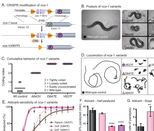
Validation of pathogenicity of NACLN mutation in C elegans.To test this model, we turned to C elegans, in which both loss-of-function and gain-of-function alleles have been characterized in the orthologous NCA channels.19 There are 2 redundant NALCN family members expressed in C elegans, nca-1 and nca-2. Loss-of-function mutations in both homologs causes a recessive phenotype: animals have normal body posture but when stimulated by touch, crawl away then suddenly halt in a stereotyped manner termed “fainting.” By contrast, gain-of-function variants in nca-1 are dominant and result in hypertonia, smaller body size, and curly posture (figure 3B).19,20
We engineered the patient’sNALCNmutation (L590F) into the orthologous position in the C elegans gene using the CRISPR-Cas9 system to generate NCA-1(V637F) in the native locus (fig-ure 3A). Animals with this single residue change resembled the characterized gain-of-function mutation NCA-1(A643V), which results in a con-stitutively open channel.19 Moreover, the strain with the human mutation was indistinguishable from the very severe gain-of-function mutant NCA-1(D647E). All 3 of these missense muta-tions led to small body size and curly posture com-pared to the wild-type control (figure 3B). We made the control strain by the same strategy as the mutant; however, no mutation was intro-duced. The mutants are easily distinguishable from the wild-type control in a blinded behavioral scor-ing assay (figure 3C). Given 3 minutes on fresh food, wild-type animals will explore a distance greater than 20 mm. In contrast, animals with the A643V, D647E, or V637F mutation display dramatically reduced locomotion (figure 3D). All 3 variants exhibit semidominant inheritance; het-erozygous animals exhibit an intermediate pheno-type (figures 3, B and D).
redundant. The D647E and V637E mutations are especially severe, and the time on aldicarb required to paralyze 50% of the worms (t1/2 for paralysis) is significantly shorter than the wild-type control (figure 3F). The A643V strain displayed a relatively weak hypersensitivity; however, the rate of paralysis (slope) was significantly greater than the wild-type control (figure 3G). Together, these findings support the clas-sification of L590F in humanNALCNas a dominant gain-of-function, pathogenic mutation.
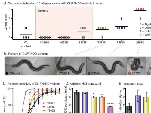
Different CLIFAHDD mutations cause gain or loss of channel function in C elegans. To test whether all CLIFAHDD mutations cause a gain-of-function channel, we engineered 6 previously described missense changes intoC elegans nca-1(figure 2A).9 We used the same CRISPR-Cas9 mutagenesis strategy. Including our patient’s mutation, 7 of the 14 previously described CLIFAHDD mutations were tested. By scoring the cumulative behavior of the animals, we observed a spectrum of phenotypes
ranging from wild-type to severely coiled (figure 4A). When thesenca-1mutations were crossed into thenca-2(2)null background, 3 strains that appeared wild-type (V359G, E373K, Y625S) proved to be fainters (genotype nca-1[*/*] nca-2[2/2]). The fainting behavior was also observed when the mutations were heterozygous (genotype nca-1[*/1] nca-2[2/2]), indicating that they are antimorphs. Therefore, these mutations disrupt NCA-1 channel function in a dominant manner and should be classified as loss-of-function. The remaining 4 mutations displayed no fainting behavior in the nca-2(2)null background; rather, the coiling behavior became subjectively more dramatic. The spectrum of postures observed in the strains with a noticeable phenotype is displayed (figure 4B). When assayed for aldicarb sensitivity, this subset of CLIFAHDD mutations caused a spectrum of hypersensitivity. The strains form an allelic series of severity based on the t1/2 and the rate (slope) of paralysis, which resembles that determined by behavior
Figure 2 Dominant pathogenic missense mutations in humans andCaenorhabditis elegansmap to the S5 and S6 segments
(WT,F559V,T580N,L556S,V637F) (figure 4, C–E). In contrast, the mutations identified as fainters had no effect on aldicarb sensitivity (figure e-2). In summary, our results suggest that mutations responsible for CLIFAHDD syndrome
may disrupt NALCN channel function by increasing or decreasing ion channel function.
DISCUSSION One problem emerging from diagnos-tic next-generation sequencing is the classification of
Figure 3 The proband’sNALCNvariant is pathogenic inCaenorhabditis elegansand phenocopies a known gain-of-function allele
(A) CRISPR modification ofnca-1. The V to F missense mutation was made in the nativenca-1gene withunc-119rescue for positive selection. Cre recombinase was injected to remove the loxP flankedunc-119selection cassette. (B) Posture ofnca-1variants. The wild-type control moves in a shallow sinusoidal wave. Animals engineered with the proband’s variant (V637F, fuchsia circle) exhibit a characteristic coiled posture identical to animals with previously identified gain-of-function alleles (D647E, dark pink and A643V, light pink). All 3 variants are semidominant and move with deeper body bends than the wild type. The color and symbol prompts are consistent throughout the subsequent panels. (C) Cumulative behavior ofnca-1variants. Ten L4 larvae were assayed per plate with 6 replicate plates per strain. The plates were scored blind after 24 hours. Each vertical bar represents a plate; the horizontal bars represent the mean. Scores of 0 to 3 are described in the panel. (D) Locomotion ofnca-1variants. Animals were placed on fresh plates with food for 3 minutes. Wild-type animals explore large areas quickly, while the V637F, D647E, and A643V strains cover very little area with uncoordinated locomotion. (E) Aldicarb sensitivity ofnca-1variants. Aldicarb blocks the degradation of acetylcholine in the synaptic cleft. Animals expressing the human variant (V637F) and the characterized gain-of-function variants (D647E and A643V) are paralyzed more quickly in aldicarb. All strains in this study were scored together, blinded, in 6 replicate experiments. Means are the average of the 6 replicates. N(plates)56; n(animals)560. (F) Aldicarb—time to one-half animals paralyzed (t1/2). For the gain-of-function mutation (D647E) and the proband’s mutation (V637F), the increase in sensitivity to aldicarb resulted in a 50% decrease in the t1/2 compared to the wild-type control (wild-type mean t1/2570.864.9 minutes, n572; D647E mean t1/2533.560.6 minutes, n572,p,0.0001; V637F mean t1/2535.860.7 minutes, n572,p,
0.0001; ordinary 1-way ANOVA with Dunnett multiple comparison correction). (G) Aldicarb—slope. Strains with the gain-of-function mutation (A643V) and the proband’s mutation (V637F) displayed an increase in the rate of paralysis as measured by the slope of the line compared to the wild-type control (wild-type slope
novel genetic variants. This difficulty is compounded by phenotypic variation due to genetic background. A possible solution is to compare novel variants in a model organism with a fixed genetic background. Here, we used the nematode C elegansto test 7 de novo mutations in the human NALCN gene responsible for CLIFAHDD syndrome. We found that all 7 variants were deleterious with dominantly inherited phenotypes. Thus, the worm appears to be a good model for CLIFAHDD—all mutations result in measurable phenotypes with the same inheritance pattern observed in humans.
Pathogenic mutations inNALCNcan be catego-rized by different modes of inheritance. IHPRF syndrome is recessive and appears to be caused by null mutations since they truncate the NALCN protein.6–8CLIFAHDD syndrome is dominantly in-herited and caused by pathogenic missense mutations inNALCN.9Previous reports disagree on the mech-anism of CLIFAHDD suggesting it may result from protein loss- or gain-of-function.9–11 Our functional assays inC elegans demonstrate that both outcomes are possible. We identified a subset of CLIFAHDD mutations with well-characterized gain-of-function
Figure 4 CLIFAHDD mutations result in gain and loss of channel function inCaenorhabditis elegans
(A) Cumulative behavior of CLIFAHDD variants. Scores of 0 to 3 demonstrate the pathogenic severity of each mutation as indicated. When crossed into thenca-2(2)null background, the mutations highlighted in light pink displayed classic“fainting”behavior. (B) Posture of CLIFAHDD variants. The NCA-1 (T560N) and (F559V) strains have a subtle change in body posture with slightly deeper body bends than the wild type. The NCA-1(L556S) and (V637F) strains are strongly coiled. (C) Aldicarb sensitivity of CLIFAHDD variants. The uncoordinated/coiled subset of CLIFAHDD mutations causes aldicarb hypersensitivity with a spectrum of severity. The mutations determined to be loss-of-function by fainting behavior are not hypersensitive to aldicarb (figure e-2). (D) Aldicarb—time to one-half animals paralyzed (t1/2). The spectrum of aldicarb hypersensitivity is evident in the t1/2. The F559V mutant (t1/2571.062.8 minutes, n572) was indistinguishable from the wild-type control (t1/2570.864.9 minutes, n572). The T580N (t1/2559.56
phenotypes. These mutations cluster around the gate of the ion channel and likely increase current. By contrast, we also observed a class of mutations that act as dominant-negatives (antimorphs) in the nem-atode. Animals with these mutations display a stereo-typed “fainting” behavior. Antimorphic channels likely cause the aggregation and degradation of wild-type NALCN, similar to mutations described in related sodium and calcium channels.22,23Thus, 3 mechanisms give rise to NALCN channelopathies: (1) IHPRF— recessive loss-of-function, (2) CLIFAHDD—dominant gain-of-function, and (3) CLIFAHDD—dominant antimorphic.
Unfortunately, the human phenotypes that result from the different genetic mechanisms of NALCN pathology are not so easily placed into these categories. IHPRF and CLIFAHDD “syndromes” are characterized by dysmorphic features and neuro-developmental disease with a significant number of
shared features. Both IHPRF and CLIFAHDD pa-tients display regional hypotonia and intellectual disability, and members of each group experience seizures. The most consistent feature attributed exclusively to CLIFAHDD syndrome is distal ar-throgryposis (reported in 18 of 19 patients described to date) (figure 1).9–11 Distal contractures were not noted in the 11 patients described with IHPRF.6–8 Our results complicate this picture further, since it appears that gain- or loss-of-function mutations in NALCNcan lead to a CLIFAHDD diagnosis. The-se results are consistent with the overlap between CLIFAHDD and IHPRF but at odds with the dis-tinctive feature such as arthrogryposis. Given that NALCN is expressed in excitatory and inhibitory neurons, both sides of a balanced circuit will be affected by functional changes to the channel. Therefore, the motor output of a given circuit is difficult to predict and likely reflects homeostatic limits to the system. Therefore, the degree of phe-notypic overlap between these syndromes may not be surprising.
The phenotypes observed in CLIFAHDD and IHPRF do suggest that the NALCN ion channel functions very broadly in the nervous system. Sus-tained muscular hyperactivity and failure of relaxa-tion of limb and cranially innervated muscles, in conjunction with the observed rhythmic cycling activity of the limbs, suggest that this ion channel functions in both upper and lower motor neurons and associated circuitry in the peripheral nervous system as well as the CNS. Hyperexcitability of the motor unit and the resulting overactivity of muscles innervating both limbs and cranial struc-tures, if present during fetal development, help to explain the characteristic dysmorphology and dis-tally predominant congenital contractures observed in these patients.
Redefining the mechanisms of CLIFAHDD syn-drome changes the approach for treating these pa-tients. Gain-of-function variants could be targeted with ion channel blockers to decrease cellular excit-ability. Further functional studies are needed to iden-tify specific blockers of NALCN. However, current medications in use for epilepsy or other indications may prove to be valuable candidates. Alternatively, NALCN variants identified with a loss-of-function mechanism may benefit from a global increase in cel-lular excitability. Unfortunately, both avenues will require extensive investigation and clinical trials. What is critical is that care providers practice caution before delivering such drugs because different patients with CLIFAHDD may respond in dramatically dif-ferent ways. In particular, testing specific variants in C elegans may lead to more accurate diagnoses and drug treatments.
Comment:
Genotype–phenotype correlation with CRISPR-Cas9—
Bedside to bench
Technological improvements and decreasing costs have led to increased use of next-generation sequencing as an a priori approach to clinical diagnosis. This approach lends itself to important discoveries of novel genotypic etiologies and phenotypic associations.
In the current report, the authors present a case of congenital arthrogryposis in an infant with a de novo missense mutation in theNALCNgene identified with
whole-exome sequencing.1Two groups originally reportedNALCNmutations in
2013 in association with congenital contractures of the limbs and face with hypo-tonia and developmental delay (CLIFAHDD) syndrome.2,3In contrast to earlier reports, Bend et al. describe in their patient the clinical electrophysiologic features of peripheral motor system hyperexcitability, thus expanding the phenotypic spec-trum ofNALCN-related disorders.
These findings are further investigated by probing the functional consequen-ces of the orthologous missenseNALCNmutation from their patient, as well as
other previously reportedNALCNmutations, using the CRISPR-Cas9 system in
the model organismCaenorhabditis elegans. Consistent with their patient’s clin-ical features of peripheral motor system overactivity, the authors nicely demon-strate inC elegansa gain of function as a consequence of the patient’s mutation. Furthermore, other mutations previously reported in association with CLIFAHDD also had either loss- or gain-of-function consequences.
The authors’approach is an excellent example of how to use the CRISPR-Cas9 in a model system to investigate the functional consequences of missense mutations. The authors’use of motor behavior ofC elegansas a straightforward
readout and the conserved nature of theNALCNgene makes the studies more
easily interpreted. However, this paradigm may be less suited to the study of other disorders with more complex phenotypic–genotypic relationships, or in the study of less well conserved genes.
1. Bend EG, Si Y, Stevenson DA, et al. NALCN channelopathies: distinguishing gain-of-function and loss-gain-of-function mutations. Neurology 2016;87:1131–1139. 2. Köroglu Ç, Seven M, Tolun A. Recessive truncating NALCN mutation in infantile
neuroaxonal dystrophy with facial dysmorphism. J Med Genet 2013;50:515–520. 3. Al-Sayed MD, Al-Zaidan H, Albakheet A, et al. Mutations in NALCN cause an
autosomal-recessive syndrome with severe hypotonia, speech impairment, and cogni-tive delay. Am J Hum Genet 2013;93:721–726.
W. David Arnold, MD
From the Department of Neurology, Division of Neuromuscular Disorders, Department of PM&R, and Department of Neuroscience, The Ohio State University Wexner Medical Center, Columbus, OH. Study funding: No targeted funding reported.
AUTHOR CONTRIBUTIONS
Eric G. Bend: design and conceptualization of the animal model studies, acquisition of data, analysis or interpretation of data, drafting/revising manuscript for content. Yue Si: acquisition of data, analysis or interpre-tation of data, drafting/revising manuscript for content. David A. Steven-son: acquisition of data, analysis or interpretation of data, drafting/ revising manuscript for content. Pinar Bayrak-Toydemir: acquisition of data, analysis or interpretation of data, drafting/revising manuscript for content. Tara M. Newcomb: drafting/revising the manuscript for con-tent, study coordination. Erik M. Jorgensen: design and conceptualiza-tion of the animal model studies, analysis or interpretaconceptualiza-tion of data, drafting/revising manuscript for content. Kathryn J. Swoboda: design and conceptualization of clinical investigation, data acquisition, draft-ing/revising the manuscript for content, analysis or interpretation of data, obtaining funding.
ACKNOWLEDGMENT
The authors thank the family for allowing their story and photos of their daughter to be shared. They also thank Rob Hobson for scoring fainting phenotypes and Pin-An Chen for the gift of NCA-1(D647E).
STUDY FUNDING
The National Center for Research Resources award ULRR025764 to the University of Utah Clinical Center for Translational Science provided core resources to help support this work, including the DNA extraction and sequencing core facilities.
DISCLOSURE
E. Bend, Y. Si, D. Stevenson, P. Bayrak-Toydemir, and T. Newcomb report no disclosures relevant to the manuscript. E. Jorgensen is a Howard Hughes Medical Institute Investigator. K. Swoboda is funded via research grants from the NIH (NICHD) (R01-HD69045 [NICHD]) and the CDC (U01-DD001108). Go to Neurology.org for full disclosures.
Received January 21, 2016. Accepted in final form May 4, 2016.
REFERENCES
1. Lu B, Su Y, Das S, Liu J, Xia J, Ren D. The neuronal channel NALCN contributes resting sodium permeability and is required for normal respiratory rhythm. Cell 2007; 129:371–383.
2. Gao S, Xie L, Kawano T, Po MD, Guan S, Zhen M. The NCA sodium leak channel is required for persistent motor circuit activity that sustains locomotion. Nat Commun 2015;6:6323.
3. Lear BC, Lin JM, Keath JR, McGill JJ, Raman IM, Allada R. The ion channel narrow abdomen is critical for neural output of the Drosophila circadian pacemaker. Neuron 2005;48:965–976.
4. Flourakis M, Kula-Eversole E, Hutchison AL, et al. A conserved bicycle model for circadian clock control of membrane excitability. Cell 2015;162:836–848. 5. Lu TZ, Feng ZP. A sodium leak current regulates
pace-maker activity of adult central pattern generator neurons in Lymnaea stagnalis. PLoS One 2011;6:e18745.
6. Al-Sayed MD, Al-Zaidan H, Albakheet A, et al. Mutations in NALCN cause an autosomal-recessive syndrome with severe hypotonia, speech impairment, and cognitive delay. Am J Hum Genet 2013;93:721–726.
7. Köroglu Ç, Seven M, Tolun A. Recessive truncating NALCN mutation in infantile neuroaxonal dystrophy with facial dysmorphism. J Med Genet 2013;50:515–520.
8. Gal M, Megen D, Zahran Y, et al. A novel homozygous splice site mutation in NALCN identified in siblings with cachexia, strabismus, severe intellectual disability, epilepsy and abnormal respiratory rhythm. Eur J Med Genet 2016; 59:204–209.
9. Chong JX, McMillin MJ, Shively KM, et al. De novo mu-tations in NALCN cause a syndrome characterized by con-genital contractures of the limbs and face, hypotonia, and developmental delay. Am J Hum Genet 2015;96:462–473. 10. Fukai R, Saitsu H, Okamoto N, et al. De novo missense mutations in NALCN cause developmental and intellec-tual impairment with hypotonia. J Hum Genet 2016;61: 451–455.
11. Aoyagi K, Rossignol E, Hamdan FF, et al. A gain-of-function mutation inNALCNin a child with intellectual disability, ataxia, and arthrogryposis. Hum Mutat 2015; 36:753–757.
12. Brenner S. The genetics of Caenorhabditis elegans. Genet-ics 1974;77:71–94.
13. Gibson DG, Benders GA, Andrews-Pfannkoch C, et al. Complete chemical synthesis, assembly, and cloning of a Mycoplasma genitalium genome. Science 2008;319: 1215–1220.
14. Dickinson DJ, Ward JD, Reiner DJ, Goldstein B. Engi-neering the Caenorhabditis elegans genome using cas9-triggered homologous recombination. Nat Methods 2013;10:1028–1034.
15. Toydemir RM, Rutherford A, Whitby FG, Jorde LB, Carey JC, Bamshad MJ. Mutations in embryonic myo-sin heavy chain (MYH3) cause Freeman-Sheldon syn-drome and Sheldon-Hall synsyn-drome. Nat Genet 2006; 38:561–565.
16. Dagoneau N, Scheffer D, Huber C, et al. Null leukemia inhibitory factor receptor (LIFR) mutations in Stüve-Wie-demann/Schwartz-Jampel type 2 syndrome. Am J Hum Genet 2004;74:298–305.
17. Payandeh J, Scheuer T, Zheng N, Catterall WA. The crystal structure of a voltage-gated sodium channel. Nature 2011;475:353–358.
18. Oelstrom K, Goldschen-Ohm MP, Holmgren M, Chanda B. Evolutionarily conserved intracellular gate of voltage-dependent sodium channels. Nat Commun 2014; 5:3420.
19. Yeh E, Ng S, Zhang M, et al. A putative cation channel, NCA-1, and a novel protein, UNC-80, transmit neuronal activity in C. elegans. PLoS Biol 2008;6:e55.
20. Xie L, Gao S, Alcaire SM, et al. NLF-1 delivers a sodium leak channel to regulate neuronal excitability and modulate rhythmic locomotion. Neuron 2013;77:1069–1082. 21. Miller KG, Alfonso A, Nguyen M, Crowell JA,
Johnson CD, Rand JB. A genetic selection for Caenorhab-ditis elegans synaptic transmission mutants. Proc Natl Acad Sci USA 1996;93:12593–12598.
22. Kazusaku K, Kaneda M, Sugawara T, et al. A nonsense mutation of the sodium channel geneSCN2Ain a patient with intractable epilepsy and mental decline. J Neurosci 2004;24:2690–2698.
DOI 10.1212/WNL.0000000000003095
2016;87;1131-1139 Published Online before print August 24, 2016
Neurology
Eric G. Bend, Yue Si, David A. Stevenson, et al.
mutations
NALCN channelopathies: Distinguishing gain-of-function and loss-of-function
This information is current as of August 24, 2016
rights reserved. Print ISSN: 0028-3878. Online ISSN: 1526-632X.
1951, it is now a weekly with 48 issues per year. Copyright © 2016 American Academy of Neurology. All ® is the official journal of the American Academy of Neurology. Published continuously since
Services
Updated Information &
http://n.neurology.org/content/87/11/1131.full including high resolution figures, can be found at:
Supplementary Material
095.DC1
http://n.neurology.org/content/suppl/2016/08/24/WNL.0000000000003 Supplementary material can be found at:
References
http://n.neurology.org/content/87/11/1131.full#ref-list-1
This article cites 23 articles, 5 of which you can access for free at:
Citations
http://n.neurology.org/content/87/11/1131.full##otherarticles This article has been cited by 1 HighWire-hosted articles:
Subspecialty Collections
http://n.neurology.org/cgi/collection/neonatal_seizures Neonatal seizures
http://n.neurology.org/cgi/collection/neonatal Neonatal
http://n.neurology.org/cgi/collection/ion_channel_gene_defects Ion channel gene defects
http://n.neurology.org/cgi/collection/all_clinical_neurophysiology All clinical neurophysiology
http://n.neurology.org/cgi/collection/all_clinical_neurology All Clinical Neurology
following collection(s):
This article, along with others on similar topics, appears in the
Permissions & Licensing
http://www.neurology.org/about/about_the_journal#permissions its entirety can be found online at:
Information about reproducing this article in parts (figures,tables) or in
Reprints
http://n.neurology.org/subscribers/advertise
Information about ordering reprints can be found online:
rights reserved. Print ISSN: 0028-3878. Online ISSN: 1526-632X.
1951, it is now a weekly with 48 issues per year. Copyright © 2016 American Academy of Neurology. All ® is the official journal of the American Academy of Neurology. Published continuously since