University of South Carolina
Scholar Commons
Theses and Dissertations
2017
Elucidation Of Protein Interactions And The
Re-Tuning Of Porphyrin Electronics For Hydroxylases
Steven Charles Ratigan
University of South Carolina
Follow this and additional works at:https://scholarcommons.sc.edu/etd Part of theChemistry Commons
This Open Access Dissertation is brought to you by Scholar Commons. It has been accepted for inclusion in Theses and Dissertations by an authorized administrator of Scholar Commons. For more information, please [email protected].
Recommended Citation
E
LUCIDATION OFP
ROTEINI
NTERACTIONS AND THER
E-T
UNING OFP
ORPHYRINE
LECTRONICS FORH
YDROXYLASESby
Steven Charles Ratigan
Bachelor of Arts The Citadel, 2005
Bachelor of Science
University of South Carolina, 2009
Master of Arts The Citadel, 2012
Submitted in Partial Fulfillment of the Requirements
For the Degree of Doctor of Philosophy in
Chemistry
College of Arts and Sciences
University of South Carolina
2017
Accepted by:
Thomas Makris, Major Professor
F. Wayne Outten, Committee Member
John Lavigne, Committee Member
Zhengqing Fu, Committee Member
ii
iii
D
EDICATIONI dedicate this work to my family, who have always been there for me. Without
their support, my time in academia would have been much more difficult.
I would also like to thank Dr. David Donnell of The Citadel Dept. of Biology for
teaching me everything I know about molecular biology, which proved to be a huge
iv
A
CKNOWLEDGEMENTSI would like to thank my mentor Dr. Makris for attempting to patiently deal
with me for six years in this program. While I swore I would never be one of those
people to sum things up with a generic quote, I’m going to do it anyway with “It was
the best of times, it was the worst of times” - Charles Dickens
I would also like to thank my fellow labmates. My early years were spent
with two very interesting science personalities in Job Grant and Jason Hseih. Job
nailed down a framework for analyzing high-valent intermediates in wild-type
P450olet and Jason’s aminoacyl-CoA was the only batch anybody has been able
to make since. Next came Jose Amaya who brought the chill to the lab and
basically nailed down a framework for turnover analysis for my GC work. Courtney
Wise, one of my former undergrads, followed Jose and dutifully wrote down, in
excruciating detail, the procedures for doing anything in the lab. Finally, Olivia
Manley arrived a little too late for knowledge to pass either way, but I think she’ll
figure her stuff out.
I would also like to thank Julia Bian, the only other undergrad that I mentored
v
A
BSTRACTNon-Ribosomal Peptide Synthetases (NRPSs) and their exogenous
tailoring partners have been heavily studied but not in the context of non-cognate
systems. Orf78, a dinuclear iron β-hydroxylase from the lysobacter pathway and
homologous to CmlA from the chloramphenicol pathway, is used to test affinities
for one native and two non-native T-domains. Results indicate that there is
enough difference between Type I and Type II NRPS systems to disfavor
common recognition motifs. Additionally, the β-hydroxylase P450sky, from the
skyllamycin biosynthetic pathway, is used in conjunction with the NRPS
AT-domain NikP1AT from the nikkomycin biosynthetic pathway, in lieu of the
homolog P450nikQ. The second portion of the thesis will discuss the creation of
a sustainable source of biodiesel products are currently a goal of ‘green’
programs. The fatty acid decarboxylase P450olet uses the cheaply obtainable
hydrogen peroxide as the oxidant to achieve a high-valent iron species.
Substitution of the iron for manganese in the porphyrin scaffold raises the redox
potential and forms a Mn(IV)-oxo complex that is too weak to perform C-H bond
abstractions. Alternatively, substitution of the protoporphyrin IX with
iron-mesoporphyrin IX lowers the redox potential, increasing the amount of
hydroxylated fatty acid at lower carbon chain lengths but also leads to a
vi
T
ABLE OFC
ONTENTSDEDICATION ... iii
ACKNOWLEDGEMENTS ... iv
ABSTRACT ... v
LIST OF TABLES ... viii
LIST OF FIGURES ... ix
CHAPTER 1:ACOMPARISON OF PCPDOMAINS AND THEIR RECOGNITION WITH A DINUCLEAR IRON HYDROXYLASE FROM THE LYSOBACTIN BIOSYNTHETIC PATHWAY ... 1
1.1INTRODUCTION ... 1
1.2MATERIALS AND METHODS ... 6
1.3RESULTS ... 13
1.4DISCUSSION ... 22
1.5CONCLUSION ... 25
1.6REFERENCES ... 26
CHAPTER 2:DETERMINATION OF THE ROLE OF A NON-RIBOSOMAL PEPTIDE SYNTHETASE IN THE RECOGNITION OF A NON-COGNATE TAILORING ENZYME .. 41
2.1INTRODUCTION ... 41
2.2MATERIALS AND METHODS ... 51
2.3RESULTS ... 60
2.4DISCUSSION ... 65
2.6REFERENCES ... 69
CHAPTER 3:THE SUBSTITUTION OF MANGANESE-PROTOPORPHYRIN IX INTO CYTOCHROME P450OLET ... 93
3.1INTRODUCTION ... 93
3.2MATERIALS AND METHODS ... 97
3.3RESULTS AND DISCUSSION ... 104
3.4CONCLUSION ... 115
3.5REFERENCES ... 115
CHAPTER 4:RE-TUNING THE REACTIVITY OF THE FERRYL-OXO INTERMEDIATES IN P450OLET THROUGH SUBSTITUTION WITH IRON-MESOPORPHYRIN IX ... 137
4.1 Introduction ... 137
4.2 Materials and Methods ... 145
4.3 Results ... 151
4.4 Discussion ... 157
4.5 Conclusion ... 159
L
IST OFT
ABLESTable 1.1 Sequence identity of 3 cognate and 2 non-cognate PCPs for
P450sky ... 40
Table 3.1 Rates of Mn(IV)-oxo formation reacted with differing concentrations of peracetic acid ... 133
Table 4.1 Kinetic rates associated with PPolet and MPolet for Cpd. I and
Cpd. II decay ... 177
ix
L
IST OFF
IGURESFigure 1.1 Diagrams of the hydroxylation reaction with P450sky and the
lysobactin antibiotic……….………...…...34
Figure 1.2 Diagrams of Orf78 endogenous ligands and the active-site structure ... 34
Figure 1.3 Size-exclusion chromatography plot of Orf78 ... 35
Figure 1.4 Reductive titration of Orf78 ... 35
Figure 1.5 Optical spectrum of Orf78 incubated with sodium azide ... 36
Figure 1.6 EPR spectra of mixed-valent Orf78, dithionite saturation, power saturation, and temperature saturation ... 37
Figure 1.7Power saturation curve for Orf78 at 10K ... 38
Figure 1.8 Molecular models for PCPAsn10, PCPTyr6, and CmlPT ... 38
Figure 1.9 Sequence alignment for PCPs from lysobactin, teicoplanin, and chloramphenicol pathways ... 39
Figure 1.10 Oxidation of reduced Orf78 with aminoacylated PCPs ... 39
Figure 2.1 Optical characterization of P450sky ... 83
Figure 2.2 Determination of the KD of imidazole through the optical change of the Soret of P450sky ... 83
Figure 2.3 Determination of the KD of imidazole in the presence of P450sky and L-his-NikP1AT through the optical change of the Soret of P450sky ... 84
Figure 2.4 Determination of the KD of cyanide through the optical change of the Soret of P450sky ... 84
Figure 2.5 Optical spectra of P450sky in the presence of L-his-NikP1AT ... 85
Figure 2.7 Decay of the P450sky oxy complex ... 87
Figure 2.8 EPR of ferric, low-spin P450sky ... 88
Figure 2.9 Size-exclusion chromatograph of the P450nikQ/L-his-NikP1AT
complex ... 89
Figure 2.10 Size-exclusion chromatograph of the P450sky/L-his-NikP1AT complex... 90
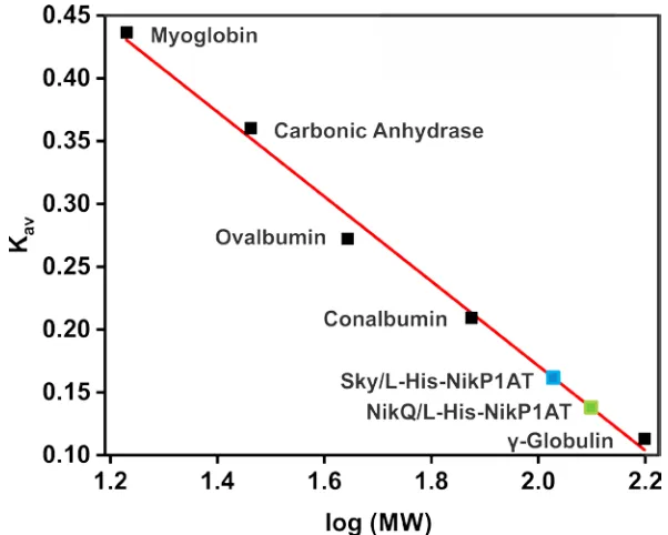
Figure 2.11 Chromatographic partition coefficients (Kav) of the hydroxylase/NRPS
complexes ... 91
Figure 2.12 Sequence alignment for PCPs of skyllamycin and nikkomycin
pathways ... 91
Figure 2.13 Sequence alignment for P450sky and P450nikQ ... 91
Figure 3.1 Diagram of non-native porphyrin incorporation into the cell with
ChuA ... 123
Figure 3.2 Pyridine hemochromagen assay for Mn-Olet and Fe-Olet ... 124
Figure 3.3 CO difference spectrum for Mn-Olet ... 125
Figure 3.4 Determination of KD using the optical changes at 463 nm for
Mn-Olet ... 126
Figure 3.5 Optical characterization of Mn-Olet ... 127
Figure 3.6 High-valent oxo species formed by Mn-Olet utilizing different
oxidants ... 128
Figure 3.7 Stopped-flow spectra of high-valent oxo formation in Mn-Olet ... 129
Figure 3.8 Proposed reduction pathway from the Mn(V)=O to Mn(IV)=O ... 129
Figure 3.9 Molecular orbitals for proposed oxidation state transitions for Mn-Olet upon addition of oxidants ... 130
Figure 3.10 Optical spectra of Mn(IV)=O formation with reduced Mn-Olet and hydrogen peroxide ... 131
xi
Figure 3.12 SVD spectra of the proposed pure Mn(IV)=O species with the
speciation plot ... 134
Figure 3.13 Stopped-flow spectra of Mn(IV)=O formation of Mn-Olet with (right) and without (left) eicosanoic acid ... 135
Figure 3.14 Hammett plot for Mn-Olet with phenol derivatives ... 136
Figure 4.1 Structures of mesoporphyrin IX, protoporphyrin IX, and
diformyldeuteroporphyrin IX ... 171
Figure 4.2 12% SDS-PAGE gel of pure P450olet after dfDPIX, MPIX, and PPIX incorporation ... 171
Figure 4.3 Optical characterization of PPolet ... 172
Figure 4.4 Optical characterization of MPolet ... 172
Figure 4.5 Determination of KD using the optical changes at 417 nm and 396 nm
for MPolet ... 173
Figure 4.6 Pyridine hemochromagen assay for MPolet and PPolet ... 173
Figure 4.7 CD spectra of purified MPolet and PPolet ... 174
Figure 4.8 Stopped-flow spectra of 10 µM MPolet rapidly mixed against 10 mM hydrogen peroxide ... 174
Figure 4.9 Plot of Cpd. I decay as a function of hydrogen peroxide
concentration ... 175
Figure 4.10 Arrhenius plot for Cpd. I and Cpd. II decay ... 175
1
C
HAPTER1
A
C
OMPARISON OFPCP
D
OMAINS AND THEIRR
ECOGNITION WITH AD
INUCLEARI
RONH
YDROXYLASE FROM THEL
YSOBACTINB
IOSYNTHETICP
ATHWAY1.1
I
NTRODUCTIONThe necessity to diversify the antibiotic arsenal has never been as crucial as it is
now with the current level of antibiotic resistant bacteria causing problematic
issues in the realm of public health, particularly Streptococcal and
Staphylococcal species 1-3. Although attempts are currently being pursued
through synthetic means 4-6, the production of more complex antibiotics may be
cost-prohibitive, if not synthetically unachievable. One potentially viable route is
through modification of the antibiotic machinery, known as non-ribosomal peptide
synthetases (NRPS). Such efforts may create opportunities to synthesize a
library of compounds built around a structural scaffold that can be subsequently
screened for antimicrobial activity. Specifically, the use of a pre-existing NRPS
complex, a protein megastructure that produces peptide-based antibiotics
through an assembly-line process, may provide a useful tool for the incorporation
of non-cognate tailoring enzymes as a means for producing modified versions of
current antibiotics. This method has not gained widespread popularity due to its
2
entire operons have been transplanted from one organism to another with
successful production of antibiotic. Although providing new biological platforms
for synthesis, pathogenic bacteria may have already developed resistance to
these natural products 8. There has been very little research involving the
transplantation of enzymes from one antibiotic biosynthetic pathway to another to
generate new products. Homologous enzymes must be identified and
characterized to determine their suitability for a non-ribosomal peptide
synthetase (NRPS) system that is not natively recognized.
Reviews by Marahiel group give an in-depth explanations of how an
NRPS is formed and how they function to create an antibiotic product 9, 10.
Briefly, a NRPS is a modular complex with each module involved with the
identification and activation of a particular amino acid that ultimately becomes
incorporated into a nascent peptide chain. These modules are further divided
into individual domains which are each responsible for a specific aspect of the
identification, modification, and transfer of the amino acid into the nascent
peptide chain. In a canonical multimodular NRPS system, a single module
contains three domains: the Adenylation (A) domain identifies and activates a
specific amino acid through an adenylation reaction, the Thiolation (T or PCP)
domain tethers the activated amino acid through a thioester bond onto a
post-translationally transferred phosphopanthetheine (ppant) and transports the
substrate between domains, and the Condensation (C) domain which extends
the nascent polypeptide chain. One interesting deviation from this operation that
3
that operate in trans to the NRPS, such as the β-hydroxylases CmlA and
P450sky, providing a method to diversify the final product.
The recognition event may depend on either ionic or hydrophobic binding
domains present on the surfaces of the enzyme and the T-domain, enzyme
specificity for the tethered amino acid, or a combination of both. P450
hydroxylases that operate in this fashion, such as P450NikQ and P450sky, are
reported to rely on hydrophobic interactions to properly recognize the binding
motif of the NRPS, although these residues have yet to be fully elucidated 11, 12.
However, there is potential for greater product diversity if these binding motifs
can be solved and incorporated into modules of an NRPS that incorporate
non-modified amino acids, allowing for the creation of non-modified products. The β
-hydroxylase Orf78, associated with the lysobactin biosynthetic pathway, is
homologous with CmlA from the chloramphenicol biosynthetic pathway.
However, the uniqueness of Orf78 lies with its ability to recognize and
hydroxylate substrate attached to three individual NRPS modules instead of the
singular modification conducted by CmlA. Study of the similarities and
recognition flexibilities between the hydroxylase binding motifs will be conducted
through comparisons of interactions between Orf78 and native and non-native
T-domains and gathering information necessary to successfully modify an NRPS
pathway to create a familiar but unique antibiotic-like compound.
Lysobactin is a depsipeptide-based antibiotic isolated from Lysobacter sp.
SC 14067 (ATCC 53042) 13, 14 (Figure 1.1, bottom). In its biologically active form,
4
peptidoglycan and inhibits the synthesis of new cell wall in gram-positive bacteria
15. The inability for the bacteria to synthesize a new cell wall decreases its
overall robustness, allowing turgor pressure to weaken the cell from within and
ruptures in the cell membrane. This mechanism of bacteriocide places
lysobactin in the same category as vancomycin 16 and similar derivatives, such
as teixobactin 17 and balhimycin 18. However, the importance of lysobactin lies in
in the fact that its minimum inhibitory concentration is one quarter of
vancomycin’s on gram-positive bacteria 13, with its strength leading to lower
dosages and the hope of a prolonged lifetime prior to eventual ineffectiveness
due to the evolution of antibiotic-resistant strains of gram-positive bacteria. Due
to the inhibitory effects of the drug to Lipid II, there is no activity towards
mammalian cells, making the drug a possible candidate for use against
troublesome gram-positive bacteria common in hospitals, such as
Staphylococcus aureus and Streptococcus pneumoniae.
The lysobactin NRPS biosynthesis pathway is organized into eleven
modules segregated into two genes, with hydroxylation of two modules on LybA
associated with hydrophobic substrates (Phe3 and Leu4) and one module on
LybB associated with a weakly acidic substrate (Asn10) 19 (Figure 1.1, top). This
contrasts with CmlA, which solely hydroxylates the hydrophobic non-native
amino acid L-para-aminobenzoic acid (L-PABA) 20. This also differs from
P450sky, a hydroxylase with a heme cofactor that hydroxylates three modules on
the skyllamycin biosynthetic pathway associated with three hydrophobic
5
recognized charges on the substrate side-chain may allow for greater flexibility
for recognition than with other β-hydroxylases; however, the reasons why some
amino acids are hydroxylated while others are not is unclear. The structure of
the antibiotic shows the β-hydroxyl group on PCPPhe3 is necessary to form an
intermolecular lactone with the C-terminal carboxylate of the nascent peptide
chain. However, it is unknown what functions the β-OH groups on PCPLeu4 and
PCPAsn10 serve, except at the very least assisting to solubilize the active
compound, which is mostly hydrophobic in nature.
Makris et al. showed that oxidation kinetics on Type I NRPS systems
(CmlA/CmlPAT) can be used as an indirect measurement to determine binding of
the substrate carrier to the hydroxylase, with quicker rates indicative of greater
oxygen access to the active site of the enzyme 20. For heme-bound
hydroxylases, the dissociation constants with Type II NRPS systems
(P450sky/PCP7O-Me-Tyr) 12, 20 were used to show the differences between bound
and unbound substrate carriers. However, there is no information concerning
cross-recognition for a Type II-associated hydroxylase with non-native Type I or
II NRPS systems. Following basic characterization of Orf78, to include iron
quantitation and structural features, a comparative analysis of oxidation kinetics
between Orf78 and modular components from three different systems will be
performed, utilizing the following T-domains: PCPAsn10 from the lysobactin
system, CmlPT from the chloramphenicol system, and PCPPhe6 from the
6
1.2
M
ATERIALS ANDM
ETHODSCloning
The orf78 gene was cloned from the genomic template from Lysobacter
sp. 53042 (ATCC). The following primers were utilized for the amplification of the
orf78 gene with Taq polymerase (NEB), where the capitalized regions are the
restriction sites for SfaAI and MssI:
forward - 5’-ccatatgGCGATCGCcaataccactca-3’
reverse - 5’-ccatatgGTTTAAACtcacagaagcag-3’
The amplicon and pVP91A vector were both digested with SfaAI and MssI
(Thermo Fisher Scientific), gel-purified, and ligated with T4 DNA ligase (NEB).
The insert was verified using the facilities at ACGT, Inc.
The PCPAsn10 gene was similarly cloned from the genomic template of
Lysobacter sp. 53042 (ATCC). The following primers were utilized for the
amplification of the A10T gene with Q5 polymerase (NEB), where the capitalized
regions are the restriction sites EcoRI and HindIII:
forward - 5'- cgtatcCATATGctgctgtcgccgccgcagcgcgag -3'
reverse - 5'- cgtatcAAGCTTcgccggcaacgccgccccgc -3'
The amplicon and pET21b(+) vector were both digested with EcoRI and HindIII
(NEB), gel-purified, and ligated with T4 DNA ligase (NEB). The sequence was
7
Protein Expression and Purification
The Orf78 plasmid was chemically transformed into BL21(Dε3) cells. A
single colony was used to inoculate a starter culture which were used as
inoculant for 1 liter flasks of M9 minimal media. The components of the M9
minimal media consists of: 200 mL M9 salts, 2 mL of 1 M MgSO4, 20 mL of 20%
glucose, 100 µL of 1 M CaCl2, 12 mL overnight culture, and 1 mL of 100 mg/mL
Ampicillin. The culture was induced at an OD600=0.7 with the addition of 150 µM
isopropyl β-D-1-thiogalactopyranoside (IPTG; RPI corp.) supplemented with 50
µM FeCl3 with further incubation for 16 hours at 18oC. The cells were lysed,
cleared, and purified with Ni-NTA resin equilibrated with 50 mM sodium
phosphate, monobasic, 300 mM sodium chloride, pH=7.5, 10 mM imidazole
(Buffer A), washed with Buffer A + 10 mM imidazole, and eluted with Buffer
A+240 mM imidazole. Following purification, the eluent was dialyzed overnight in
50 mM HEPES, pH=7.5 + 10% glycerol. The protein was concentrated,
aliquoted, and stored at -80oC.
The PCPAsn10 plasmid was chemically transformed into BL21(Dε3) cells.
A single colony was used to inoculate a starter culture which were used as
inoculant for 1 liter flasks of Luria-Bertani media. The culture was induced at
OD600=0.6 with 150 µM IPTG followed by incubation for an additional 16 hours at
37oC. The cells were lysed, cleared, and purified with Ni-NTA column
equilibrated with Buffer A, washed with Buffer A + 10 mM imidazole, and eluted
with Buffer A + 240 mM imidazole followed by dialysis overnight in 25 mM
8
equilibrated with 25 mM HEPES, pH=7.5. The flow-through, which contains the
properly-folded protein, was dialyzed in 100 mM HEPES, pH=7.5, 30% glycerol
prior to concentrating, aliquoting, and storage at -80oC.
Size Exclusion Chromatography
Nine milligrams of Orf78 were loaded onto an S-200 size-exclusion
column attached to an ATKA FPLC equilibrated with 50 mM HEPES, pH=7.5 and
eluted at 0.3 mL/min. The elution profile was matched with a set of protein
standards with known molecular weight to determine the oligomeric state of the
enzyme.
UV-Vis
All UV-Vis measurements were obtained on an Agilent 8453 UV-Vis
spectrophotometer and analyzed on Agilent proprietary software or the OriginPro
software package. The concentration of Orf78 was calculated using Beer’s Law
and the theoretical ε280 = 58.8 mM-1cm-1. An azide assay was used to determine
the presence of any diiron cluster within the enzyme. 50 µM of Orf78 was
incubated in 1 mL of 50 mM HEPES, pH=7.5, 8 M sodium azide for 20 minutes at
4oC. The solution was cleared by centrifugation at 4oC and spectrum taken of
the supernatant at 340 nm and 420 nm.
A reductive titration was performed to determine the number of electrons
necessary to fully reduce the enzyme from the fully oxidized state. All solutions
were buffered in 50 mM HEPES, pH=7.5 degassed with N2. A 2 mM solution of
sodium dithionite was utilized as a reductant. Separately, a solution of 50 uM
9
equivalents of methyl viologen. Sodium dithionite was titrated with a gas-tight
syringe in 30 µM increments until the charge-transfer band at 340 nm completely
bleached and the methyl viologen absorption peaks from 350-395 nm became
apparent, indicating full reduction of the enzyme. The data was analyzed on
OriginPro 2016 as a plot showing number of electrons against total absorbance
at 340 nm.
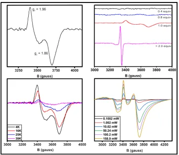
Electron Paramagnetic Resonance (EPR)
Spectra of the mixed-valent enzyme were collected on an EMXplus
X-band EPR (Bruker) at 10K with 5 mW power and a microwave frequency of 9.834
GHz. Each spectrum is a composite of 15 averaged scans per sample. The
data were collected and analyzed on either the Bruker Xenon software suite or
the SpinCount software package. Determination of the g-values were performed
by the use of the equation g = (71.4484v) / B, where magnetic field, B (mT) and
the microwave frequency, v (GHz). All solutions were buffered in 50 mM
HEPES, pH=7.5 and degassed with N2. Sample preparation was performed
anaerobically with O2 levels less than 1 ppm.
The visualization of the mixed-valent species involved the preparation of
four samples. The sample containing one electron-equivalent to create the
mixed-valent species involved the addition of 250 µM sodium dithionite to 400 µL
of 400 µM degassed Orf78 + 200 µM methyl viologen and incubated until the
solution transformed from purple to clear. The solution was transferred to a
quartz cuvette, capped, and frozen in liquid nitrogen for storage. For the
electron-10
equivalents, 200 µM of sodium dithionite was added to 400 µM Orf78 + 200 µM
methyl viologen and stored as above. For samples containing no mixed-valent
species from 0.4 electron-equivalents, 400 µM Orf78 + 200 µM methyl viologen
were immediately frozen and stored. For samples containing no mixed-valent
species from two electron equivalents, 400 µM sodium dithionite was added to
400 µM Orf78 + 200 µM methyl viologen and frozen off for storage.
Alteration of the temperature can determine the protonation state of the
oxygen bridge between the two iron molecules. The mixed-valent species from
the fully mixed-valent sample above was measured at a constant power of 5 mW
and all spectra are an average of 15 scans. Measurements were taken at
temperature points at 4K, 16K, 25K, and 39K.
The sample with highest content of the mixed-valent form was
subsequently used for the power saturation experiment. The temperature was
kept at a constant temperature of 8K ± 0.75K and the power was increased from
0.07942 mW to 158.9 mW, with each spectrum representing the average of 15
scans. The distance, from peak to trough, of the second g-value was used as a
measure of the signal intensity (I) at any given power. The power at
half-saturation (P1/2) was determined using the following equation:
I = Io*P1/2/(1+P/P1/2))b/2
, where Io is the intensity under unsaturated conditions and b is a homogeneity
value assumed to be equal to 1. The data was plotted as log(I/√(P)) as a
11
Synthesis of Coenzyme-A derivatives
Bodipy-CoA was synthesized by dissolving 110 nanograms each of
Bodipy-FL (Life Technologies) and Coenzyme A (CoA; RPI corp.) in 300 µL
DMSO + 1.8 mL 75 mM MES, pH=6 + 100 mM magnesium acetate. The solution
incubated on ice in a dark environment for 10 minutes followed by incubation at
room temperature in a dark environment for 2 hours while rapidly stirring. Pure
product was isolated with several extractions of ice-cold diethyl ether. Excess
ether was evaporated under N2 prior to aliquoting and storage at -20oC.
Synthesis of Phe-CoA involved dissolving 5 mg of Boc-Amino Acid (AA), 4
mg dicyclohexylcarbodiimide (DCC, Sigma Aldrich), and 3 mg
N-hydroxysuccinimide (NHS, Sigma Aldrich) with 1 mL acetonitrile and incubating
on ice for 10 minutes prior to stirring at room temperature for 24 hours under a
nitrogen atmosphere. Following the incubation time, 15 mg of CoA dissolved in
1.5 mL of 40 mM lithium carbonate were added to the solution and incubated on
ice for 5 minutes prior to further incubation at room temperature for an additional
2 hours. The addition of H2O:TIPS:TFA at a ratio of 2.5:2.5:95 was added to
deprotect the amine group of the Boc-AA-CoA and the solution was allowed to
stir at room temperature for 2 additional hours. The Phe-CoA was purified by
several extraction steps with ice-cold diethyl ether, resulting in the precipitation of
the desired product. The precipitated product was isolated via centrifugation,
dried under N2, and resuspended in dH2O to a concentration of 2.5 mM,
12
SfP recognition of the T-domain
The Phe-PPant is transferred to the T-domain with a 4´-
phosphopantetheinyl transferase (SfP) derived from Bacillus subtilis and
expressed in E. coli. After the T-domain is purified, Bodipy-PPant is transferred
to test for SfP recognition of the serine residue, as part of the GGxS motif. The
reaction is set up with 30 mM magnesium chloride, 60 mM Tris, pH=8.0, 700 mM
AA-CoA, 5 µM SfP, and 200 µM apo-PCPAsn10 followed by incubation at room
temperature for 1 hour and desalting in 50 mM HEPES, pH=7.5. The sample is
then run on a 12% SDS-PAGE gel and, upon completion, viewed under a
transilluminator to verify its presence at ~10kDa. If the SfP recognizes the
T-domain, then the same procedure is used to transfer Phe-PPant for use in
Stopped-flow experiments.
Stopped-flow
Binding between Orf78 and any T-domain was determined by the rate of
rebound for the iron-oxo charge-transfer band at 340 nm from the bleached
reduced state to its re-emergence when oxidized. The measurement of the
oxidation rates for the hydroxylase/NRPS complex was performed on an SX20
Stopped-flow spectrophotometer (Applied Photophysics) for accurate
visualization and rate measurements. The temperature of the experiment was
set to 4oC and data was collected utilizing a PMT detector set at 340nm with
5000 time points over 500 seconds per measurement.
Syringe A contained 50 µM Orf78 + 25 µM methyl viologen that was fully
13
small molecules. The solution was then transported anaerobically and loaded
into the stopped-flow pre-chilled at 4oC. Syringe B contained 50 mM HEPES, 2
mM O2, and one of the following: apo- PCPAsn10, holo- PCPAsn10, Phe-PCPAsn10,
Phe-CmlPAT from the chloramphenicol NRPS, or Phe-PCPTyr6 from the
teichoplanin NRPS. The collected data was an average of triplicate traces for
each sample and fit to single exponential functions to determine the final rates in
Pro-Data Viewer (Applied Photophysics).
1.3
R
ESULTSOrf78 contains a dinuclear iron cluster
An analysis of the sequence alignment shows the endogenous ligands
present in CmlA are fully conserved in Orf78 (Figure 1.2). A model of Orf78,
created using SWISS-MODEL 22 and the published structure of CmlA (PDBID:
4JO0) as a template, putatively confirms an overlap for all residues involved in
the ligation of the metal atoms. The first coordination sphere consists of three
His, two Glu, and two Asp residues, with one of the Asp forming a µ-1,1
carboxylate bridge between the two irons in the cluster. The presence of a
carboxylate bridge is not uncommon, but the bridge is not usually a µ-1,1
configuration, but rather a µ-1,3 carboxylate linkage. Crystallographic studies of
other diiron enzymes show at least one µ-1,3 carboxylate bridge, such as the
case with Methane Monooxygenase (MMO), Purple Acid Phosphatase (PAP),
and Ribonucleotide Reductase (RNR) 23-25. Specifically, the crystal structure of CmlA confirms the presence of residue D403 forming a μ-1,1-carboxylate bridge
14
an adventitious acetate anion present from the mother-liquor that may be
displacing D403 into a conformation forming a mono-dentate rather than
bi-dentate bridge. It is worth noting that this same acetate anion may be
responsible for displacing residue D430, which is a viable ligand in range of an
iron atom. However, it is unknown whether a similar bridge or solvent ligation
pattern is present in Orf78, as attempts to crystallize the protein have been futile.
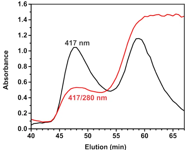
In similar fashion to that proposed for CmlA, Orf78 purifies as a dimer with
two joined 60.8 kDa units (Figure 1.3). Size-Exclusion Chromatography shows
the protein elution at around 125 kDa with no peaks associated with monomeric
or other oligomeric forms of the enzyme. The crystal structure for CmlA suggests
the dimer interaction is derived from a dimerization arm that electrostatically
interacts with the same domain on the opposing monomer in a hook-like fashion
separate from the active site region of the jelly-roll motif. Similar residues are
present at the dimer interface of the model for Orf78, inferring the nature of the
dimerization is electrostatic in nature as well.
Iron quantitation with the ferrozine assay consistently yields 1.7-2.3 irons
per dimer. These results were confirmed with ICP-MS and show only trace
amounts of manganese, zinc, and copper present in the sample. However, the
fully-loaded metal count for the dimer should be around four irons per dimer
since each active site in the dimer requires occupancy by two iron molecules.
Given the minimal media growth conditions supplemented with ample iron (III)
chloride, it is not known why complete loading does not occur. Metal
15
been unsuccessful. Nonetheless, the lack of an appreciable high-spin Fe3+
signal in the as-isolated enzyme, reductive titration of Orf78 with sodium
dithionite, and the presence of a mixed-valent signal in EPR demonstrates that
almost all of the irons are bound as a cluster.
The UV-Vis spectrum of Orf78 consist of a broad spectral region with
weak extinction coefficients extending from 300-450 nm, culminating in a major
peak maximum at 340 nm and a minor peak maximum at 420 nm. Similar peaks
are observed in other hydroxo-bridged diiron enzymes like CmlA 20, and
Hemerythrin (Hr) 26 when in a low-spin, diferric state. The peak at 340 nm
manifests through the electronic charge-transfer exchange by the Fe2-O, which is
created by splitting the π-orbital electrons resonating between the two iron atoms
via an oxo-bridge 27. The intensity of the bands is less significant than those of
proteins with bridges, indicating that resonance is weaker due to the
oxo-bridge being protonated. An Fe2-O bridge that forms in a linear fashion will
usually absorb at 420 nm and 500 nm. However, at the more acute angles of
114o to 130o, the lowest Fe
2-O π-transition state at 500 nm blends with the
second lowest state at 420 nm, indicating a less-obtuse bridge form is present
with Orf78 28.
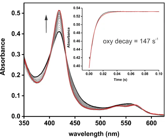
Titrating sodium dithionite to the protein, and subsequent reduction of the
diferric cluster, decreases the intensity of the 340 nm and 420 nm peaks, leading
to bleaching of the charge-transfer signal through the quenching of its π-orbital
resonance effects (Figure 1.4). Reduction is quick, inferring minimal difficulty for
16
diferrous state mimics CmlA and human dehydroxypusine hydroxylase by fully
oxidizing to the diferric state as observed by a rebound of the bleached 340 nm
and 420 nm charge-transfer bands 29. However, Stopped-flow data shows that
oxidation of Orf78 from the diferrous state in the presence of molecular oxygen is
a slow process, suggesting difficulty for the molecular oxygen cofactor to gain
access to the active site. This, in addition to an increased rate in the presence of
Phe-PCPAsn10, suggests the presence of necessary events prior to the activation
of O2 by the reduced enzyme.
The introduction of sodium azide to diferric Orf78 forms an additional π
-stabilized bridge, leading to an increase in absorbance for the 340 nm and 420
nm bands (Figure 1.5). Similar spectra are observed with biological non-heme
diiron enzymes, such as metHr and Hr 27, and synthetic constructs 30. However,
the concentration requirements of sodium azide necessary for bridge formation is
four-fold higher than required for CmlA and binding is not immediate, requiring
incubation at 4oC for longer periods to time to observe maximum peak
amplitudes. Two possible reasons for this is that access to the active site for
azide is restrictive or the distance between the two iron molecules is not optimal
to facilitate formation of the azide bridge.
The EPR spectrum of the diferric state does not induce a signal, which
confirms that Orf78 is a S=5/2-5/2 anti-ferromagnetically spin-coupled system
(Figure 1.6, top right). The absence of a signal at g=4.3 shows that there is both
little free iron in the sample and also that the two irons are not separate S=5/2
17
a S=5/2-2 spin-coupled system with an orthorhombic mixed-valent signal and
observable values of gz=1.96 and gy=1.86 at low temperature (Figure 1.6, top
left). These values are observed in other anti-ferromagnetically spin-coupled
systems, like RNR, when one-electron equivalent is added to the diferric enzyme
32. The gx value is not observed, presumably blending into an extended gy
signal. This behavior is also observed in the mixed-valent signal for CmlA.
There is no signal when the sample is reduced with two-electron equivalents to a
diferrous state, indicating a S=2-2 spin-couple. This reinforces the conclusion
that the cluster is anti-ferromagnetically coupled since it is also observed in
similarly spin-coupled systems, such as MMO and Hr 33, 34.
The intensity of the mixed-valent EPR signal for Orf78 shows a
temperature-dependent that suggests the presence of a hydroxo-bridge between
the two iron molecules (Figure 1.6, bottom left). Increasing the temperature from
4K to 18K does not significantly alter the amplitude of the signal. However, the
signal decreases as the temperature is increased above 18K and is ultimately
lost beyond 39K. Similar tendencies are observed in other hydroxo-bridged
diiron proteins, like CmlA, MMO, and PAP, which have decreased relaxation
times due to the increased acidity of the associated irons 35. The presence of an
oxo-bridge would increase the relaxation time of the π-electron transfer between
the two iron atoms, preventing decay of the mixed-valent signal until higher scan
temperatures (>100K) are achieved 36.
Changes to the local environment, such as the presence of substrate or
18
diiron species and observed through power saturation studies 37. As the power
increases, the relaxation state of the electrons sit at a higher energy level and the
signal decreases. Power saturation is also informative for determining what
power should be used to measure any EPR spectra so there is a reduction in
unwanted g-value broadening and signal reduction. For Orf78, a plot of the
power saturation curve shows a P1/2 equal to 93.2mW ± 8.6mW (Figure 1.7), so
a high amount of power is needed to saturate the signal at 10K and overcome
the relaxation times of the mixed-valent signal. Plotting ln(P1/2) for this value as a
function of temperature (K-1) gives a value of 4.53 at 10K, which is slightly higher
than CmlA (~3.8) and the hydroxo-bridged diiron Mitochondrial Alternative
Oxidase (~4.9) 20, 38. The values are assumed to be close enough to associate
similar power saturation values of Orf78 for all temperature ranges. Ideally, more
points would be preferred, but the slope, and the corresponding J value, of the
plot can be inferred due to the homology of the two enzymes and is assignable
as a hydroxo-bridge in the mixed-valent form.
Similarity of PCP domains
Three PCPs for the lysobactin biosynthetic pathway, PCPPhe3, PCPLeu4,
and PCPAsn10, provide substrate that are all β-hydroxylated prior to their
incorporation into the nascent peptide chain. A protein sequence alignment
(Clustal Omega) of the three contain a sequence identity of 40%-60% (Table
1.1). It has been theorized that this is due to gene duplication followed by
mutation of pre-existing modules, creating a useful and cheap mechanism to
19
facilitate β-hydroxylation of amino acids in the skyllamycin biosynthetic pathway
(incorporated in sky30 and sky31) have a sequence identity of 38%-60% 21.
Although the identity range is wider than the lysobactin pathway, variations for
both systems are mostly localized in the flanking regions of the four-helix bundle
of the PCPs. It is unclear what effects these flanking regions have on the overall
function of the module or their role in recruiting their respective β-hydroxylases.
Incorporation of PCPPhe6, the only PCP in the teicoplanin biosynthetic pathway
(incorporated in dbv17) that is β-hydroxylated by the diiron hydroxylase Orf12,
into the sequence alignment with the lysobactin PCPs lowers the sequence
identity to 30% 41. However, a strong conservation of residues exists within the core regions containing helices α2 and α3, which are presumed to contain the
binding motifs to the hydroxylase. Modeling the three PCPs from lysobactin to
the PCP from teichoplanin illuminate overlaps across all four helices, suggesting
that the PCPs for both systems adopt similar four-helix bundles in their active
forms (Figure 1.8).
Insertion of CmlPT from the Chloramphenicol biosynthetic pathway
(incorporated in orf8) 42, a Type II NRPS which contains a β-OH L-PAPA, into the
sequence alignment with the four Type I PCPs drops the sequence identity to
20%. The homology among all five T-domains also exemplifies this, as there is a
large drop-off in similarity upon the addition of the CmlPT PCP (Figure 1.9). A
model of CmlPT with any of the Type I PCPs above show incompatibility with the
Type II systems, as the conformation adopts a two-helix bundle in lieu of four.
20
the outfitted model, so does not conform to the four helix motif for the Type I
systems. This may infer that hydroxylases from Type I NRPS systems may not
recognize Type II NRPS systems and vise versa.
Despite the poor conservation between all five PCPs, certain aspects of
the core motif, mainly Lx[5]Gx[5]Fx[2]GGxSx[4]Q, remain consistent. In all cases,
residues for SfP recognition are similar enough on all five PCPs to allow the
transfer of Bodipy-PPant 43. The majority of conserved residues are also
hydrophobic, suggesting the interaction between the T-domain and the
hydroxylase are driven by Van der Waals forces or are less reliant on
electrostatics for their direct connection with each other. The PCP TycC3 from
the tyrocidine biosynthetic pathway contains a leucine residue that is presumably
associated with the interaction between the itself and Sfp 44, although there are
PCPs where the SfP recognition site could not be elucidated 45. Mutation of this
leucine from TycC3 inhibited recognition of SfP for the T-domain, but it is
unknown whether similar behavior occurs with hydroxylase/T-domain
interactions. Residues on the flanking ends of the PCPs may assist with
protein-protein interaction or they may provide subtle positioning of the four-helix bundle
so residues near the SfP recognition site can interact with the hydroxylase.
Site-directed mutations demonstrate that alterations of the α-helices of the PCP will
21
Oxidation rates of PCPs with Orf78
A study of diferrous CmlA and its NRPS system shows an association
between the rate of oxidation of the enzyme and the presence of aminoacylated-
or non-amionoacylated NRPS and molecular oxygen, which conclude that a
notable difference exists concerning oxygen-gating by the enzyme 20. In the
absence of NRPS, the rate of oxidation for CmlA is slow at 0.005 s-1. The rate is
relatively unchanged upon the introduction of apo- or holo-NRPS, with an
observed rate of 0.01 s-1. The rate of oxidation increases to 12 s-1 in the
presence of aminoacylated NRPS, suggesting that proper binding and channel
access for O2 in CmlA occurs only in the presence of aminoacylated-NRPS.
This experiment was reproduced with Orf78 to determine the rate of
oxidation for the enzyme in the presence of native and non-native PCPs. Since
phenylalanine is one of the native substrates recognized by Orf78 and the
aromatic ring is similar to the native tyrosine substrate for PCPTyr6 and the native
L-PABA substrate for CmlP, recognition is assumed to be driven by the binding
motifs on the PCPs and not the tethered amino acid. Poor expression and
solubility of PCPAsn10 prevents these experiments from being performed under
pseudo-first order conditions so the molar excess of PCP was kept at no more
than four times greater than Orf78 for all three T-domains.
All rate measurements are shown in Figure 1.10. The oxidation rates for
Orf78 in the presence of any apo-PCP and holo-PCP results in a slow rate of
oxidation of 0.004 s-1, similar to Orf78 in the absence of PCP and the rates of
22
indicates that either the apo- nor holo- forms of the PCP do not bind or that that
they bind but do not properly gate O2 in the absence of tethered amino acid.
Orf78 in the presence of Phe-PCPAsn10 shows fits with a double exponential, with
an initial phase of 0.2 s-1 followed by a second phase of 0.005 s-1. The initial
phase may be a mixture of the PCP binding event and quick oxidation and a slow
oxidation rate due to unbound enzyme, and artifact of the experiment not
adhering to pseudo-first order conditions. The second phase is likely O2 gating
into Orf78 unbound to PCPAsn10. The rates of Orf78 in the presence of
Phe-PCPTyr6 and Phe-CmlPT is monophasic with an oxidation rate of 0.004 s-1,
matching the oxidation rate of Orf78 in the absence of PCP. This suggests there
is another factor other than substrate recognition that is inhibiting binding of the
hydroxylase to the PCP, such as the proposed incompatibility with CmlPT
proposed earlier or the presence of higher dissociation constants between Orf78
and non-native T-domains.
1.4
D
ISCUSSIONIt is necessary to study monooxygenases homologous to CmlA enhance
the flexibility of enzyme choice when engineering a biosynthetic gene cluster.
This choice may allow for the recognition of certain modules in a semi-specific
manner and is important when attempting to build libraries of antibiotic-like
compounds. Ideally, the selection of a hydroxylase for an engineered NRPS
system would be dependent on its solubility in-vivo, redox partners, and its ability
to properly recognize and utilize cofactors required to perform the necessary
23
combining the use of an enzyme from one pathway with PCP components from
non-native systems in an effort to discern the specificity of these interactions.
However, the results do not favor the use of non-native enzymes with NRPS
systems without modifying their components to promote a higher degree of
recognition between the two. Although there is an interaction between Orf78 and
PCPAsn10, there is no interaction with the non-native aminoacylated-PCPs.
The oxidation rate of the Orf78/Phe-PCPAsn10 complex does not reach a
level to be considered biologically relevant, as bacterial defenses would be
overwhelmed with such slow production. This contrasts with the oxidation rate
for CmlA in the presence of L-PAPA-CmlPAT at 12 s-1, rising two orders of
magnitude higher than any of the rates associated with Orf78. One argument is
that the dissociation constant of the interaction is much higher than published for
other hydroxylase/PCP systems, such as P450sky or P450nikQ, and ratio of PCP
to Orf78 is too low to visualize quick oxidation. This led to a slower rate for the
first phase than would otherwise be observed. It is currently unknown if there are
additional interactions between the hydroxylase and the A-domain that assist
with recognition and helps to guide the interaction. Unfortunately, the rate for
CmlA/L-PAPA-CmlPT is unknown, so a true comparison between Orf78/PCPAsn10
and CmlA/CmlPAT cannot be obtained. The inability to clone out any soluble
forms of the AT domains from lysobactin system prevented testing that
hypothesis with any complexes with Orf78.
The inability to properly model CmlPT based off of published NMR
24
CmlPT, explaining the absence of reactivity during oxidation experiments with
Orf78. However, is should be noted that the Phe-PPant may not sit correctly on
the PCP if the para-amino group is influences the interaction of substrate stability
on the T-domain and destabilizing its binding to the hydroxylase. In addition, the
NRPS from the chloramphenicol pathway is unimodular and may not have
sufficient complexity compared to their multimodular counterparts, meaning the
modular components of CmlPAT may not be homologous with Type II NRPS
systems and cannot interact with Orf78. However, Phe-PCPTyr6 displays a
monophasic rate comparable to Orf78 alone with O2. This is surprising since its
sequence aligns the alpha-helical regions well for PCPTyr6 and PCPAsn10.
Although the similarity between these two PCPs appear high enough to assume
some interaction between the hydroxylase and the Phe-PCPTyr6 domain, this
illustrates the subtlety of the structural differences between the PCPs from these
two systems and represent the fine-tuning that must occur to properly lock the
complex into place.
Although the enzyme purifies as a dimer, the issues concerning the
protein-protein interaction between an enzyme with a singly-loaded cluster per
dimer an the PCP domain may affect the kinetic rates, although these effects are
not known. The incompleteness of iron-loading may be due to iron lability in the
unloaded monomer. Alternatively, the unloaded monomer may be forced into a
different structural conformation by the loaded monomer and inherently does not
load upon dimerization. One example involves the incomplete loading of the
25
but only partially loads the second cluster in the second monomer due to the
loaded monomer exerting conformation dominance over the partially loaded
monomer 47. However, the result for Orf78 differs from CmlA, which purifies with
two complete clusters per dimer; so nuances concerning the monomeric
influences for the dimeric form may exist for Orf78. If the enzyme solely operates
on the NRPS system, as its placement in the lysobactin operon suggests, then
there is no need for a two fully-loaded clusters per dimer to do the necessary
chemistry, as only one monomer could bind at a time to the larger, multimodular
complex. Since the unloaded monomer is presumably distorted in a manner that
prevents iron-loading, it is unknown whether PCPAsn10 will prioritize the loaded
monomer over the unloaded one. Additionally, any PCP bound to the unloaded,
non-catalytic monomer will lower the rate of oxidation since the effective
concentration of PCP will be lower than previously calculated. The PCP
concentration is already poised for sub-pseudo first order kinetics with only a
four-fold excess to Orf78, so the effective concentration will be halved at two-fold
to Orf78, assuming the PCP binds with equal preference for each monomer.
Alternatively, PCP binding to one monomer may inhibit binding to the unbound
monomer.
1.5
C
ONCLUSIONOrf78, the hydroxylase from the lysobactin biosynthetic pathway, was
purified and characterized as a dimeric, diiron enzyme. The hydroxylated PCP
domains from the lysobactin pathway are compared to view similarities in
26
Type I NRPS teicoplanin and Type II NRPS chloramphenicol biosynthetic
pathways. Oxidation experiments were performed to test for protein-protein
interactions between these PCPs and Orf78, with the only interaction recorded
with Orf78’s native T-domain, PCPAsn10. The reasoning for some T-domains
being hydroxylated and not others is still an aspect of ongoing research by both
our and other labs.
1.6
R
EFERENCES[1] Chambers, H. F., and Deleo, F. R. (2009) Waves of resistance:
Staphylococcus aureus in the antibiotic era, Nat. Rev. Microbiol.7,
629-641.
[2] Dohrmann, S., Anik, S., Etesami, N., No, H., Olson, J., Nizet, V., and
Okumura, C. Y. (2013) Streptococcal Collagen-Like Protein 1 (Scl-1)
Protects Group A Streptococcus From Antimicrobial Molecules, Mol Biol
Cell24, 4011-4020.
[3] Shimamoto, J., Doehrmann, S., Etesami, N., Martin, G., Nizet, V., and
Okumura, C. (2014) Streptococcal collagen-like protein 1 shields group A
Streptococcus from antimicrobial molecules, FASEB J.28.
[4] Dexter, H. L., Williams, H. E. L., Lewis, W., and Moody, C. J. (2017) Total
Synthesis of the Post-translationally Modified Polyazole Peptide Antibiotic
Goadsporin, Angew. Chem.56, 3069-3073.
[5] Koteva, K., King, A. M., Capretta, A., and Wright, G. D. (2016) Total Synthesis
and Activity of the Metallo--lactamase Inhibitor AspergillomarasmineA,
27
[6] Wojtas, K. P., Riedrich, M., Lu, J. Y., Winter, P., Winkler, T., Walter, S., and
Arndt, H. D. (2016) Total Synthesis of Nosiheptide, Angew. Chem. Int. Ed.
55, 9772-9776.
[7] Winn, M., Fyans, J. K., Zhuo, Y., and Micklefield, J. (2016) Recent advances
in engineering nonribosomal peptide assembly lines, Nat. Prod. Rep. 33,
317-347.
[8] Awan, A. R., Blount, B. A., Bell, D. J., Shaw, W. M., Ho, J. C. H., McKiernan,
R. M., and Ellis, T. (2017) Biosynthesis of the antibiotic nonribosomal
peptide penicillin in baker's yeast, Nat Commun8, 1-7.
[9] Finking, R., and Marahiel, M. A. (2004) Biosynthesis of nonribosomal
peptides, Annu. Rev. Microbiol.58, 453-488.
[10] Strieker, M., Tanovic, A., and Marahiel, M. A. (2010) Nonribosomal peptide
synthetases: structures and dynamics, Curr. Opin. Struct. Biol.20,
234-240.
[11] Wise, C. E., and Makris, T. M. (2017) Recruitment and Regulation of the
Non-ribosomal Peptide Synthetase Modifying Cytochrome P450 Involved
in Nikkomycin Biosynthesis, Acs Chem Biol12, 1316-1326.
[12] Uhlmann, S., Sussmuth, R. D., and Cryle, M. J. (2013) Cytochrome p450sky
interacts directly with the nonribosomal peptide synthetase to generate
three amino acid precursors in skyllamycin biosynthesis, Acs Chem Biol8,
28
[13] Bonner, D. P., O'Sullivan, J., Tanaka, S. K., Clark, J. M., and Whitney, R. R.
(1988) Lysobactin, a novel antibacterial agent produced by Lysobacter sp.
II. Biological properties, J Antibiot (Tokyo)41, 1745-1751.
[14] O'Sullivan, J., McCullough, J. E., Tymiak, A. A., Kirsch, D. R., Trejo, W. H.,
and Principe, P. A. (1988) Lysobactin, a novel antibacterial agent
produced by Lysobacter sp. I. Taxonomy, isolation and partial
characterization, J Antibiot (Tokyo)41, 1740-1744.
[15] Lee, W., Schaefer, K., Qiao, Y., Srisuknimit, V., Steinmetz, H., Muller, R.,
Kahne, D., and Walker, S. (2016) The Mechanism of Action of Lysobactin,
J. Am. Chem. Soc.138, 100-103.
[16] Gale E.F, C. E., Reynolds P.E., Richmond M.H., Waring M.J. (1981) The
Molecular Basis of Antibiotic Action, 2nd ed., John Wiley & Sons Ltd, New
York, NY.
[17] Ling, L. L., Schneider, T., Peoples, A. J., Spoering, A. L., Engels, I., Conlon,
B. P., Mueller, A., Schaberle, T. F., Hughes, D. E., Epstein, S., Jones, M.,
Lazarides, L., Steadman, V. A., Cohen, D. R., Felix, C. R., Fetterman, K.
A., Millett, W. P., Nitti, A. G., Zullo, A. M., Chen, C., and Lewis, K. (2015)
A new antibiotic kills pathogens without detectable resistance, Nature517,
455-459.
[18] Frasch, H. J., Kalan, L., Kilian, R., Martin, T., Wright, G. D., and Stegmann,
E. (2015) Alternative Pathway to a Glycopeptide-Resistant Cell Wall in the
29
[19] Hou, J., Robbel, L., and Marahiell, M. A. (2011) Identification and
Characterization of the Lysobactin Biosynthetic Gene Cluster Reveals
Mechanistic Insights into an Unusual Termination Module Architecture,
Chem. Biol.18, 655-664.
[20] Makris, T. M., Chakrabarti, M., Munck, E., and Lipscomb, J. D. (2010) A
family of diiron monooxygenases catalyzing amino acid beta-hydroxylation
in antibiotic biosynthesis, Proc. Natl. Acad. Sci. USA107, 15391-15396.
[21] Pohle, S., Appelt, C., Roux, M., Fiedler, H. P., and Sussmuth, R. D. (2011)
Biosynthetic Gene Cluster of the Non-ribosomally Synthesized
Cyclodepsipeptide Skyllamycin: Deciphering Unprecedented Ways of
Unusual Hydroxylation Reactions, J. Am. Chem. Soc.133, 6194-6205.
[22] Schwede, T., Kopp, J., Guex, N., and Peitsch, M. C. (2003) SWISS-MODEL:
an automated protein homology-modeling server, Nucleic Acids Res.31,
3381-3385.
[23] Rosenzweig, A. C., Frederick, C. A., Lippard, S. J., and Nordlund, P. (1993)
Crystal-Structure of a Bacterial Nonheme Iron Hydroxylase That Catalyzes
the Biological Oxidation of Methane, Nature366, 537-543.
[24] Lindqvist, Y., Johansson, E., Kaija, H., Vihko, P., and Schneider, G. (1999)
Three-dimensional structure of a mammalian purple acid phosphatase at
2.2 A resolution with a mu-(hydr)oxo bridged di-iron center, J. Mol. Biol.
30
[25] Nordlund, P., and Eklund, H. (1993) Structure and Function of the
Escherichia-Coli Ribonucleotide Reductase Protein R2, J. Mol. Biol.232,
123-164.
[26] Kao, W. C., Wang, V. C., Huang, Y. C., Yu, S. S., Chang, T. C., and Chan,
S. I. (2008) Isolation, purification and characterization of hemerythrin from
Methylococcus capsulatus (Bath), J. Inorg. Biochem.102, 1607-1614.
[27] Reem, R. C., Mccormick, J. M., Richardson, D. E., Devlin, F. J., Stephens,
P. J., Musselman, R. L., and Solomon, E. I. (1989) Spectroscopic Studies
of the Coupled Binuclear Ferric Active-Site in Methemerythrins and
Oxyhemerythrin - the Electronic-Structure of Each Iron Center and the
Iron-Oxo and Iron Peroxide Bonds, J. Am. Chem. Soc.111, 4688-4704.
[28] Kurtz, D. M. (1990) Oxo-Bridged and Hydroxo-Bridged Diiron Complexes - a
Chemical Perspective on a Biological Unit, Chem. Rev.90, 585-606.
[29] Vu, V. V., Emerson, J. P., Martinho, M., Kim, Y. S., Munck, E., Park, M. H.,
and Que, L., Jr. (2009) Human deoxyhypusine hydroxylase, an enzyme
involved in regulating cell growth, activates O2 with a nonheme diiron
center, Proc Natl Acad Sci U S A106, 14814-14819.
[30] Chino, M., Maglio, O., Nastri, F., Pavone, V., DeGrado, W. F., and Lombardi,
A. (2015) Artificial Diiron Enzymes with a De Novo Designed Four-Helix
Bundle Structure, Eur. J. Inorg. Chem., 3371-3390.
[31] Kamat, S. S., Holmes-Hampton, G. P., Bagaria, A., Kumaran, D., Tichy, S.
E., Gheyi, T., Zheng, X., Bain, K., Groshong, C., Emtage, S., Sauder, J.
31
(2011) The catalase activity of diiron adenine deaminase, Protein Sci.20,
2080-2094.
[32] Davydov, R., Kuprin, S., Graslund, A., and Ehrenberg, A. (1994)
Electron-Paramagnetic-Resonance Study of the Mixed-Valent Diiron Center in
Escherichia-Coli Ribonucleotide Reductase Produced by Reduction of
Radical-Free Protein-R2 at 77-K, J. Am. Chem. Soc.116, 11120-11128.
[33] Fox, B. G., Surerus, K. K., Munck, E., and Lipscomb, J. D. (1988) Evidence
for a mu-oxo-bridged binuclear iron cluster in the hydroxylase component
of methane monooxygenase. Mossbauer and EPR studies, J. Biol. Chem.
263, 10553-10556.
[34] Mccormick, J. M., Reem, R. C., and Solomon, E. I. (1991) Chemical and
Spectroscopic Studies of the Mixed-Valent Derivatives of the Nonheme
Iron Protein Hemerythrin, J. Am. Chem. Soc.113, 9066-9079.
[35] Davydov, R. M., Smieja, J., Dikanov, S. A., Zang, Y., Que, L., Jr., and
Bowman, M. K. (1999) EPR properties of mixed-valent oxo and
mu-hydroxo dinuclear iron complexes produced by radiolytic reduction at 77
K, J. Biol. Inorg. Chem.4, 292-301.
[36] Davydov, R., Behrouzian, B., Smoukov, S., Stubbe, J., Hoffman, B. M., and
Shanklin, J. (2005) Effect of substrate on the diiron(III) site in stearoyl acyl
carrier protein Delta(9)-desaturase as disclosed by cryoreduction electron
paramagnetic resonance/electron nuclear double resonance
32
[37] Hirsh, D. J., and Brudvig, G. W. (2007) Measuring distances in proteins by
saturation-recovery EPR, Nat Protoc2, 1770-1781.
[38] Berthold, D. A., Voevodskaya, N., Stenmark, P., Graslund, A., and Nordlund,
P. (2002) EPR studies of the mitochondrial alternative oxidase - Evidence
for a diiron carboxylate center, J. Biol. Chem.277, 43608-43614.
[39] Bushley, K. E., Ripoll, D. R., and Turgeon, B. G. (2008) Module evolution
and substrate specificity of fungal nonribosomal peptide synthetases
involved in siderophore biosynthesis, BMC Evol. Biol.8, 328.
[40] Bushley, K. E., and Turgeon, B. G. (2010) Phylogenomics reveals
subfamilies of fungal nonribosomal peptide synthetases and their
evolutionary relationships, BMC Evol. Biol.10, 1-23.
[41] Li, T. L., Huang, F. L., Haydock, S. F., Mironenko, T., Leadlay, P. F., and
Spencer, J. B. (2004) Biosynthetic gene cluster of the glycopeptide
antibiotic teicoplanin: Characterization of two glycosyltransferases and the
key acyltransferase, Chem. Biol.11, 107-119.
[42] He, J., Magarvey, N., Piraee, M., and Vining, L. C. (2001) The gene cluster
for chloramphenicol biosynthesis in Streptomyces venezuelae ISP5230
includes novel shikimate pathway homologues and a monomodular
non-ribosomal peptide synthetase gene, Microbiology-Sgm147, 2817-2829.
[43] Reuter, K., Mofid, M. R., Marahiel, M. A., and Ficner, R. (1999) Crystal
structure of the surfactin synthetase-activating enzyme sfp: a prototype of
the 4'-phosphopantetheinyl transferase superfamily, EMBO J.18,
33
[44] Tufar, P., Rahighi, S., Kraas, F. I., Kirchner, D. K., Lohr, F., Henrich, E.,
Kopke, J., Dikic, I., Guntert, P., Marahiel, M. A., and Dotsch, V. (2014)
Crystal Structure of a PCP/Sfp Complex Reveals the Structural Basis for
Carrier Protein Posttranslational Modification, Chem. Biol.21, 552-562.
[45] Mofid, M. R., Finking, R., Essen, L. O., and Marahiel, M. A. (2004)
Structure-based mutational analysis of the 4'-phosphopantetheinyl transferases Sfp
from Bacillus subtilis: carrier protein recognition and reaction mechanism,
Biochemistry-Us43, 4128-4136.
[46] Quadri, L. E., Weinreb, P. H., Lei, M., Nakano, M. M., Zuber, P., and Walsh,
C. T. (1998) Characterization of Sfp, a Bacillus subtilis
phosphopantetheinyl transferase for peptidyl carrier protein domains in
peptide synthetases, Biochemistry-Us37, 1585-1595.
[47] Zhou, B., Su, L., Yuan, Y. C., Un, F., Wang, N., Patel, M., Xi, B., Hu, S., and
Yen, Y. (2010) Structural basis on the dityrosyl-diiron radical cluster and
the functional differences of human ribonucleotide reductase small
34
Figures and Tables
Figure 1.1: Diagrams of the hydroxylation reaction with P450sky and the
lysobactin antibiotic. Top panel - a diagram of the pre- and post-hydroxylated PCP with affected amino acids. Bottom panel – a diagram of the depsipeptide antibiotic lysobactin. Red lettering indicates the locations of hydroxylation by Orf78.
Figure 1.2: Diagrams of Orf78 endogenous ligands and the active-site
35
Figure 1.3: Size-exclusion chromatography plot of Orf78. The elution profile is
present in the insert. 25 µM protein was directly injected onto the column following purification. The elution profile shows a dimeric form of the enzyme with no peaks suggesting monomeric or other oligomeric forms of the protein, with extraneous peaks eluting earlier and later being contaminants present during purification.
Figure 1.4: Reductive titration of Orf78. The plot shows the bleaching of the
band at 340 nm as sodium dithionite reduces the enzyme from the diferric to the diferrous state. The inset shows a plot of the titration points and show the
36
Figure 1.5: Optical spectrum of Orf78 incubated with sodium azide. 3.5
37
Figure 1.6: EPR spectra of mixed-valent Orf78, dithionite saturation,
power saturation, and temperature saturation. Top left – mixed-valent EPR spectrum of 200 µM Orf78. Top right – EPR spectra of Orf78 with 0.4, 0.8, 1.0, and 2.5 molar equivalents of electrons, demonstrating the transition from the diferric to mixed-valent to diferrous state. Bottom left – temperature saturation of the mixed-valent signal. The signal bleaches as temperatures reach 40K,
38
Figure 1.7: Power saturation curve for Orf78 at 10K. The sample
consists of 200 µM mixed-valent Orf78. The half-saturation value is 2 mW higher than CmlA, but reasonably similar to justify assignment of the same J coupling constant.
Figure 1.8: Molecular models for PCPAsn10, PCPTyr6, and CmlPT. Models
of PCPTyr6 from the teicoplanin pathway and CmlPT from the chloramphenicol
pathway based off of a model of PCPAsn10 from the lysobactin pathway (derived
from PDBID# 1DNY). The panel on the right duplicates the models on the left but with a 90o rotation. Note the absence of two terminal helices for CmlPT that
39
Figure 1.9: Sequence alignment for PCPs from lysobactin, teicoplanin,
and chloramphenicol pathways. Sequence alignment showing homology among the three hydroxylated PCPs for lysobactin (green), lysobactin PCPs with the addition of the PCP from teicoplanin (blue), and also lysobactin and teicoplanin PCPs with the addition of the PCP from chloramphenicol (red). There is slight drop in homology between the lysobactin and teicoplanin PCPs, both derived from Type I NRPSs. The homology heavily decreases upon the addition of the chloramphenicol PCP, derived from a Type II NRPS.
Figure 1.10: Oxidation of reduced Orf78 with aminoacylated PCPs. 25
µM reduced Orf78 was shot against either a four-fold excess of L-phe-PCPAsn10,
ten-fold excess of L-phe-PCPTyr6, or ten-fold excess of L-phe-CmlPT and 1 mM
O2. The results are biphasic for Orf78’s native PCP, but monophasic for the two
40
Table 1.1: Sequence identity of 3 cognate and 2 non-cognate PCPs for P450sky
41
C
HAPTER2
D
ETERMINATION OF THER
OLE OF AN
ON-R
IBOSOMALP
EPTIDES
YNTHETASE IN THER
ECOGNITION OF AN
ON-C
OGNATET
AILORINGE
NZYME2.1
I
NTRODUCTIONBacterial synthesis of antibiotics has been intensively studied over the
past three decades, but have not ranged beyond the analysis of individual
biosynthetic pathways. As more of the individual components of the machinery
are elucidated, the creation of novel synthetic pathways that are transformable
into a bacterial host for the mass production of new products are providing a
natural change in direction for future research. Alteration of currently understood
non-ribosomal peptide synthetase (NRPS) systems provides a basis for this
research; however, their use in conjunction with cognate tailoring enzymes may
enhance the range of possible products. Although these cognate enzymes have
been elucidated in several systems 1-7, their interactions with the NRPS subunits
are unclear, as well as their potential interactions with non-native systems. An
understanding of how these processes behave may assist in the choice of gene
combinations, potentially from multiple pathways, necessary to form
newly-designed operons that are capable of synthesizing a new natural product.
The number of biosynthetically derived antibiotics vary greatly due to the