R E S E A R C H
Open Access
Progesterone-mediated effects on gene
expression and oocyte-cumulus complex
transport in the mouse fallopian tube
Anna Bylander
1, Lina Gunnarsson
1, Ruijin Shao
2, Håkan Billig
2and DG Joakim Larsson
1*Abstract
Background:The fallopian tube transports the gametes to the fertilization site and delivers the embryo to the uterus at the optimal time for implantation. Progesterone and the classical progesterone receptor are involved in regulating both tubal ciliary beating and muscular contractions, likely via both genomic and non-genomic actions.
Methods:To provide more details of the underlying mechanisms, we investigated the effect of progesterone on gene expression in mice fallopian tubesin vitroat 20 min, 2 h and 8 h post progesterone treatment using microarray and/or quantitative PCR. In parallel, oocyte cumulus complex transport was investigated in ovulating mice that were injected with one of the progesterone receptor antagonists, Org 31710 or CDB2194.
Results:Microarray analyses did not reveal any apparently regulated genes 20 min after progesterone treatment, consistent with the proposed non-genomic action of progesterone controlling ciliary beating. After 2 h, 11 genes were identified as up-regulated. Analyses using quantitative PCR at 2 h and 8 h showed a consistent up-regulation of endothelin1 and a down-regulation of its receptor Endothelin receptor A by progesterone. We also confirmed that treatment with progesterone receptor antagonists before ovulation accelerates the transport of the oocyte cumulus complex.
Conclusions:This is the first study showing that progesterone regulates the expression of endothelin1 and endothelin receptor A in the fallopian tube. Together with previous studies of the effects of endothelin on muscular contractions in the fallopian tube, the results from this study suggest that endothelin is a mediator of the progesterone-controlled effects on muscular contraction and eventually gamete transport in the fallopian tube.
Keywords:Fallopian tube, progesterone, gene expression, endothelin, gamete transport
Background
The fundamental steps in female reproduction include the transport of the oocyte cumulus complex (OCC) from the site of ovulation to the site of fertilization and further transport of the fertilized embryo to the site of implant-ation. The fallopian tube provides the ways and means of this process by muscular contraction, ciliary movement and flow of secretions [1], which are influenced and affected by progesterone and the classical progesterone re-ceptor (PGR) [2–4]. The roles of progesterone in the
ovary, uterus and mammary gland have been well de-scribed, but its role and detailed mode of action in the fal-lopian tube are not as well defined. Overall, progesterone affects processes that influence and optimize the transport of the OCC to the site of fertilization as well as nourish and transport the embryo to the uterus. Because proges-terone acts to increase the chance of a successful implant-ation, it is important to also reveal the mechanisms behind its actions in the fallopian tube [5–8].
Progesterone is involved in the regulation of the fre-quency of muscle contractions in human fallopian tubes in vitro in a dose-dependent manner [9]. The regulation of muscle contractions is complex and influenced by several factors, such as sex steroids, prostaglandins and * Correspondence:[email protected]
1Department of Infectious Diseases, Institute of Biomedicine, The Sahlgrenska Academy, University of Gothenburg, Guldhedsgatan 10, SE–413 46 Gothenburg, Sweden
Full list of author information is available at the end of the article
endothelins [9–15]. Estrogen promotes muscular con-traction, and progesterone promotes relaxation [10, 15]. Prostaglandins seem to have a dual effect on contractility where the E-series (PGE) relaxes muscle activity and the F-series (PGF) induces muscle activity. This response ap-pears to be influenced by ovarian steroids, as progesterone increases the response to PGE1 and decreases the response to PGF2alpha [16]. Research have shown that the expres-sion of PGE and PGF2alpha receptors are affected by mife-pristone (RU486), an antagonist to both corticoid and progesterone receptors, suggesting that prostaglandin re-ceptors in the fallopian tube are directly or indirectly regu-lated by progesterone [17]. The role of endothelins in the fallopian tube is relatively well established. Both endothe-lin 1 (EDN1) and endotheendothe-lin 2 (EDN2) have been shown to increase muscular contractility in the fallopian tube [11–13]. One in vitro study by Wanggren et al. demon-strated the effects of 100 nM progesterone on muscle con-tractions in the fallopian tube within 20 min [9]. Such a rapid time course supports the hypothesis that progester-one regulates muscular activity through both transcrip-tional and non-transcriptranscrip-tional pathways. In the study by Wanggren, the effect of progesterone was not blocked by the antagonist mifepristone (RU486), which could indicate the involvement of a receptor other than the PGR. How-ever, progesterone and RU486 was administered at the same time and at equal concentrations, which may not be sufficient for efficient blockage [18, 19].
The exact location of the PGR in the fallopian tube is under debate. Immunohistochemistry data suggest a lo-cation in the lower half of the cilia in both mouse and human fallopian tubes [20]. Another study suggests a PGR location in the nuclear compartments of the lu-minal, epithelial and smooth muscle cells in the mouse fallopian tube [21]. In addition to the PGR, there are several candidate receptors that could potentially medi-ate the non-transcriptional effects of progesterone, in-cluding the membrane progesterone receptors (mPRs) [22]. Two isoforms of mPRs are present in the fallopian tube in both humans and mice, and both isoforms are regulated by progesterone [23, 24].
It is now well documented that progesterone decreases the ciliary beat frequency (CBF) of the fallopian tube. In humans, a high concentration of progesterone (10μmol/L) reduced the CBF by 40–50 % 24 h after exposure [2]. In a similar study on ciliary cells from bovine oviduct, 20 μmol/L progesterone caused a reduction in the CBF within 30 min after exposure [19]. Studies in our labora-tory on ciliary cells from mice have demonstrated an even faster (10–20 min) significant reduction of CBF by proges-terone, using physiologically relevant concentrations of progesterone in the range of 10–100 nmol/L [4, 18]. We have previously established the involvement of the clas-sical PGR in these rapid effects of progesterone on CBF.
The reduction in CBF was induced when ciliary cells were exposed both to progesterone and promegestone, a specific agonist to PGR, and the reduction in CBF was blocked when ciliary cells were pre-treated with excess RU486 before exposure to progesterone. In mice lacking a functionalPgr,there was no reduction in CBF after pro-gesterone administration [18]. Taken together, these find-ings indicate that PGR mediate the rapid CBF response to progesterone in the fallopian tube. The rapid effects sug-gest a non-genomic response may be involved, but data on the potential rapid changes in gene transcription in the fallopian tube following progesterone treatment are not known.
Studies have also suggested that PGR directly affects OCC and embryo transport of the fallopian tubein vivo. In one study, rats were treated with RU486 from the day of ovulation after which the fallopian tubes and uteri were flushed at different time points. In rats treated with RU486, the eggs arrived approximately 11 h earlier to the uterus than in the control rats. In another study, mice were administered a daily subcutaneous injection of two different progesterone receptor antagonists, RU486 and ZK98734, or vehicle during the 3 first days of pregnancy, and the uteri were flushed on the afternoon of day 3. Both antagonists significantly increased the number of embryos in the uterus compared to the control i.e., stimulated pre-mature entry to the uterus [25, 26].
We hypothesize that PGR-mediated gene expression in the fallopian tube is part of the short and/or long-term regulation of OCC transport. We have started to address this by investigating global gene expression in the mouse fallopian tube after progesterone treatment in vitro to exclude indirect effects mediated by modulating the se-cretion of other hormones within the hypothalamus-pituitary-gonadal axis. The timing of regulation as well as the identity of these genes could provide insight into the mechanisms of progesterone-regulated OCC transport. Furthermore, the functional role of progesterone and PGR in the transport of the OCC in the fallopian tube was con-firmed. The distribution of the PGR in the mouse fallopian tube was also investigated.
Methods
Animals
In vivotreatment with progesterone antagonists and assessment of OCC transport
To investigate the role of PGR in OCC transport, ovulation was stimulated in 105 immature mice by intraperitoneal injection (i.p) of 5 IU Pregnant mare's serum gonadotropin (PMSG) (N .V. Organon, Oss, Holland) followed 48 h later by i.p injection of 5 IU of human chorionic gonadotropin (hCG) (N.V. Organon, Oss, Holland). In rodents, ovula-tion occurs 12–14 h after PMSG/hCG treatment. To de-termine whether a single dose of a PGR antagonist influences ovum transport, 40 of the PMSG/hCG-treated mice received an i.p. injection of Org31710 (8μg/g) 6 h after the hCG injection, 25 were instead treated with an-other more specific PGR antagonist CDB2194 (8 μg/g) [27–29], whereas 40 control mice were administered hu-man chorionic gonadotropin vehicle only. The time point for the antagonist injection was chosen because we wanted to administer the antagonist before the OCCs were released into the fallopian tube but not too early to risk blocking the ovulation process. Both antagonists were dissolved in ethanol and suspended in sesame oil, whereas control mice were administered ethanol and sesame oil only (i.p. injection volume 100 μl). Both Org31710 and CDB2194 are more potent and show less anti glucocortic-oid activity than other PGR antagonists such as RU486 and ZK 98299 [30]. We use two antagonists to show with higher certainty that the effect is indeed through the PGR. The doses of Org31710 and CDB2194 were chosen based on previous studies [28, 30]. All drug solutions were freshly prepared before each experiment. Mice from each treatment group were killed by cervical dislocation, and their fallopian tubes were removed, dissected and exam-ined under a dissecting microscope at 24, 27, 30, 36, and 48 h after hCG injection. The presence of OCCs in the fal-lopian tubes were determined by flushing the falfal-lopian tubes and observing if any OCCs were retrieved under the microscope. Please note that the females were kept in cages without males, and thus, there were no fertilized eggs present.
Tissue collection and progesterone exposurein vitro
Immature female mice (not treated with any hormones) were anesthetized by inhalation of isoflourane and then killed by inhalation of carbon dioxide followed by dissec-tion. Both fallopian tubes were immediately removed and washed in pre-warmed (37 °C) G-MOPS™(Vitrolife, Gothenburg, Sweden). G-MOPS is a medium specially developed for handling and manipulation of oocytes and embryos under normal air. After the removal of the broad ligaments, fat and blood vessels, the whole fallo-pian tubes, including all segments (total length approxi-mately 18 mm) were longitudinally opened, cut into smaller pieces and then transferred to a new petri dish containing 2 ml pre-warmed G-MOPS™. In fallopian
tubes from mice in this stage of development, ciliary cells and secretory cells are clearly visible [23]. Proges-terone (>99 % purity, Sigma-Aldrich, St Louis, MO) was first dissolved in ethanol and then further dissolved in G-MOPS™. To add progesterone to the fallopian tubes, half of the medium in the petri dish was removed and replaced with an equal amount of medium containing progesterone and ethanol resulting in a final concentra-tion of 100 nmol/L progesterone and 0.01 % (v/v) ethanol. The fallopian tubes in the control samples were exposed to ethanol (0.01 % (v/v)) only. The concentration of pro-gesterone was based on results from our previous studies demonstrating clear effects on ciliary motility in this in vitro model [4, 18]. From each mouse, one fallopian tube was exposed to progesterone and one was used as the control. A total number of 48 mice and 96 fallopian tubes were used. The tissue samples were exposed for 20 min or 2 h, and immediately after exposure, they were snap-frozen in liquid nitrogen and stored at−80 °C until RNA extraction. To obtain sufficient quantities of RNA for the microarray analyses, six fallopian tubes (three ani-mals) were pooled in one petri dish to form one sample. For each time point, we had four sample pairs resulting in a total of 16 samples. For additional gene expression ana-lysis with quantitative PCR (qPCR), the fallopian tubes from an additional 36 mice were exposed to 100 nmol/L progesterone or control medium for 2 h or 8 h, resulting in four groups. For this analysis, less RNA was required, and the fallopian tube tissue from one mouse was suffi-cient to analyze as an independent sample. Thus, in total, nine samples from nine separate mice from each treat-ment group were analysed using qPCR. Immediately after exposure, the samples were snap−frozen in liquid nitrogen and stored at−80 °C.
RNA extraction and gene expression analysis
Fallopian tube samples were removed from liquid nitrogen storage and homogenized using a Tissue Lyser (Qiagen, Hilden, Germany). Total RNA was extracted using the RNeasy Plus micro Kit (Qiagen) according to the manufac-turer’s protocol. RNA quality and quantity were evaluated based on spectrophotometric measurements (Nanodrop 1000, NanoDrop Technologies, USA) and possible RNA degradation was measured with an RNA StdSens ana-lysis kit and Experion (Experion™Electrophoresis Station, Bio-Rad laboratories, Hercules, CA).
Microarray analyses
RevD, (Illumina) according to the manufacturer’s instruc-tions to generate double stranded cDNA. The cDNA was used as a template to produce biotin-labeled cRNA ac-cording to the manufacturer’s specifications. The cRNA was then hybridized on to MouseRef-8 v2 Expression BeadChip using Whole-Genome Gene Expression Direct Hybridization Assay (Illumina) following the manufac-turer’s instructions. The Beadchips were scanned in the iScan system using the Standard Illumina iScan N240 scanning protocol. Experimental quality analyses, probe summarization and quantile normalization [31] were per-formed using the GenomeStudio software V2011.1. Probe sets without signal intensity, i.e., intensity above the me-dian of negative control intensity signals, in 80 % of the samples were excluded. Probe sets with no gene annota-tions or expired annotaannota-tions were filtered. A paired (four sample pairs exposed for 20 min and four sample pairs ex-posed for 2 h) Significance Analysis of Microarrays (SAM) [32] was preformed to identify differently expressed genes between the groups using the TMEV v4.0 software [33]. For each gene, a q-value was reported. The q-value is similar to the more well-known p-value but adapted to multiple testing situations. Genes with a q-value≤20 were considered differentially expressed. Data from the micro-array experiment is available with accession number GSE61407 at the NCBI Gene Expression Omnibus public repository (GEO) according to MIAME guidelines. The time point of 20 min was chosen to be able to identify possible genes involved in regulating the rapid reduc-tion in CBF observed in our previous papers within that time period. We choose two hours as our second time point because we had previously established that the ciliary activity remains stable for at least two h of incuba-tion [4, 18].
Quantitative PCR
Four genes (Edn1, Amigo2, Arlf4andRasd1)that tended to be differentially expressed after 2 h of progesterone exposure, as measured by the microarray analysis, were further assessed with quantitative PCR analysis. The genes were selected based on their fold change and a connection to progesterone or PGR in the literature. However, no PCR products were obtained forArlf4(data not shown). The gene expression of five additional genes (Pgr,Edn2,Edn3, Ednra, Ednrb)was also assessed. These genes were not regulated according to the microarray but were selected based on a potential role in gamete transport. The analysis was performed on new samples from 36 mouse fallopian tubes exposed to progesterone (or control) for 2 h or 8 h in vitro. From each sample 1 μg of RNA was reverse-transcribed with a mixture of random hexamers and oligo (dt) primers, using the iScript cDNA Synthesis Kit (Bio-Rad). cDNA synthesis was performed according to the manufacturer’s protocol.
The template cDNA, TaqMan Gene Expression master mix and pre-formulated primers and probes purchased as TaqMan Gene Expression assays (Life Technologies) were mixed and run in accordance with the manufac-turer’s instructions. Taqman assay-identities are presented in Additional file 1: Table S1. Amplification reactions were carried out in triplicate with and started with a 10 min denaturation step at 95 °C followed by 40 cycles of 15 s at 95 °C and 1 minute at 60 °C. All amplification reactions were carried out in 385-well plates using the QuantStudio 12 K Flex (Applied Biosystems/Life Technologies; Stockholm, Sweden) and analysed with QuantStudio software. Technical replicates that evidently differed from the others were excluded from further ana-lysis. The qPCR results were analysed by normalizing the median CTvalue of the three technical replicates with the
average of the median CTvalues from two reference genes,
and the resultingΔCTvalues were used for statistical
ana-lyses.Rpl19andHprt1were used as housekeeping genes, both of which had stable mRNA expression levels in all samples and between groups (2 h p= 0.91, 8 h p= 0.55 and 2 hp= 0.91, 8 hp= 0.91, respectively) (two-sided stu-dent’s t-test).
LacZ staining
To identify cells that express the PGR, we used fallopian tubes from PgrLacZ (+/−) mice. The LacZ mice were adult females who were kept without males, and we did not assess the stage of the cycle at the time of sampling. The fallopian tubes were dissected out and cleaned be-fore they were frozen in OTC medium (Tissue-Tek O.T.C. Sakura Finetek Europe B.V). Then, they were cut into pieces (5–10 μM) in a cryostat (Leica CM3050S, Leica Microsystems AB) and placed on glass slides. To determine the β-galactosidase activity in the tissue we used a LacZ staining kit (InvivoGen, San Diego, USA). The glass slides were prepared according to the manu-facturer’s protocol. In cells expressing the LacZ gene, β-galactosidase catalyzes the hydrolysis of X-Gal, pro-ducing a blue precipitate that can be visualized under a microscope.
Statistical analyses
Results
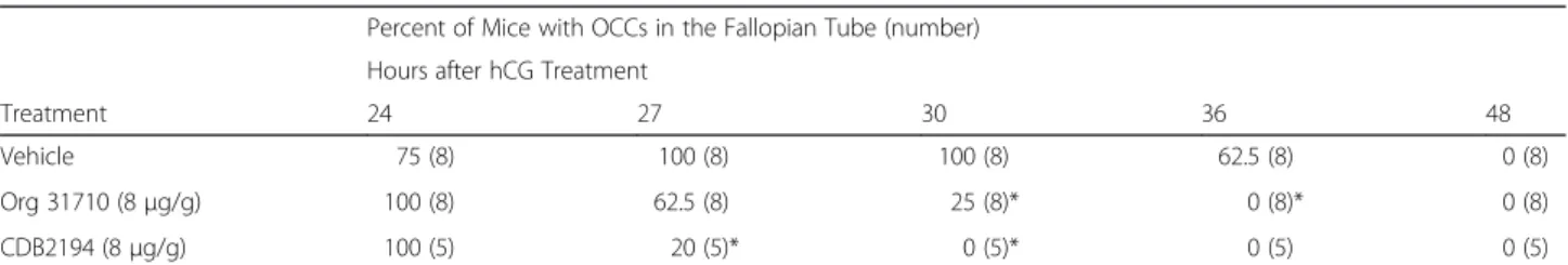
Table 1 presents the percent of mice with OCC present in the fallopian tube after treatment with two PGR an-tagonists or vehicle at different time points. In mice treated with Org 31710, a significant decrease in the ra-tio of mice with OCC present in the fallopian tube was found both at 30 h (p= 0.007) and 36 h (p= 0.0256) after hCG treatment. For mice treated with CDB2194, a simi-lar significant decrease in the ratio of mice with OCC present was found at 27 h (p= 0.007) and 30 h (p= 0.0008) after hCG injection. At the doses given, there were no significant differences at any time point between the effects of the two antagonists. These results are con-sistent with the hypothesis that blockage of the PGR ac-celerates OCC transport.
To identify possible progesterone-dependent genes in-volved in gamete transport and in rapid reduction of CBF, we performed a microarray analysis on fallopian tubes iso-lated from mice and exposedin vitroto 100 nmol/L pro-gesterone for 20 min and 2 h. Exposure to propro-gesterone for 20 min did not induce any significant change in gene expression in the mouse fallopian tube as assessed by the microarray analysis.
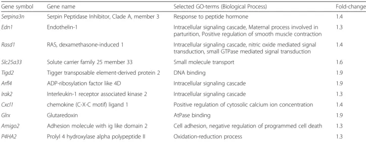
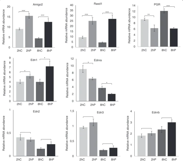
Table 2 provides a list of 11 genes that were differently expressed in mouse fallopian tubes after exposure to progesterone for 2 h compared to control fallopian tubes. Nine of these genes (Arlf4, Rasd1, Edn1, Irak2, Amigo2, Cxcl1, Edn1, P4ha2 and Serpina3n) produce a gene product that is involved in signalling processes. The progesterone induced gene expression changes of three of these genes (Edn1, Amigo2 and Rasd1) were confirmed with qPCR analysis (Fig. 1). The mRNA abundance of Amigo2 was significantly higher after 2 and 8 h of expos-ure to progesterone (p<0.0001 for both time points, respectively) compared to the controls. The mRNA levels of Rasd1 (p<0.0001 for both time points) and Edn1 (p= 0.043 andp= 0.012) were also significantly increased at both time points. EDN1 is known to be involved in muscular contraction of the human and bovine fallopian tube [12–14], and thus, the two other isoforms of endothelin (Edn2 and Edn3) and its receptors (Ednra and Ednrb) were included in the analysis even though
they were not identified as regulated by the microarray. We also includedPgrin the analysis because earlier studies have shown a regulation by progesterone in the fallopian tube [21]. Ednra(p= 0.01 andp= 0.004, 2 h and 8 h, re-spectively) andPgr(p= 0.004 and p<0.0001) had a lower mRNA abundance after progesterone exposure, whereas there was no significant treatment effect forEdn2(p= 0.63 and 0.10) andEdn3(p= 0.25 and 0.29).
To investigate the location of PGR in the mouse fallo-pian tube, we used fallofallo-pian tubes from PGRLacZ mice and a staining kit to detect β-galactosidase activity. As shown in Fig. 2, positive staining was observed in both epithelial and muscle cells.
Discussion
Previous studies reporting the localization of PGR in the fallopian tube has been based on immunostaining [20, 21]. Here, we applied a method that does not depend on the specificity of antibodies. The results show that a sub-population of both epithelial cells and stromal cells ex-press the PGR. This is consistent with the hypothesis that progesterone regulates both ciliary activity and muscle contractions via the PGR.
We also show that a single dose of two different PGR antagonists causes a rapid disappearance of the OCCs in the mouse fallopian tube. This is consistent with earlier studies showing that in rats and mice treated continu-ously with antagonists for PGR, the embryos arrived pre-maturely to the uterus [25, 26].
In a previous study from our laboratory, we showed that the rapid reduction of CBF, which was manifested within 20 min after progesterone administration, was inhibited if the ciliary cells were pre-treated with RU486 and in ciliary cells from mice lacking a functional Pgr [18]. This shows that PGR is most likely involved in this rapid regulation of the CBF. Combining these data with the effect of progesterone antagonists on OCC transport in vivo further indicates an important role of progester-one and the classical nuclear progesterprogester-one receptor in this process, which is at least partly mediated by changes in ciliary activity. In this study, we were not able to iden-tify any changes in transcription abundance 20 min after
Table 1Progesterone receptor antagonists accelerate the transport of the oocyte cumulus complex (OCC) in mouse fallopian tube
Percent of Mice with OCCs in the Fallopian Tube (number)
Hours after hCG Treatment
Treatment 24 27 30 36 48
Vehicle 75 (8) 100 (8) 100 (8) 62.5 (8) 0 (8)
Org 31710 (8μg/g) 100 (8) 62.5 (8) 25 (8)* 0 (8)* 0 (8)
CDB2194 (8μg/g) 100 (5) 20 (5)* 0 (5)* 0 (5) 0 (5)
progesterone treatment. Although it cannot be ruled out whether there are genes regulated after 20 min of pro-gesterone exposure that were not discovered with the microarray analyses, our finding is consistent with the hypothesis that PGR regulates ciliary function in the fal-lopian tube via a non-genomic mechanism. Experiments with blockers of transcription or translation may provide more evidence for this hypothesis.
Transcriptional effects by progesterone were detectable after 2 h, as measured by microarray and qPCR. Nine of the genes that tended to be differentially expressed (Arlf4, Rasd1, Edn1, Irak2, Amigo2, Cxcl1, Edn1, P4ha2 and Serpina3n) produce gene products that are involved in different signalling processes, and some of the genes are regulated by progesterone and Pgr in different cell lines in other studies [34–40].Amigo2plays a role in cell-adhesion and cell migration.Rasd1encodes for a protein that is an activator of G-protein signalling. This gene is thought to be involved in nitric oxide signal transduction, which in the fallopian tube has been shown to affect muscle contractility [41, 42].
Most interestingly, Edn1 was differentially expressed, both after 2 and 8 h of progesterone exposure. The ab-sence of apparent regulation at 20 min suggests that progesterone-activation of the endothelin system via gene transcription is not a prerequisite for regulating cil-iary function, but its regulation at later time points in the fallopian tube suggests that the endothelin system could be involved in other processes related to gamete transport. Indeed, EDN1 is known to induce muscle contractility in the fallopian tube [11, 12, 43]. Earlier stud-ies show that estrogen and estrogen in combination with progesterone and luteinizing hormone significantly stimu-lates the production and secretion of EDN1 from bovine oviduct epithelial cells in culture. However, progesterone
alone at concentrations ranging from 30 nmol/L to 3 μmol/L caused no significant increase in either produc-tion or secreproduc-tion of EDN1 [44, 45]. In the present study progesterone induced the expression of the Edn1 gene while causing a down-regulation of its receptor,Ednra. The reason for these somewhat contradictory results between the studies might be related to the fact that they are per-formed in different species using different methodologies. We measured the mRNA abundance in fallopian tubes from immature mice, afterin vitroprogesterone exposure. The other study measured the cell content and secretion of EDN1 from cultured oviductal epithelial cells from cows sampled during the estrous cycle. Consistent with our results, it has been previously demonstrated that Edn1 is induced by Pgr-A in mouse granulosa cell culture [46].
EDN1 binds to two receptors EDNRA and EDNRB, with equal affinity, but the majority of its effect is medi-ated through EDNRA [47]. The localization of EDN1 within the human fallopian tube is suggested to be mainly within the tubal epithelium but also to a lesser extent in the muscle layer, while EDNRA is suggested to be dominant in the muscle layer [48]. The Edn1, Edn2 and Edn3 as well as the Ednra and Ednrb genes are re-ported to be expressed in bovine and mice oviducts during the estrous cycle [14, 49]. Consistently, we detected mRNA from all three endothelin isoforms and both receptors using qPCR. The expression of Edn2, Edn3 and Ednrb was, however, very low compared toEdn1andEdnra, and only Edn1 and Ednra were differently expressed after exposure to progesterone. In the bovine oviduct, the ex-pressionof EDN1, EDNRAandEDNRBmRNA was high-est during the peri-ovulatory period. During this period, EDN1 also caused the highest increase in oviduct con-traction. In bovine oviduct epithelial cells, EDN1 stimu-lates the production and release of prostaglandins. This
Table 2Genes regulated by progesterone after 2 h in mouse fallopian tubes
Gene symbol Gene name Selected GO-terms (Biological Process) Fold-change
Serpina3n Serpin Peptidase Inhibitor, Clade A, member 3 Response to peptide hormone 1.4
Edn1 Endothelin-1 Intracellular signaling cascade, Maternal process involved in parturition, Positive regulation of smooth muscle contraction
1.3
Rasd1 RAS, dexamethasone-induced 1 Intracellular signaling cascade, nitric oxide mediated signal transduction, small GTPase mediated signal transduction
1.4
Slc25a33 Solute carrier family 25 member 33 Small molecule transport 1.6
Tigd2 Tigger transposable element-derived protein 2 DNA binding 1.9
Arfl4 ADP-ribosylation factor like 4D Intracellular signaling cascade 1.9
Irak2 Interleukin-1 receptor associated kinase 2 Intracellular signaling cascade 1.3
Cxcl1 chemokine (C-X-C motif) ligand 1 Positive regulation of cytosolic calcium ion concentration 1.4
Glrx Glutaredoxin AtPase binding 1.9
Amigo2 Adhesion molecule with ig like domain 2 Cell adhesion, negative regulation of programmed cell death 1.3
P4HA2 Prolyl 4 hydroxylase alpha polypeptide II Oxidation-reduction process 1.3
Rasd1
0 2 4 6 8 10 12 14
Amigo2 PGR
0 5 10 15 20 25 30 35 40
0 5 10 15 20
Relative mRNA abundance
2hC 2hP 8hC 8hP 2hC 2hP
Relative mRNA abundance
8hC 8hP
Relative mRNA abundance
0 0,5 1
Edn2
0 0,5 1
1,5 Edn3
2hC 2hP 8hC
2hP 2hC
8hP
8hP 8hC 2hP 2hC 8hP
8hC ***
*** ***
***
**
***
0 1 2 3 4 5 6 7 8 9
Edn1
8hP 8hC 2hP 2hC
*
*
Relative mRNA abundance
0 2 4 6 8 10
12 Ednra
2hC 2hP 8hC 8hP
*
*
Relative mRNA abundance
0 1 2 3
4 Ednrb
2hC 2hP 8hC 8hP
Relative mRNA abundance
Relative mRNA abundance
Relative mRNA abundance
Fig. 1Progesterone-regulated gene expression in mouse fallopian tubes. Quantitative PCR analyses of 8 transcripts in mouse fallopian tubes treated with ethanol (EtOH) 0.01 % (v/v) as control (C) or progesterone (P; 100 nmol/L) in vitro for 2 h or 8 h. The relative mRNA abundance in the figure is presented as the normalized value (normalized to the housekeeping genes Hprt and Rpl19). Error bars represent standard error of mean. Significantly different expression between control and exposed fallopian tubes at the two time points is indicated by * (p < 0.05), ** (p < 0.01) or *** (p < 0.001)
suggests that in the bovine oviduct, EDN1 produced by the oviduct has a major role in the control of local con-tractions during the transport of gametes/embryos [14]. Our results indicate that the endothelin signalling is likely regulated, in turn, by progesterone.
We are not aware of any previous studies of Ednra regulation in the fallopian tube. Zhang et al. showed that Ednrawas up-regulated in different tissues after proges-terone treatment as well as in pregnant mice [46]. We found a down regulation of Ednrain the fallopian tube, suggesting that progesterone might regulate Ednra -ex-pression in different directions depending on the tissue. When the controls for Ednra were compared, a decrease in mRNA abundance between the 2 h and 8 h samples was discovered. This decrease in abundance could either be due to less transcription and/or decreased stability of the mRNA.
In both the mouse and human fallopian tube, both iso-forms of PGR are highly expressed [20]. In humans, the expression seems to vary across the menstrual cycle and differ between the different parts of the fallopian tube [50]. In our study,Pgr mRNAwas down-regulated by pro-gesterone, which is consistent with results from an earlier study showing that there was a time-dependent reduction of PGRA and PGRB protein expression in immature mice treated with a single dose of progesterone [21].
Ednraand Pgrwere not markedly affected by the pro-gesterone treatment as measured by the microarray ana-lysis at 20 min or 2 h. However, with qPCR, significantly lowered mRNA levels after progesterone treatment were identified at both 2 h and 8 h in a separate experiment. One reason for this apparent difference at 2 h could be that both are receptors, and therefore, their expression is low compared to many other types of mRNA. A micro-array has a considerably higher detection limit than a qPCR assay; thus, changes in genes that are hardly de-tectable by a microarray are much more subjected to noise than more highly expressed genes. A second rea-son might be that a gene might only be present and reg-ulated in a small subset of the cells analysed, making it difficult to find small changes because they can drown in the noise from other cells. A third explanation could also be that the arrays were only performed with a repli-cation of four per group, while the smaller amount of RNA required for the qPCR allowed us to analyze 9 bio-logical replicates per treatment group, providing much higher statistical power.
A recent study investigated the differences in gene ex-pression in fallopian tubes of mice completely lacking a functional Pgr (PgrLacZ mice) and heterozygous mice (PgrLacZ (+/−)) carrying one copy of thePgrgene using both microarray and qPCR [51]. However, the overlap of the differentially expressed genes in that study and the genes found to be regulated directly by progesterone in
the present study was small. Indeed, comparing the dif-ference in gene expression between the two genotypes will reveal differences that are also indirectly affected by the lack of the receptor. Indeed, of the 9 genes investi-gated by qPCR after stimulation of ovulation in imma-ture mice by hCG (thus inducing a progesterone-surge), 6 genes differed before treatment between the two geno-types. The authors identified an up-regulation of Edn3 in heterozygous mice 8 hours after hCG treatment, while we did not observe any changes of Edn3 either at 2 or 8 h after progesterone treatment. This suggests that the up-regulation of Edn3 found during induced ovulation in the fallopian tube in the other study is not a direct ef-fect of PGR activation.
Conclusions
To our best knowledge, this is the first study demon-strating that Edn1 and Ednra are directly regulated by progesterone in the mouse fallopian tube. We also con-firm that PGR antagonists accelerate the transport of the OCC through the fallopian tube. Our results further sup-port that progesterone and PGR plays an essential role in OCC transport that involves both non-genomic and gen-omic mechanisms. It is important to reveal the mecha-nisms and regulation of gamete transport in the fallopian tube because it is an essential step in natural reproduction and may contribute to a better understanding of the causes of infertility in women and the development of new and better contraceptives. To better understand the role of pro-gesterone in the fallopian tube, studies on gene regulation in vivo would be important to perform in the future.
Additional file
Additional file 1: Table S1.Taqman assay-id for primers and probes used for quantitative PCR analysis.
Abbreviations
CBF:Ciliary beat frequency; EDN1: Endothelin 1; EDN2: Endothelin 2; EDN3: Endothelin 3; EDNRA: Endothelin receptor A; EDNRB: Endothelin receptor B; hCG: Human chorionic gonadotropin; mPRs: Membrane progesterone receptors; OCC: Oocyte cumulus complex; PGE: Prostaglandins E-series; PGF: Prostaglandins F-series; PGR: Progesterone receptor;
PMSG: Pregnant mare's serum gonadotropin; qPCR: Quantitative PCR; RU486: Mifepristone.
Competing interests
The authors declare that they have no competing interests.
Authors’contributions
Acknowledgements
We thank the Swegene Center for Integrative Biology at Lund University for help with the microarray laboratory work and analysis and the Genomics Core Facility platform at the Sahlgrenska Academy, University of Gothenburg for help with the qPCR analysis. We thank American Journal Experts (www.aje.com) for assistance with language and writing style. We also thank Dr Carolin Rutgersson for assistance with tissue sampling. This work was supported by the Swedish Research Council Formas and the Swedish Research Council VR.
Author details 1
Department of Infectious Diseases, Institute of Biomedicine, The Sahlgrenska Academy, University of Gothenburg, Guldhedsgatan 10, SE–413 46 Gothenburg, Sweden.2Institute of Neuroscience and Physiology, the Sahlgrenska Academy, University of Gothenburg, Box 454, SE–405 30 Gothenburg, Sweden.
Received: 21 November 2014 Accepted: 30 April 2015
References
1. Croxatto HB, Ortiz ME. Egg transport in the fallopian tube. Gynecol Invest. 1975;6(3–4):215–25.
2. Mahmood T, Saridogan E, Smutna S, Habib AM, Djahanbakhch O. The effect of ovarian steroids on epithelial ciliary beat frequency in the human Fallopian tube. Hum Reprod. 1998;13(11):2991–4.
3. Lyons RA, Saridogan E, Djahanbakhch O. The effect of ovarian follicular fluid and peritoneal fluid on Fallopian tube ciliary beat frequency. Hum Reprod. 2006;21(1):52–6.
4. Bylander A, Nutu M, Wellander R, Goksor M, Billig H, Larsson DGJ. Rapid effects of progesterone on ciliary beat frequency in the mouse fallopian tube. Reprod Biol Endocrinol. 2010;8:48.
5. Graham JD, Clarke CL. Physiological action of progesterone in target tissues. Endocr Rev. 1997;18(4):502–19.
6. Bishop CV, Stormshak F. Non-genomic actions of progesterone and estrogens in regulating reproductive events in domestic animals. Vet J. 2008;176(3):270–80.
7. Frye CA. Steroids, reproductive endocrine function, and cognition: A review. Minerva Ginecol. 2009;61(6):563–85.
8. Akison LK, Robker RL. The critical roles of progesterone receptor (PGR) in ovulation, oocyte developmental competence and oviductal transport in mammalian reproduction. Reprod Domest Anim. 2012;47 Suppl 4:288–96. 9. Wanggren K, Stavreus-Evers A, Olsson C, Andersson E, Gemzell-Danielsson K.
Regulation of muscular contractions in the human Fallopian tube through prostaglandins and progestagens. Hum Reprod. 2008;23(10):2359–68. 10. Helm G, Owman C, Sjoberg NO, Walles B. Motor activity of the human
Fallopian tube in vitro in relation to plasma concentration of oestradiol and progesterone, and the influence of noradrenaline. J Reprod Fertil. 1982;64(1):233–42.
11. Al-Alem L, Bridges PJ, Su W, Gong MC, Iglarz M, Ko C. Endothelin-2 induces oviductal contraction via endothelin receptor subtype A in rats. J Endocrinol. 2007;193(3):383–91.
12. Jankovic SM, Jankovic SV, Lukic G, Canovic D, Folic M. The contractile effects of endothelins on isolated isthmic segment of human oviduct at the luteal phase of the menstrual cycle. Methods Find Exp Clin Pharmacol. 2010;32(2):91–5.
13. Jankovic SM, Jankovic SV, Lukic G, Radonjic V, Cupara S, Stefanovic S. Contractile effects of endothelins on isolated ampullar segment of human oviduct in luteal phase of menstrual cycle. Pharmacol Res. 2009;59(1):69–73. 14. Priyadarsana M, Wijayagunawardane B, Miyamoto A. Endothelin-1 system in the bovine oviduct: a regulator of local contraction and gamete transport. J Cardiovasc Pharmacol. 2004;44 Suppl 1:S248–51.
15. Lindblom B, Hamberger L, Ljung B. Contractile patterns of isolated oviductal smooth muscle under different hormonal conditions. Fertil Steril. 1980;33(3):283–7.
16. Spilman CH, Harper MJ. Effects of prostaglandins on oviductal motility and egg transport. Gynecol Invest. 1975;6(3–4):186–205.
17. Wanggren K, Lalitkumar PG, Stavreus-Evers A, Stabi B, Gemzell-Danielsson K. Prostaglandin E2 and F2alpha receptors in the human Fallopian tube before and after mifepristone treatment. Mol Hum Reprod. 2006;12(9):577–85.
18. Bylander A, Lind K, Goksor M, Billig H, Larsson DGJ. The classical
progesterone receptor mediates the rapid reduction of fallopian tube ciliary beat frequency by progesterone. Reprod Biol Endocrinol. 2013;11(1):33. 19. Wessel T, Schuchter U, Walt H. Ciliary motility in bovine oviducts for sensing
rapid non-genomic reactions upon exposure to progesterone. Horm Metab Res. 2004;36(3):136–41.
20. Teilmann SC, Clement CA, Thorup J, Byskov AG, Christensen ST. Expression and localization of the progesterone receptor in mouse and human reproductive organs. J Endocrinol. 2006;191(3):525–35.
21. Shao R, Weijdegard B, Ljungstrom K, Friberg A, Zhu C, Wang X, et al. Nuclear progesterone receptor A and B isoforms in mouse fallopian tube and uterus: implications for expression, regulation, and cellular function. Am J Physiol Endocrinol Metab. 2006;291(1):E59–72.
22. Zhu Y, Hanna RN, Schaaf MJ, Spaink HP, Thomas P. Candidates for membrane progestin receptors-Past approaches and future challenges. Comp Biochem Physiol C Toxicol Pharmacol. 2008;148(4):381–9. 23. Nutu M, Weijdegård B, Thomas P, Bergh C, Thurin-Kjellberg A, Pang Y, et al.
Membrane progesterone receptor gamma: tissue distribution and expression in ciliated cells in the fallopian tube. Mol Reprod Dev. 2007;74(7):843–50. 24. Nutu M, Weijdegård B, Thomas P, Thurin-Kjellberg A, Billig H, Larsson DGJ.
Distribution and hormonal regulation of membrane progesterone receptors beta and gamma in ciliated epithelial cells of mouse and human fallopian tubes. Reprod Biol Endocrinol. 2009;7:89.
25. Fuentealba B, Nieto M, Croxatto HB. Ovum transport in pregnant rats is little affected by RU486 and exogenous progesterone as compared to cycling rats. Biol Reprod. 1987;37(4):768–74.
26. Vinijsanun A, Martin L. Effects of progesterone antagonists RU486 and ZK98734 on embryo transport, development and implantation in laboratory mice. Reprod Fertil Dev. 1990;2(6):713–27.
27. Gainer EE, Ulmann A. Pharmacologic properties of CDB(VA)-2914. Steroids. 2003;68(10–13):1005–11.
28. Hild SA, Reel JR, Hoffman LH, Blye RP. CDB-2914: anti-progestational/ anti-glucocorticoid profile and post-coital anti-fertility activity in rats and rabbits. Hum Reprod. 2000;15(4):822–9.
29. Reel JR, Hild-Petito S, Blye RP. Antiovulatory and postcoital antifertility activity of the antiprogestin CDB-2914 when administered as single, multiple, or continuous doses to rats. Contraception. 1998;58(2):129–36. 30. Kloosterboer HJ, Deckers GH, Schoonen WG. Pharmacology of two new
very selective antiprogestagens: Org 31710 and Org 31806. Hum Reprod. 1994;9 Suppl 1:47–52.
31. Irizarry RA, Hobbs B, Collin F, Beazer-Barclay YD, Antonellis KJ, Scherf U, et al. Exploration, normalization, and summaries of high density oligonucleotide array probe level data. Biostatistics. 2003;4(2):249–64.
32. Tusher VG, Tibshirani R, Chu G. Significance analysis of microarrays applied to the ionizing radiation response. Proc Natl Acad Sci U S A.
2001;98(9):5116–21.
33. Saeed AI, Sharov V, White J, Li J, Liang W, Bhagabati N, et al. TM4: a free, open-source system for microarray data management and analysis. Biotechniques. 2003;34(2):374–8.
34. Orbo A, Moe BT, Gronaas H, Paulssen RH. Early effects of high
concentrations of progesterone and mifepristone A gene expression study of endometrial cancer cells (Ishikawa). J Steroid Biochem Mol Biol. 2009;113(1–2):139–49.
35. Marquez RT, Baggerly KA, Patterson AP, Liu J, Broaddus R, Frumovitz M, et al. Patterns of gene expression in different histotypes of epithelial ovarian cancer correlate with those in normal fallopian tube, endometrium, and colon. Clin Cancer Res. 2005;11(17):6116–26.
36. Sriraman V, Sinha M, Richards JS. Progesterone receptor-induced gene expression in primary mouse granulosa cell cultures. Biol Reprod. 2010;82(2):402–12.
37. Srivastava MD, Thomas A, Srivastava BI, Check JH. Expression and modulation of progesterone induced blocking factor (PIBF) and innate immune factors in human leukemia cell lines by progesterone and mifepristone. Leukemia Lymphoma. 2007;48(8):1610–7.
38. Bray JD, Jelinsky S, Ghatge R, Bray JA, Tunkey C, Saraf K, et al. Quantitative analysis of gene regulation by seven clinically relevant progestins suggests a highly similar mechanism of action through progesterone receptors in T47D breast cancer cells. J Steroid Biochem Mol Biol. 2005;97(4):328–41.
in regulating gene expression in pregnant human uterine myocytes. J Cell Mol Med. 2012;16(10):2487–503.
40. Su S, Blackwelder AJ, Grossman G, Minges JT, Yuan L, Young SL, et al. Primate-specific melanoma antigen-A11 regulates isoform-specific human progesterone receptor-B transactivation. J Biol Chem. 2012;287(41):34809–24. 41. Ekerhovd E, Brannstrom M, Alexandersson M, Norstrom A. Evidence for
nitric oxide mediation of contractile activity in isolated strips of the human Fallopian tube. Hum Reprod. 1997;12(2):301–5.
42. Ekerhovd E, Brannstrom M, Weijdegard B, Norstrom A. Localization of nitric oxide synthase and effects of nitric oxide donors on the human Fallopian tube. Mol Hum Reprod. 1999;5(11):1040–7.
43. Wijayagunawardane MP, Miyamoto A, Cerbito WA, Acosta TJ, Takagi M, Sato K. Local distributions of oviductal estradiol, progesterone, prostaglandins, oxytocin and endothelin-1 in the cyclic cow. Theriogenology. 1998;49(3):607–18.
44. Wijayagunawardane MP, Choi YH, Miyamoto A, Kamishita H, Fujimoto S, Takagi M, et al. Effect of ovarian steroids and oxytocin on the production of prostaglandin E2, prostaglandin F2alpha and endothelin-1 from cow oviductal epithelial cell monolayers in vitro. Anim Reprod Sci. 1999;56(1):11–7.
45. Wijayagunawardane MP, Miyamoto A, Sato K. Prostaglandin E2, prostaglandin F2 alpha and endothelin-1 production by cow oviductal epithelial cell monolayers: effect of progesterone, estradiol 17 beta, oxytocin and luteinizing hormone. Theriogenology. 1999;52(5):791–801. 46. Zhang Y, Knutsen GR, Brown MD, Ruest LB. Control of endothelin-a receptor
expression by progesterone is enhanced by synergy with Gata2. Mol Endocrinol. 2013;27(6):892–908.
47. Apa R, Miceli F, de Feo D, Mastrandrea ML, Mancuso S, Napolitano M, et al. Endothelin-1 inhibits basal and human chorionic gonadotrophin-stimulated progesterone production. Hum Reprod. 1998;13(9):2425–9.
48. Sakamoto M, Sakamoto S, Kubota T, Aso T, Azuma H. Localization and role of endothelin-1 and endothelin receptors in the human Fallopian tube. Mol Hum Reprod. 2001;7(11):1057–63.
49. Jeoung M, Lee S, Hawng HK, Cheon YP, Jeong YK, Gye MC, et al. Identification of a novel role for endothelins within the oviduct. Endocrinology. 2010;151(6):2858–67.
50. Pollow K, Inthraphuvasak J, Manz B, Grill HJ, Pollow B. A comparison of cytoplasmic and nuclear estradiol and progesterone receptors in human fallopian tube and endometrial tissue. Fertil Steril. 1981;36(5):615–22. 51. Akison LK, Boden MJ, Kennaway DJ, Russell DL, Robker RL. Progesterone
receptor (PGR) dependent regulation of genes in the oviducts of female mice. Physiol Genomics. 2014;46(16):583–92.
Submit your next manuscript to BioMed Central and take full advantage of:
• Convenient online submission
• Thorough peer review
• No space constraints or color figure charges
• Immediate publication on acceptance
• Inclusion in PubMed, CAS, Scopus and Google Scholar
• Research which is freely available for redistribution