Page 1 of 61
Managing the Challenge of Drug Induced Liver Injury:
A roadmap for the
development and deployment of preclinical predictive models
AUTHORS
Richard J Weaver1, Eric A Blomme2, Amy E Chadwick3, Ian M Copple3, Helga HJ Gerets4, Christopher E Goldring3, Andre Guillouzo5 Philip G Hewitt6, Magnus Ingelman-Sundberg7, Klaus Gjervig Jensen8, Satu Juhila9, Ursula Klingmüller10, Gilles Labbe11, Michael J Liguori2, Cerys A Lovatt12, Paul Morgan13, Dean J Naisbitt3, Raymond HH Pieters14, Jan Snoeys15, Bob van de Water16, Dominic P Williams13, B Kevin Park3
1. Institute de Recherches Internationales Servier, Biopharmacy, rue Carnot, 92284, Suresnes, France
2. Abbvie, Global Preclinical Safety, 1 North Waukegan Road, R46G/AP13A-3, North Chicago, IL 60064
3. MRC Center for Drug Safety Science, Department of Molecular and Clinical Pharmacology , University of Liverpool , Sherrington Building, Ashton Street , Liverpool , L69 3GE , England. 4. UCB BioPharma SPRL, Developmental Sciences, Chemin du Foriest, 1420 Braine-l'Alleud,
Belgium
5. University Rennes, Inserm, Inra, Institut NUMECAN (Nutrition Metabolisms and Cancer), UMR_S 1241, 35000 Rennes, France
6. Merck KGaA, Non-Clinical Safety, Darmstadt, Germany
7. Karolinska Institutet, Department of Physiology and Pharmacology, Biomedicum 5B, Stockholm, Sweden
8. H Lundbeck, Drug ADME Research, H. Lundbeck A/S, 9 Ottiliavej, Valby, Denmark. 9. Orion Pharma, In Vitro Biology, Espoo , Finland
10. Deutsches Krebsforschungszentrum (DKFZ), Systems Biology of Signal Transduction, German Cancer Research Center, Heidelberg, Germany
11. Sanofi, Investigative Toxicology, Preclinical Safety, Sanofi R&D, Paris, France. 12.GlaxoSmithKline, R&D, Ware, Hertfordshire, UK
13.AstraZeneca, Drug Safety and Metabolism, IMED Biotech Unit, Cambridge Science Park, Cambridge, CB4 0WG, UK.
14. Institute for Risk Assessment Sciences, Immunotoxicology, Faculty of Veterinary Medicines, Utrecht University, Utrecht, The Netherlands
15. Drug Metabolism and Pharmacokinetics, Janssen Research and Development, Turnhoutseweg 30, Beerse, Belgium
Page 2 of 61
Authors for Correspondence
B Kevin Park
MRC Centre for Drug Safety Science, Department of Molecular and Clinical Pharmacology, University of Liverpool, Sherrington Building, Ashton Street, Liverpool, L69 3GE, England.
Richard J Weaver
Page 3 of 61
Glossary
2D, 2-dimensional; 3D, 3-dimensional; ADME, Absorption, Distribution, Metabolism Excretion; ADR, Adverse Drug Reaction; ADRs, Adverse Drug Reactions; AE, Adverse event; AEs, Adverse events;
APAP, acetaminophen; ATF6, Activating transcription factor 6; ATP, adenosine triphosphate; BA, bile acid; BAC, bacterial artificial chromosome; BC, bile canaliculi; BCRP, breast cancer resistance protein;
Bip, Binding immunoglobulin protein; BSEP, bile salt export pump; CsA, cyclosporine A; CA, cholic acid; CADs, Cationic amphiphilic drugs; CDCA, chenodeoxycholate; CHOP, C/EBP homologous protein; ClogP, The logP value of a compound; Cmax, maximum concentration; CPT-1, carnitine palmitoyltransferase-1; CRM, chemically reactive metabolites; Css, Concentration at Steady State; CTLA-4, cytotoxic T-lymphocyte-associated protein 4; CYP, cytochrome P450; DAMPs, damage-associated molecular patterns; DCA, deoxycholate; DIC, diclofenac; DILI, drug-induced liver injury;
DILI-sym, drug-induced liver injury modelling software; EFPIA, European Federation of Pharmaceutical Industries and Associations; ENT, equilibrative nucleoside transporter;ER,
endoplasmic reticulum; GCDCA, glycochenodeoxycholate; GFP, green fluorescent protein; Glu-Gal, glucose-galactose; GSH, glutathione; HCA, high-content analysis; hENT1, human endonucleoside transporter-1; HLA, human leukocyte antigen; HMGB1, high mobility group box protein-1; ICAM1, intercellular Adhesion Molecule 1; IFN-, interferon-; IL-1β, interleukin-1 β; IL-8, interleukin-8; iPS, induced pluripotent stem cells; IRE1α, inositol-requiring enzyme 1 α; IVIVE, in vitro-in vivo; LCA, lithocholic acid; LPS, lipopolysaccharide; LSEC, liver sinusoidal endothelial cell; luc, luciferase; MATE, Multidrug and toxin extrusion protein; MICS, microphysiological systems; MIP-DILI, Mechanism-Based Integrated Systems for the Prediction of Drug-Induced Liver Injury; MLC, myosin light chain; MLCK, myosin light chain kinase; MRP, multidrug resistance-associated protein; mtDNA,
mitochondrial DNA; NCE, new chemical entity; NCEs, new chemical entities; NFkB, nuclear factor-kappa B; NK, natural killer; NPCs, non-parenchymal cells; Nrf2, nuclear factor erythroid 2–related factor 2; NTCP, Na+-taurocholate co-transporting polypeptide; OATP, organic anion transporting polypeptide; OAT, Organic Anion Transporter; OCT, organic cation transporter; OST, Organic solute transporter; PBMC, peripheral blood mononuclear cells; PD1, Programmed cell death protein 1; PERK, protein kinase RNA-like endoplasmic reticulum kinase; PHH, primary human hepatocyte;
PKPD, Pharmacokinetics & pharmacodynamics; PL, phospholipidosis ; R&D, research and development; RelA, REL-associated protein; ROCK, Rho kinase; ROS, reactive oxygen species; S100A9, S100 calcium-binding protein A9; SCHH, sandwich-cultured human hepatocyte; SOS, sinusoidal obstructive syndrome; SRXN1, sulfiredoxin 1; TCA, taurocholic acid; TCR, T-cell receptor;
Page 4 of 61
Abstract
Page 5 of 61
Introduction
Adverse drug reactions (ADRs) are a major clinical problem in terms of patient morbidity, patient mortality, cost to healthcare systems and failure of drugs in development. The liver is one of the organs most susceptible to drug toxicity, and in the clinic, drug-induced liver injury (DILI) has accounted for more than 50% of acute liver failure cases1. Excluding acetaminophen, DILI accounts for approximately 14% of acute liver failures with a mortality rate of up to 10%2. DILI is also a major cause of drug attrition, leading to withdrawal of potentially valuable therapies, both during preclinical testing, clinical trials and post-marketing3,4. Importantly, the FDA has annotated 750 drugs that have some degree of DILI risk5. However, it is clear that current preclinical testing paradigms based on a combination of various in vitro and in vivo models are poorly predictive, at a quantitative and mechanistic level, of the potential of a new drug candidate to cause DILI in humans, in particular those drugs that show poorly defined dose-response relationships and/or human specific mechanisms of toxicity.
There is an emerging body of evidence that DILI, as it occurs in humans, can be a multi-step and multicellular disease process with a diverse range of chemical aetiologies (Figure 1)6-8. This means that prediction of all forms of DILI may be inherently intractable to simple solutions, such as single cell culture screening strategies. Thus, a much better understanding of the mechanisms underlying DILI is essential in order to a) evaluate the strengths and weaknesses of currently available test systems and b) inform the design and construction of new improved predictive models. Crucially, we must define not only whether a test system is fit-for-purpose, but more critically “what particular purpose is a test system fit for”. In addition, we need to be aware of which forms of DILI we can predict and which forms of DILI we cannot predict, when there is no human experience with the drug.
Page 6 of 61
mechanism-based, pragmatic and sufficiently adaptable to be of practical application to 1) influence drug design early enough in the discovery phase and 2) manage risk assessment in drug development, which is amenable to evaluation by drug regulators, but cognisant of current limitations in our understanding of DILI. This is particularly important when human risk factors dominate over chemical risk factors, e.g. for idiosyncratic drug toxicity.
It is clear from a review of the literature, that no single system is fit-for-purpose as a universal test for DILI in humans, which is a patient-specific, temporal, multifactorial pathophysiological process. Therefore, what is required is a portfolio of robust and well-characterised predictive DILI platforms that have their purpose well-defined, and acceptable in a theoretical and practical sense to academic, industry and regulatory agencies.
Page 7 of 61
Proposed DILI Roadmap
Potential strategies to de-risk DILI in drug discovery have previously been published. These
approaches, whether presented as in vitro hazard/liability matrices9,10 or tiered testing cascades, are typically those of a single institution or focus only on single mechanisms, proprietary test systems 11-14, legacy compound datasets15, and/or technology platforms16,17. For this reason, there has
remained little or no consensus on the use or approach to de-risk DILI. Moreover, many of these strategies may not be readily amenable for adoption by small enterprises and start-ups where in-house capacity and availability of proprietary compounds to validate such approaches are lacking.
The DILI Roadmap discussed in this Perspective was established by nine medium-large EFPIA
pharmaceutical companies, SMEs and academic partners as a holistic strategy to manage human DILI risk. Our approach delivers an in-depth inter-laboratory evaluation of the fundamental and
reproducible performance of assays, to identify chemical liabilities in drug discovery and to manage DILI risk in development. Our roadmap (Figure 2) integrates established and emerging test systems, and is based on a 3-tiered approach, whereby the complexity of the model increases progressively from single cell 2D to multi-cell 3D systems through to systems that incorporate human individual-specific factors, such as genetic- or disease-related factors.
As the Roadmap incorporates assays and cell models which are well-characterized and have proven their utility18-20 as well as emerging novel systems with anticipated future benefit, this enables the versatility of the Roadmap to evolve with the advent of future qualified complex DILI models
Cell Models
Page 8 of 61
exert a relevant phenotype, mimicking the functionality of hepatocytes in vivo and, since many drugs exert a delayed DILI response, the system must exhibit long-term stability.
Much attention has been placed on the refinement of 2D cell systems and development of a 3D spheroid system based largely on primary hepatocytes, where the transcriptomic, proteomic and metabonomic profile as well as the functionality of the spheroids are very similar to the specific donor liver18,21-23. Such spheroids have also been shown to imitate liver disease induced by altered physiological conditions24. When challenged in long-term cultures with 122 drugs with or without direct implication in clinical DILI, this spheroid system successfully detected 69% of all hepatotoxic compounds without producing any false positive results (100% specificity), thereby exceeding all previously published in vitro assays at substantially lower drug concentration levels, that approach Cmax or Css in blood with drugs known to cause human DILI23. Single cell models, whether primary or immortalised, nevertheless lack the intricacies of a multicellular environment, and whilst a range of inventive technologies have been developed to build complex 3-D structural models, none published so far has been able to recapitulate the complex physiology of the intact liver.
Much effort is now being directed to develop different kinds of more physiologically-relevant in vitro
systems such as 2D and 3D multi-cellular tissue chip and microfluidic systems which can provide a relevant cellular milieu for studies of liver function and DILI25,26. In addition, microfluidic microphysiological (organ-on-chip) systems are being built in a multi-organ fashion which in the future may be able to further integrate complex mechanisms relevant for the production of idiosyncratic human DILI27.
However, despite significant efforts towards developing the next generation of advanced in vitro -based liver models with greater physiological relevance, none are presently in routine use by industry.
This in part is due to remaining technological challenges and a required demonstrable ‘paradigm shift’
Page 9 of 61
common practice for screening due to their ease of use, availability and suitability for HTS28,31, despite their reported limitations of physiological relevance8,32.
It may not be possible to combine all features of a fully functioning liver in microphysiological tissue-based systems (MPS) models, thereby requiring the integration of input from more than a single test system. Nevertheless, there is an opportunity to capitalize on the use of established 2D cell models and assays in a tiered and congruent manner that applies physiological and pharmacological knowledge, advantages and limitations of these 2D cell models, with more advanced models as they become available and qualified for use. Cell-based 2D models currently comprise traditional liver derived cell-lines and primary hepatocytes. Features facilitating use of 2D cell models such as HepG2, HepaRG, and primary hepatocytes are summarized elsewhere33 and in Table 1, and represent the most commonly available and widely used cell models by industry. Given this, along with the breath of available literature on the characterization of HepG2 and HepaRG, this prioritizes the use of these cell models in a tiered approach for screening of key mechanisms associated with human DILI.
Page 10 of 61
Physicochemical characteristics associated with DILI risk
High daily doses and high systemic exposures to drugs are associated with increased risk of DILI34,35. It is therefore important to account for the intended pharmacological drug exposure (concentration) and ADME data alongside the use of the in vitro DILI roadmap (Figure 2), to help integrate concentration-effect relationships for the prediction of DILI. Low doses are desirable and ideally doses < 100 mg/day should be targeted for oral drugs36. For example, evaluation of the top 200 oral drugs in 2009 in the USA and of 68 drugs recalled or associated with a black box warning due to idiosyncratic toxicity, indicated that the major differentiating factor between the two groups of drugs was the total daily dose37. Likewise, the vast majority of oral drugs with reported idiosyncratic liver toxicity are administered at high clinical doses38.
Not surprisingly, given their influence on efficacious doses, some physicochemical properties (e.g. lipophilicity, polar surface area, number of hydrogen bond donors and acceptors) have also been shown to be associated with an increased risk of DILI. For this reason, determining the physiochemical properties of a compound is important. Computational tools are available as TIER 1 assays to guide early drug design by predicting physicochemical properties or sites of metabolism, metabolite structure, cytochrome P450 (CYP) binding/inhibition, and CYP induction39,40. Examples include the Meteor program, MetaSite, and ADMET Predictor39,41,42. The links between compound characteristics of interest (e.g., physicochemical properties, bioactivation, and general toxicity) and molecular
structure (i.e., ‘the similarity principle’) form the basis of these in silico applications that differ only in
terms of complexity and performance43. For example, various toxicophores or problematic substructures can be identified with this approach44,45. These computational assessments are typically conducted prior to TIER I assays in order to prevent the synthesis of compounds with low probability of success or to prioritize screening of subsets of compounds, thereby optimizing drug discovery
efforts. However, in silico tools still have some limitations and traditional “wet-lab” experimentation
Page 11 of 61
compounds to confirm predicted readouts from these in silico applications (i.e., spot-checking exercise). Several quantitative systems toxicology platforms have been proposed to predict and understand DILI. DILIsym® is an in silico approach to predict DILI based on known mechanisms46. The software relies on predetermined mechanisms, such as oxidative stress, glutathione metabolism, mitochondrial dysfunction, and ATP depletion to simulate a DILI outcome41. The DILI prediction system (DILIps) uses a quantitative structure-activity relationship (QSAR) approach to classify a compound’s
propensity to induce DILI47 by use of drugs known to cause DILI. Whilst retrospective analyses of known hepatotoxic drugs help to identify potential hazard, quantifying risk and predictive value and identification of some types of DILI, such as idiosyncratic DILI in patients is not yet achievable. The main points concerning the importance of dose and physicochemical properties are presented in BOX 1 and discussed in further detail in the supplementary section.
Intracellular perturbations associated with DILI liability
A number of intracellular chemical perturbations elicited by pharmaceutical compounds are associated with a DILI liability, including mitochondrial dysfunction, inhibition of biliary efflux, lysosomal impairment, production of reactive metabolites, endoplasmic reticulum stress, as well as involvement of the immune system (Figure 1). Below we will discuss these factors and examine how they can be evaluated using our tiered testing strategy as shown in the DILI roadmap (Figure 2).
Mitochondrial dysfunction
Page 12 of 61
Page 13 of 61
investigation to confirm classification and define mechanisms can include traditional respirometry in isolated mitochondria or the use of a Seahorse extracellular flux analyser for membrane potential. These two assays allow the measurement of both oxidative phosphorylation and glycolytic function in real-time, in whole and permeabilised cells or isolated mitochondria53,55.
Page 14 of 61
weeks, confirming the mechanism of toxicity via inhibition of mitochondrial DNA replication63. The use of this model to assess mitochondrial toxicity against a background of underlying liver pathology has also been described. In particular, steatotic HepaRG models have been developed, and used to evaluate the effects of steatosis on cellular bioenergetics and individual susceptibility to mitotoxicants61.
TIER 2 mitotoxicity testing can also encompass 3D cell systems, such as spheroids, which represent a more functionally-relevant liver system64. Work to create 3D spheroids of primary human hepatocytes (PHH) has revealed an increased sensitivity to fialuridine over extended periods, with effects beginning at 7 days and increasing over 28 days of exposure21. Such studies reveal the importance of temporal toxicodynamic events not captured in short-term assays.
Although TIER 1 and TIER 2 testing has developed to improve the detectability of mitotoxicants, these results must always be viewed as identification of chemical liabilities in the absence of quantitative IVIVE extrapolation and PKPD clinical readout. Traditionally, it has been difficult to bridge from in vitro
studies to humans using animal models of mitotoxicity. Specifically, in vivo studies do not adequately represent the clinical situation as they are often performed in young and healthy in-bred rodent models which have therefore a reduced sensitivity to mitotoxicants65. As such, TIER 3 systems for evaluating mitotoxicity remain less well-defined. However, one successful example was the susceptibility of the chimeric TK-NOG mouse with humanised liver to fialuridine-induced hepatotoxicity, at doses approximately ten times the therapeutic exposure, which presented with clinical and pathological features in line with the DILI observed in humans66.
It is clear that the current DILI roadmap for mitotoxicity testing can identify certain mitochondrial liabilities. However, there is still no clear path to predicting potential risk in patients. In silico
Page 15 of 61
been developed to aid the mechanistic understanding of clinical hepatotoxicity and was used successfully to determine retrospectively the role of mitochondria in tolvaptan hepatotoxicity and most recently macrolide antibiotics and TAK-87567-70. Overall, the last 10 years have seen significant advances in the development of models and test systems to identify mitochondrial toxicity. Practically, it is recommended that they are deployed in preclinical safety testing in a step-wise approach; TIER 1 is suitable for the risk assessment of many compounds, whilst TIER 2 and subsequent mechanistic work incorporating temporal toxicodynamic events can be used to follow-up on selected compounds of interest. TIER 3 remains a work in progress which is continuing to develop as the fundamental mechanisms directing inter-individual variation and species selectivity are discovered. Until such further progress is made, TIER 3 can function as an opportunity to retrospectively assign mechanisms and understand at-risk populations once clinical signals of hepatotoxicity have been observed.
Energy metabolism and in particular, mitochondria respiration are key processes in the build-up of fatty liver deposits as the site of fatty acid oxidation, and many of the drugs known to interact with
the mitochondria at the level of β-oxidation, electron transport chain or mtDNA are associated with hepatic steatosis71. Drugs implicated in hepatic steatosis affecting the electron transport chain (ETC)
and β-oxidation of fatty acids is metabolically linked, such that drugs affecting ETC pathway invariably
inhibit β-oxidation. The rate-limiting step for fatty acid oxidation is the mitochondrial carnitine-palmitoyl transferase-1 system; when mitochondrial β-oxidation is severely inhibited, fatty acyl-CoA
Page 16 of 61
Transporters in drug- and bile acid (BA)- induced liver injury
Drug-induced cholestatic liver disorders are among the most severe clinical manifestations of human DILI and account for almost half of the cases of drug-related hepatotoxicity73,74. These liver disorders are broad, yet are characterized by acute and chronic impaired hepatocellular secretion of bile, with accumulation of systemic BAs and their bile salts, bilirubin and cholesterol (Supplementary information).
In many cases, the disruption of BA secretion results from functional changes in the hepatobiliary transporter system. These membrane transporters facilitate uptake and efflux of endogenous substances and drugs between the basolateral, sinusoidal and apical (bile canalicular) membranes of the hepatocyte. Many drugs are also identified as inhibitors of these membrane transporters (Figure 3). The localization of membrane transporters, expression of family and sub-family members, and polarity of transporter proteins convey the highly-regulated physiological and pharmacological role in the hepatobiliary transport of BAs (supplemental text).
Page 17 of 61
DILI severity assessment, published BSEP inhibition datasets were recently re-examined, and it was concluded that the inverted vesicle assay is not predictive of the toxic potential of drugs80.
Inverse membrane vesicles expressing human transporters allow us to detect only direct interactions with drugs. The influence of cellular factors that can be critical in the occurrence of DILI can be investigated only with polarized functional hepatocytes as part of a TIER 1 testing strategy. Sandwich-cultured human hepatocytes have been widely used to evaluate BA disposition; indeed over time in sandwich configuration, hepatocytes develop functional bile canalicular networks and express hepatic transport proteins on the correct membrane domains. This cell model has served widely for studies on drug-induced alterations of BA disposition using taurocholate as a model BA. Effects of drugs on canalicular versus basolateral efflux, measurements on BA accumulation in cells and bile canaliculi versus medium have been extensively analyzed81-86. However, published data sets are scarce. The sandwich-cultured human hepatocyte model has allowed a better separation of BSEP inhibitors associated with severe DILI (e.g. cyclosporine, ritonavir, troglitazone) from those with no DILI or mild DILI (e.g. rifampicin)78. Despite many studies to try and relate BSEP inhibition and DILI, there is a recognized lack of causality between drug exposure and BSEP inhibition and human DILI87.
Page 18 of 61
Early BC deformation associated with deregulation of the Rho-kinase and MLCK pathways appear to represent common features induced by cholestatic drugs. Importantly, a causal relationship between drug exposure and BC deregulation appears evident for drugs with known clinical cholestatic or rare cases of cholestatic potential in human. Drugs recognized as cholestatic or involved in only rare cases of clinical cholestasis were found to cause dilatation or constriction of BC8,20,92. An additional drug, i.e. macitentan, which possesses a similar chemical structure to bosentan was also classified as cholestatic8. It is noteworthy that the first case of acute liver failure associated with macitentan treatment was only recently reported, and that histology revealed chronic hepatitis associated with a process of micronodular cholestatic cirrhosis93.
non-Page 19 of 61
cell-based transport inhibition screening studies. Another method that has been developed for the evaluation of the cholestatic potential of drugs is based on the inhibition of urea production in sandwich-cultured human hepatocytes co-exposed to drugs and an exogenous BA mixture99. Compared to the preceding method that measures cellular accumulation of toxic BAs, this drug-induced cholestatic index likely reflects BA-enhanced cellular toxicity of cholestatic drugs rather than changes in BA secretion and formation. In addition, 3D PHH spheroids have been used for predicting drug-induced changes in BA transport and hepatocellular toxicity. Highest sensitivity is seen during prolonged incubation time of the spheroids in the presence of exogenous BAs and the drug and sensitivity might be somewhat improved100,101.
The difficulties in interpreting transporter data using inverted membrane vesicles clearly warrants confirmatory studies by the use of well-characterized phenotypically-stable cell models. Sandwich-cultured PHH and HepaRG hepatocytes currently appear to be the most appropriate cell models for a TIER 1 test system. They possess both physiological and anatomical features of the native hepatocyte with correctly polarized transport proteins and bile canaliculi. Of the two cell models, HepaRG hepatocytes are easier to handle and functionally relatively stable over several weeks in 2-D configuration102. Both models can serve as a priori TIER 1 assays for early hazard identification of drugs to cause acute and even chronic changes in BA transport and secretion processes. 3-D cultures can also be obtained with PHH and HepaRG cells, and may serve as TIER 2 confirmatory test systems. In general, functions are better preserved over several weeks and co-cultures with other hepatic or non-hepatic cells can be performed. Recently, 3-D models of cholangiocytes have also been developed; they could be used to analyze extrahepatic mechanisms of toxicity induced by some compounds that cause damage to bile ducts103.
Page 20 of 61
Robust quantifiable readouts required for ranking sets of compounds by use of transporter-selective probe substrates are not yet available. Most importantly, 2D PHH models do not permit time-resolved studies over extended treatment periods of several days to assess delayed onset of drug treatment without underlying changes in the constitutive phenotypic expression of DMEs and transporters. Opportunities for the use of advanced cell-based TIER 2 assays for the study of ‘long-term/chronic’
drug treatment on transporter function, BAs and on multiple indirect and direct targets should be more fully explored. For example, CsA and chlorpromazine exhibit concentration-dependent effects on mitochondrial membrane permeability and ER stress, respectively, which in turn may have longer-term implications on hepatobiliary transporter function. Advanced TIER 2 models, with incorporation of NPCs, would also permit more comprehensive mechanistic studies on the role of innate immune function in the initiation, adaptation and progression of changes in hepatobiliary transport function and BA secretion by drugs; the association between indirect and direct effects on the role of transporters on BAs secretion and transport, and role of the immune system are well-described107,108. Furthermore, technological advances in the development of in vitro models to explore the role of innate and adaptive immunity (TIER 3) in cholestasis is still required and remains an important focus for future research efforts to understand human DILI.
Lysosomal perturbation
Drug-induced phospholipidosis (PL) is the accumulation of phospholipids in cells, characterized by lamellated, membranous deposits in lysosomes. The accumulation of phospholipids can occur in any
Page 21 of 61
leading to reduced ability to process phospholipids, or bind to phospholipids, resulting in a large complex that cannot be metabolized or processed110.
In the context of DILI, the consequences of PL remain the subject of debate. Some association between DILI and PL has been proposed for drugs like amiodarone and perhexilene maleate111; however, the literature and supporting data are not very clear, not confirmatory of a real association, and suggest that the primary mechanism of toxicity is more related to mitochondrial dysfunction, disrupted ATP production and fatty acid catabolism. For example, perhexilene is a carnitine palmitoyltransferase-1 (CPT-1) inhibitor with multiple effects on mitochondrial function and in vivo evidence of fatty acid metabolism inhibition112. This mechanism is more consistent with the histopathological appearance
of the DILI cases that have been called “pseudo-alcoholic hepatitis,” rather than a classic case of
phospholipidosis. Likewise, the histologic appearance of amiodarone DILI cases suggests that PL is an independent and non-related phenomenon113. Other hypotheses have suggested that PL is a novel protective mechanism of the cell, in which CADs are sequestered in the lysosomes preventing their migration and damage to critical organelles114.
In general, the pharmaceutical discovery strategy is to lessen the propensity of candidates to induce PL. However, in the absence of other issues or toxicity, PL has not been necessarily considered a signal sufficient to halt drug development. To optimize compounds toward reduced PL propensity, various
Page 22 of 61
the primary objectives of an in vivo evaluation are to establish a better understanding of the safety margins associated with PL (and consequently, good estimates of systemic exposures are required) and the nature of the target tissues which will determine the level of risk. These in vivo studies are typically first conducted routinely for practical reasons and since the type of molecules associated with PL usually have a high volume of distribution, dosing for more than a week is preferable. In addition to traditional histopathology evaluation, special immunohistochemical methods and biomarkers may be used to strengthen the assessment117,118. Determining reversibility of PL in toxicology studies and developing a strategy for clinical monitoring may be warranted in some situations.
Reactive metabolites and oxidative stress
Numerous experimental and clinical investigations have demonstrated that chemically reactive metabolites (CRM) are produced during the biotransformation of many pharmaceuticals, including those linked with DILI in patients7,15,119,120. The microsomal CYPs are dominant hepatic catalysts of drug bioactivation and responsible for the transformations of the many drugs developed before limiting bioactivation became a major objective of medicinal chemistry119,121,122. Whilst this strategy has not guaranteed safety for all newly-developed drugs, it has reduced the chemical liability of molecules with respect to DILI. Nevertheless, there remain complex challenges of managing CRM in the development of safe drugs, as exemplified by the case of fasiglifam (TAK-875) which was withdrawn from a Phase III clinical trial due to hepatotoxicity that has since been linked to protein adduction by a reactive acyl glucuronide metabolite123. As the role of CRM in DILI has been reviewed extensively121,122,124, it will not be detailed again here.
Page 23 of 61
lipid peroxidation, glutathione depletion) or the evaluation of key adaptive responses (i.e. increased activity of the Nrf2 stress response pathway). In the former case, there is evidence of inadequate specificity, risking misplaced biochemical perceptions, when using some of the older and technologically less demanding ROS assays127-129, thereby promoting an unproductive proliferation of
reports of oxidative stress. It is likely that the fundamental understanding of a ‘probe’ compound’s
chemistry can deliver a much more accurate assay of a ROS130 as well as more accurate biochemical understanding, leading ultimately to more accurate assessments of biological risk. Such considerations are important when probes are included in high content screening assays using simple cell models in TIER 1.
Page 24 of 61
in a fully metabolically-competent model – this is relevant for human translation as it will allow a better association with dose and PK, also enabling repeat dosing and monitoring of adaptation.
Acetaminophen is typically considered to be the paradigm compound for the study of CRM and oxidative stress in hepatotoxicity136. It is well-established that cytochrome P450 (CYP)-mediated bioactivation of acetaminophen to the CRM N-acetyl-p-benzoquinoneimine results in the depletion of hepatic glutathione and the consequent accumulation of ROS and covalent modification of numerous protein targets. From the clinical perspective, it is interesting to note that delayed idiosyncratic DILI for acetaminophen has not been described, despite the fact that thousands of people take it daily for osteoarthritis. In the context of the DILI roadmap (Figure 2), it is important to note that simple cell systems such as HepG2 generally lack metabolic competence and are therefore largely incapable of reflecting the mechanisms underlying acetaminophen hepatotoxicity in vivo (extensively reviewed in137), although genetic manipulation of cells to restore expression of selected CYP enzymes has been shown to improve sensitivity to acetaminophen and other selected hepatotoxicants11. In the future, as more CRM-forming drugs become eliminated from preclinical programs121, the metabolic deficiencies of simple cell systems could become less of a hindrance in early drug safety testing. Experimental confirmation of the value of a test system for examining the toxicity associated with acetaminophen and similar compounds can be gained by assessing the impact of CYP inhibition (e.g. with 1-aminobenzotriazole) on drug-induced changes in pertinent cellular readouts. For example, such an intervention was found to ameliorate the hepatotoxicity and localised reporter signal in acetaminophen-treated Nrf2-luc mice135, which is consistent with the role of CRM formation and oxidative stress in the activation of the Nrf2 stress response in this setting.
Page 25 of 61
day) and the very high concentration of the hepatic glutathione pool (5-10 mM) that must be depleted in order to allow CRM and/or ROS to accumulate to toxic levels.
Endoplasmic reticulum stress
Direct hepatotoxic drugs can induce oxidative stress in different organelles, such as endoplasmic reticulum (ER), leading to necrosis or apoptosis. Such stress will induce adaptive stress response pathways, including the ER unfolded protein response (UPR), which is initiated by three ER
transmembrane proteins: IRE1α, PERK, and ATF6138. Under physiological conditions, the unfolded proteins accumulated in the ER can activate these three signalling proteins either through the ER-resident chaperone Bip or through direct binding to them. The activation of these proteins is important to restore ER homeostasis by increasing expression of ER chaperones and antioxidant response through the Nrf2 pathway, decreasing mRNA translation, and enhancing degradation of misfolded proteins139,140. When the activation of the UPR fails to protect survival, the cell actively pursues the proapoptotic pathway, ultimately leading to apoptotic cell death, inflammation and/or fat accumulation. This includes hyperactivation of protein kinase R-like ER kinase (PERK)/ATF6 mediated activation of C/EBP homologous protein (CHOP), IRE1-mediated activation of TRAF2, and increased Ca2+ release from the ER141. The precise point at which this shift from adaptation to apoptosis occurs is not certain but clearly is influenced by the degree and the duration of the ER stress.
ER stress has been associated with various drug-induced liver lesions and recently a clear link between ER stress pathway activation and drug-induced cholestasis has been reported92, where penicillinase-resistant antibiotics (flucloxacillin, cloxacillin and nafcillin) caused early cholestatic effects through the induction of ER stress.
Page 26 of 61
it, and those that result from it, giving rise to a cycle in which ER stress can eventually promote inflammation, cell injury and steatosis, all of which can (subsequently) exacerbate ER stress142.
Wink et al131 have recently established a robust high-throughput imaging-based platform for the single-cell assessment of adaptive stress response pathway activation, based on a specific BAC-GFP HepG2 reporter cell line reflecting ER stress / UPR response pathways (specific biomarkers – XBP1, ATF4, BiP and CHOP). These UPR reporters have been applied together with above-mentioned Nrf2 pathway reporters to evaluate these reporter systems against a panel of >100 DILI compounds with different DILI risk132.
The immune system in DILI
Immune-mediated DILI is rare and the mechanism of tissue injury is believed to be a complex multi-cellular event. There is currently no screening strategy that can detect and eliminate candidates that may cause immune-mediated DILI. Below, we will review the evidence that implicates the innate (non-specific) and adaptive (antigen-specific) immune system in DILI at the chemical and cellular level, and critically assess what test systems might eventually be used for predicting hazard of NCEs using the DILI roadmap.
Detection of drug-specific T-cells in patients with DILI
Page 27 of 61
without evidence of liver damage145. Cloning T-cells from patients with isoniazid-induced DILI identified CD4+ T-cells that release an array of cytokines following drug stimulation146. A range of anti-drug and autoantibodies have been identified in patients with isoniazid-induced DILI147, but not detected in tolerant patients. In recent years, flucloxacillin-, amoxicillin- and clavulanic acid-responsive T-cells were isolated from patients with DILI and characterized in terms of cellular phenotype and mechanisms of drug antigen presentation148-150. When activated with the drug, the flucloxacillin-specific T-cells killed hepatocyte-like cells expressing the relevant HLA allele in an in vitro model151. These studies are clearly beginning to define an immune basis for DILI.
For an increasing number of DILI drugs (e.g., flucloxacillin152, augmentin153, lumiracoxib154, lapatinib155, ximelagatran156, isoniazid157, ticlopidine158, minocycline159, terbinafine160), genome-wide association studies have detected specific HLA alleles as important susceptibility factors. These data suggest that a highly restricted drug-derived antigen interacts with the HLA molecule to activate T-cells in susceptible patients. In fact, activation of flucloxacillin-responsive CD8+ T-cells from patients with flucloxacillin-induced liver injury has been shown to be HLA-B*57:01-restricted, which links the genetic association to the tissue injury148,151.
However, it should also be noted that the majority of individuals who carry HLA risk alleles do not develop DILI when exposed to the culprit drug, and for most HLA-associated DILI drugs, many patients that develop DILI will not carry the risk allele.
Page 28 of 61
Two hypotheses that are pertinent to the development of test systems for the assessment of new drugs are (1) liver injury inducing drugs activate innate signalling, and hence provide the surge of co-stimulatory signalling and a cytokine environment rich in inflammatory mediators, to promote the drug-specific T-lymphocyte response and (2) susceptibility relates to dysregulated expression and activity of immune regulatory pathways (cell surface receptors, Tregs, cytokines, and innate cells, including NK T-cells, neutrophils, macrophages), which are influenced by the host’s genetic makeup
alongside disease and environmental factors (including infections, diet, co-exposure to other drugs). Figure 4 summarizes our current understanding of the role of the adaptive immune system in drug-induced liver injury, focussing on flucloxacillin reactions as a model form of HLA allele-associated immunological DILI.
Involvement of innate and adaptive immune system in animal DILI models
Page 29 of 61
roles the innate and adaptive immune systems play in DILI. The main reason animal models cannot be used as a surrogate for human immunological drug reactions is that they do not contain fully integrated human innate and adaptive immune function. In the most recent advance in this field, HLA-B*57:01-transgenic mice were generated to study HLA-linked skin reactions and a single compound, abacavir, was demonstrated to activate CD8+ T cells and induce inflammation in the skin when regulatory pathways were perturbed169. Despite this, the tissue injury did not mimic that seen in humans. It is possible to consider the use of mice with humanized livers for toxicity testing purposes; however, such models have disrupted immune systems and therefore have limited application for immunological drug reactions that target liver. There is therefore a need to study human DILI in physiologically- and immunologically- relevant models using human cells, and focussing on the parameters that have been defined in patients (HLA, TCR, immune regulatory pathways).
There have been attempts to enhance the relevance of hepatocyte cell lines and co-culture systems with hepatocytes and macrophage-like cells using three-dimensional culture170. Drugs such as sulindac sulphide, chlorpromazine, diclofenac, and trovafloxacin have been shown to synergize with the cytokines TNF- and IFN- to kill hepatocytes in single cell culture162,171-173, indicating that it might be possible to develop relatively simple TIER 1 systems to explore innate immune signalling and drug-induced hepatocyte death. Monocyte-derived hepatocyte-like cells from DILI patients have been used for causality assessment of drug-specific immune-mediated reactions174, in which in vitro toxicity testing revealed that DILI patient cells are more susceptible to culprit drugs. It will be interesting to see whether this approach can be applied to a pre-clinical setting.
In vitro culture systems to explore the role of the immune system in DILI
Page 30 of 61
inflammatory genes. Using a combined score of S100 calcium-binding protein A9 (S100A9), IL-1β and
IL-8 gene expression, the authors classified test compounds as DILI-positive and DILI-negative. To progress this assay towards a predictive test for NCEs, human drug exposure levels must be considered and whether inflammatory signals from HL-60 cells are an accurate marker for human DILI must be determined. More recently, freshly-isolated PHH have been used to characterise drug-specific signalling between the liver and innate immune cells176. Drug-treated PHH released damage associated molecular patterns, particularly HMGB1, in a drug- and dose-dependent manner. Furthermore, hepatocyte-conditioned media stimulated dendritic cells to secrete pro-inflammatory cytokines. However, the construction of in vitro systems to study the interface between the immune system and parenchymal cells is beset by a number of theoretical and practical obstacles. Transcriptomic analysis177 has revealed that freshly-isolated hepatocytes closely resemble “damaged” hepatocytes, which is perhaps not surprising given the conversion of intact tissue into a suspension of
free cells. The theoretical concern is that such “damage” may inadvertently result in an undefined
“danger signal”178 that will trigger the innate response in an uncontrolled fashion. This represents an important challenge for the engineering of organ-on-chip models which must be constructed from materials that are inert with respect to immunological activation. Ultimately, this is one of the reasons why relatively long-term 3-D hepatocyte culture21 hold promise in this area, along with iPS or adult stem cells179-184 for differentiation towards hepatocytes, hepatic stellate cells and cholangiocytes as well as hepatic organoids, once these models are proven to exhibit sufficient maturity. As an important aside, the conventional inclusion of supraphysiological concentrations of steroids in hepatocyte media may dampen immune responses, and this needs to be considered carefully when conducting any hepatic cell-based assays.
drug-Page 31 of 61
protein adducts will result in an antigen-specific T-cell response186-189. Since cross-talk between hepatocytes and immune cells is likely to be critical in determining the outcome of drug exposure, it will be necessary to develop a co-culture system using immune and iPS-derived hepatocyte-like cells from the same donors with which to begin to explore the antigenic and polarizing signals released from hepatocytes, T-cell activation and whether the activated T-cells kill hepatocytes.
Page 32 of 61
studies, or animal models which are intrinsically less heterogeneous with respect to human variables so far discovered for idiosyncratic toxicity, such as the HLA immune system. Nevertheless, we can clearly reduce some of the overall risk by eliminating compounds with particular DILI mechanisms in reproducible and well-defined models as described.
Outlook
The development of predictive models of DILI has been driven by the complementary imperatives of the pharmaceutical Industry and academic research. Industry requires serviceable predictive models, suitable for routine use in the support of drug candidate selection and decision-making that can aid a deeper understanding of the various factors that culminate in the manifestation of toxicity and can be confidently used to translate to the patient setting.
From a general perspective, whilst single cell model systems are invaluable within the tiered screening process, it is unlikely in the short-term that any single system is able to incorporate physiologically-relevant aspects of all of the mechanisms of DILI that have been described in the clinic and thereby demonstrate genuine predictive clinical safety of new drugs. At the same time, novel complex model systems are being used largely to further our understanding of the mechanisms involved in DILI.
The DILI roadmap (Figure 2) proposed in this perspective has therefore been developed as a guide for early drug safety evaluation, and the selection of particular test systems, with careful reference to their pharmacological and physiological relevance to the new chemical entity under consideration,
Page 33 of 61
preclinical drug development at the present time, which is critical for drug regulation and public education.
A potential limitation of our strategy is that we focus predominantly on hepatocellular damage in the progression of events in human DILI. There are important examples of other forms of DILI, that require improved in vitro models for assessment of risk, and that could be introduced into the current roadmap strategy. In this Perspective, we have attempted to concentrate on the earliest chemico-biological signals that can represent perturbation in the most numerous and metabolically-active cells of the liver (the hepatocyte compartment) which can provide a biologically meaningful signal for interpretation in evaluating a range of chemical candidates. Other cells which are the primary target of compounds known to cause DILI manifestations such as sinusoidal obstructive syndrome, or vanishing bile duct syndrome – i.e. the liver sinusoidal endothelial cell (LSEC) and the biliary epithelial cell – have not been specifically examined using the roadmap approach. Nevertheless this approach would be feasible; there are established experimental protocols for isolation of these cells, and there are emerging protocols available for generating stem cell-derived LSECs and cholangiocytes191,192. Therefore, future work can apply the roadmap outlined here for establishing assays using these and indeed other non-hepatocyte liver cells and the suite of mechanistic targets discussed(e.g. mitochondrial stress, ER stress etc.) to assess the human relevance of these new cell models in screening for a range of manifestations of human DILI.
Page 34 of 61
integrated as a part of the drug discovery platforms in pharmaceutical R&D27,193. Several factors can be attributed to slower than expected uptake in R&D, including:
1) On-going company-specific evaluations rather than a coordinated effort across industry to unify on a common platform or agreed set of platforms and clear added value194,
2) Substantial body of work required to thoroughly evaluate the physiological and pharmacological characteristics of these models and to assess their fidelity with fresh human-derived liver tissue195,
3) Curated test-set and training set of compounds to permit evidence-based step-change and improved detection of chemical insult196 and relevance to human when compared to traditional 2D hepatocellular-based culture models.
4) Confidence in preclinical to clinical PK predictions, for plasma and liver, to enable concentration-effect understanding of DILI data.
5) Few data describing MPS responses to biologics.
Given that the immunogenic response to NCEs in idiosyncratic DILI, and also to biologics176,197,198, remains a key safety challenge, future MPS systems will necessitate bioengineering and integration of both the innate and adaptive immune biology to study mechanisms and emulate the response in hypersensitivity reactions. This must be supported by the continued development of novel safety biomarkers with mechanistic and translational value for use in drug discovery and clinical research199. Parallel valuation(s) of disease models of the liver to define changes in the safety margins of drugs in healthy tissue versus disease models must be performed.
Page 35 of 61
that have been designed by retrospective analysis of DILI in humans are also needed for prospective risk assessment, representing a strategic pathway of “Human to molecule and back again”.
Drug-induced liver injury in man is a function of the chemistry of the drug, the dose (mass & duration) and human biological variables. The roadmap provides context of use of existing test systems to mitigate against DILI in humans. The roadmap also provides a future guide where the continuing efforts must shape the distinct and focused direction of linking technologies to build-in physiologically relevant and pharmacologically phenotype required of in vitro systems, which not only mimic human exposure to drug and metabolites during therapeutic conditions but also take into account the spectrum of therapeutic modalities and ever-changing nature of therapeutic innovations.
Acknowledgements
This work was supported by the European Community [Contract MIP-DILI-115336] under the Innovative Medicines Initiative Joint Undertaking and contribution from the European Union's
Seventh Framework Programme [FP7/20072013] and EFPIA companies’. http://www.imi.europa.eu/.
Page 36 of 61
BOX 1. Role of dose and physico-chemical properties in the context of DILI
Human evidence
1. Majority of oral drugs with reported idiosyncratic liver toxicity are administered at high clinical doses
2. Lipophilic drugs have higher DILI risk, explained by the increased promiscuity of high cLogP compounds.Carboxylic acid drugs metabolized by acyl glucuronidation have been
associated with idiosyncratic, often liver toxicity Key points:
• A low daily drug dose (<100 mg/day) is a key attribute for lower DILI risk.
• Daily dose is a function of target potency, dose interval, and pharmacokinetic parameters
• The “rule of two” states that a high daily dose (>100 mg/day) and lipophilicity (LogP >3)
enhance the risk of DILI.
• Daily dose, solubility, and lipophilicity are the three most important measures of compound quality from a medicinal chemistry perspective.
• Other physico-chemical parameters useful to assess DILI risk include carbon bond saturation and acid/base characteristics.
• Reactive metabolites can be risk factors for DILI due to their ability to bind cellular macromolecules and form adducts.
Page 37 of 61
BOX 2. Role of mitochondrial perturbation in DILI
Human evidence
• Retrospective analysis showed that 50% of drugs with a black-box warning for hepatotoxicity contained a mitochondrial liability200.
• A PubMed search of “mitochondrial toxicity” + “drug induced liver injury” returns 332 publications since 2013
• Fialuridine, an example of a mitotoxicant with clear evidence in human57,201: targets mtDNA, localised to the mitochondria by hENT1 (human-specific).
Application of the tiered system
• The HepG2 glucose/galactose (glu-gal) model is based on modification of the cellular bioenergetic phenotype and can be used to define chemical entities, which have a direct effect upon mitochondrial function via the electron transport chain50,53.
• Using the glu-gal model to simultaneously assess ATP cellular content and cell death can classify compounds as 1) mitochondrial toxicants and 2) mitotoxicants that lead to cell death. The assay can be used to screen parent compounds in TIER 1 to rank compounds in terms of mitotoxic liability53,54.
• The glu-gal model is compatible with other methods to detect mitotoxicity.
• Primary human hepatocytes lack the bioenergetic flexibility essential for differentiating mitotoxicity from non-mitochondrial toxicity, although they are still useful for detailed mechanistic studies.
Page 38 of 61
BOX 3. Role of transporters and bile acids (BAs) in DILI
Human evidence
• Hepatobiliary membrane transporters play a central role in the vectoral transport of BAs and secretion of cholephilic substances required for bile formation.
• Bile canalicular membrane transporters are highly specialized proteins, which secrete bile salts, bilirubin glucuronides, GSH conjugates and sulfoconjugated BAs.
• Drugs and metabolites directly and indirectly perturb hepatobiliary function, in particular bile canalicular function of the liver.
• Impaired bile formation and excretion arrests bile flow, characterized by accumulation of BA in the liver and systemic blood202.
• Arrest in bile flow leads to liver dysfunction, clinical cholestasis and concomitant hyperbilirubinemia with chronic impairment, leading to severe liver injuries including cirrhosis and liver failure203
• Progressive familial intrahepatic cholestasis type 2 (PFIC-2) is caused by mutations in BSEP204
Application of the tiered system
• Hepatocellular-based models required to assess direct and indirect effects of drugs on hepatobiliary transporter function (TIER 1).
• Well-characterised physiological, pharmacological and phenotypically stable cell models required to study drugs and metabolites on transporter function, BA uptake and efflux205
• Key features of TIER 1 cell-models are well-formed bile canaliculi, polarized transporters and drug-metabolising enzymes, enabling the study of the effect of drugs and metabolites on BA uptake and efflux206
• Deregulation of bile canaliculi dynamics and cellular accumulation of hydrophobic BA appears a unifying feature of early events in drug-induced cholestasis.
• TIER 2 cell models would permit study of the late onset of hepatocellular dysfunction and toxicity, or adaptation, and help facilitate the extrapolation of mechanistic findings associated with hepatobiliary transport and secretion.
• TIER 3 advanced test systems would permit the study of multicellular biological
Page 39 of 61
BOX 4. The role of chemically reactive metabolites (CRM) and oxidative stress in DILI
Human evidence:
• Significant evidence indicates that reactive metabolites are formed from drugs known to cause hepatotoxicity. The best exemplar from clinical and preclinical studies is
acetaminophen.
• The molecular signatures of drug-induced oxidative stress have been detected in many test systems for many compounds, but the relevance of these signatures to liver damage in humans, remains to be determined.
Application of the tiered system:
Chemically reactive metabolites
• Microsome-based assessment of CRM formation is a useful pre- TIER 1 chemical assay, although it takes no account of the crucial cellular context in which CRM are formed.
• In TIER 1, an accurate assessment of the extent, nature and impact of CRM formation cannot be derived from hepatocyte cell models that have been shown to lack adequate CYP activity such as HepG2 and iPSC-derived differentiated hepatocytes (see Table 1).
• If fresh PHH are unavailable, HepaRG cells, which contain significantly more CYP activity (particularly CYP3A4207) than more basic cell lines can be used.
• In TIER 2, various novel cell-based models, such as 3D primary hepatocytes21 and 3D HepaRG cells are now being assessed to determine their metabolic competence.
• In TIER 3, humanised animal in vivo models may be used for mechanistic investigations on a case-by-case basis.
Oxidative stress
• In TIER 1, ROS formation can be quantified with fluorescent probes as part of a high-content screening strategy in simple cell systems such as HepG2 or PHH.
• Genetically-modified HepG2 cells expressing GFP-tagged SRXN1131,132 can be used in TIER 1 to monitor adaptive responses to oxidative stress in higher throughput using automated fluorescence microscopy.
• In TIER 2, 3D models can enable more complex biological variables, such as xenobiotic metabolism and biliary transport, and longer incubations can be used.
Page 40 of 61
BOX 5. Role of endoplasmic reticulum (ER) stress in DILI
Human evidence?
• Elevated ER stress markers reported in human liver cell lines (e.g. HepG2, HuH7, HepaRG), primary human hepatocytes and human liver slices (e.g.208) following exposure to several drugs, including acetaminophen, diclofenac, clozapine and efavirenz.
• ER stress studied to a far lesser extent than other mechanisms in DILI.
• More work is required to understand if ER stress is a direct causative mechanism for certain forms of human DILI, or merely a consequence of other perturbations that are more closely related to the mechanism of hepatotoxicity.
Application of the tiered system
• In tier 1, HepG2 cells can be used to examine the potential for compounds to cause ER stress, through measurement and imaging of components of UPR pathways131, and the use of UPR inhibitors to examine the effects on drug-induced toxicity.
• In tier 2, human liver slices have been used to demonstrate an UPR response to hepatotoxic drugs.
Page 41 of 61
BOX 6. Role of the immune system in DILI
Human evidence
• Histological evaluation reveals innate immune cell infiltration in liver sections taken during transplantation or liver biopsy of patients with DILI
• Human evidence to support the idea that the adaptive immune system is involved in DILI:
○ HLA association
○ Presence of T cells in liver biopsies (flucloxacillin, sulfasalazine)
○ Detection of anti-drug and auto-antibodies (isoniazid)
○ Isolation of drug-specific T cells (flucloxacillin, isoniazid, amoxicillin, clavulanic acid)
Application of the tiered system
• Currently no validated assays available in TIER 1 or TIER 2 to assess the role of the innate or adaptive immune system in DILI.
• TIER 1 assays are being developed to study the release of mediators direct from the hepatocyte that have the potential to activate and recruit innate immune cells such as DAMPs. HepG2 GFP-reporters for NFkB signalling (RelA and ICAM1) have been established to monitor the effect of compounds on disturbance of cytokine signalling172.
• TIER 2 assays being developed allow the investigation of cell-cell communication through direct contact or via soluble mediators. Attempts are in progress to integrate innate immune cells within 2D or 3D liver microtissues.
• Co-culture of liver cells and T cells requires an HLA-matched system. Development of stem cell technology to generate the different cell types from the liver is required before these assays can be developed.
• In TIER 3, many mouse models use supra-physiological doses and do not mimic the features of DILI in man.
• In TIER 3, in vitro T-cell assays are being developed to advance our understanding of the role of the adaptive immune system in DILI. Current applications include:
○ Examination of drug-specific T cells in patients with DILI. These assays can be used to diagnose the culpable drug.
Page 42 of 61 Figures
Figure 1. Various chemical insults can lead to diverse clinical manifestations of DILI.
Page 43 of 61
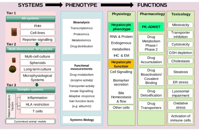
Figure 2. Roadmap for the development of ‘fit-for-purpose’ predictive models of human DILI
The proposed DILI Roadmap is a tier-based testing strategy incorporating present Test Systems and future expectations of advanced models for DILI testing.TIER ONE comprises single cell systems that report on immediate chemical/biological effects such as bioactivation, drug or bile acid accumulation due to transporter inhibition, mitotoxicity, and signalling associated with oxidative stress, endoplasmic reticulum stress and inflammatory signalling. TIER TWO comprises more complex systems containing liver cells in a more physiologic state, enabling assessment of the consequences of chronic drug exposure. TIER THREE comprises complex test systems in which a specific biological variable (e.g. HLA phenotype or inflammation) is introduced in a manner that can be used for both hazard identification and risk assessment related to idiosyncratic DILI. Underlying this philosophy, we believe it to be essential that the pharmacological and physiological phenotype of the test system is considered (Phenotype → Functions), before undertaking toxicological investigations, to ensure that the most appropriate methods are used to determine the potential DILI liability of a new drug. To integrate findings from different test systems and to dissect the multi-level impact of compounds, mathematical models will also be useful.
Multi-dimensional 3D systems
Complex systems Tier 1
2D systems
Physiology Pharmacology Toxicology
RNA & Protein
Endogenous
metabolites
IHC & EM
Hepatocyte phenotype Cell Signalling Biomarker secretion Bile Homeostasis & flow Hepatocyte function PHH Cell-lines
Long term culture Multi-cell culture Spheroids Tier 2 Tier 3
SYSTEMS
FUNCTIONS
Drug Accumulation Drug Bioactivation/ Covalent Binding Drug Detoxification Drug Metabolism Phase I Phase 2 Other cells Lysosomal impairment Transporter inhibition GSH depletion Cholestasis Cytotoxicity Mitotoxicity ER stress Oxidative stress Activation of immune cellsCustomised animal models
Functional measurements Drug-metabolism (enzyme activity) Transporter activity Innate Signalling Adaptive response
liver function tests
Page 44 of 61
Figure 3. Hepatocyte couplet illustrating the basolateral and canalicular location of transport
proteins.Bile acids (BAs): Unconjugated BA species (BA); monovalent BA (mBA), BA-G
(glucuronidated BAs); BA-S (Sulpho and sulpho-conjugated BAs). Examples of different classes of drug substrates (blue), inhibitors (red) and inducers (purple) across multiple transporters are given. Some drugs are both substrates and inhibitors of transporter proteins depending on the affinity of respective drugs. Selectivity of transporters for the different monovalent, divalent and conjugated forms of BAs (green) across the basolateral and apical membranes illustrates the multiplicity of transporters involved in bile uptake and efflux. The heterodimeric organic solute transporter
OSTα/OSTβ an efflux transporter, but also bidirectional transporter for some organic anions. Some
amphiphilic BAs passively diffuse across the basolateral membrane. Phosphatidylcholine (PC) is a physiologically important substrate for MDR3 and its inhibition may play an important role in cholestasis and vanishing bile duct syndrome (VBDS). The role of taurocholic acid (TCA) in micelles has an important role on MDR3 activity. See supplementary information for transporter protein homology, function and drug interactions.
Inhibitor Inducer Substrate
mBA: Monovalent Bile Acid dBA: Divalent Bile Acid PC: Phosphatidylcholine
Page 45 of 61
Figure 4. The role of the adaptive immune system in drug-induced liver injury
Page 46 of 61 TABLES
Table 1: Relative advantages and disadvantages of the most popular single cell hepatocyte models for industry DILI assessment, including their position in the proposed tiered testing system
Cell Type/Tier Advantages Disadvantages
HepG2/TIER 1 Human hepatic origin.
Easy to culture.
Inexpensive.
Consistent, reproducible, fast turnaround assay performance for the toxicological endpoint under investigation.
No donor variation.
Easily adaptable for specific assays – e.g. mitotoxicity assay, high-content screening. Can be cultured in 3D.
Large, publicly-available datasets for cytotoxicity & gene expression assists risk assessment. Popular for use in HTS approaches and toxigenomics
Cancer-derived.
Many clonal variants exist with different cellular characteristics.
Relative lack in expression of drug metabolizing enzymes and transporters. Majority of genes expressed in HepG2 cells are also expressed in primary hepatocytes, yet 30% of the gene expression profiles are unique to HepG2209.
HepaRG/
TIER 1 or TIER 2
Human hepatic origin. Relatively straightforward to culture.
More consistent than primary hepatocytes.
Cancer cell.