Moving towards adaptive management of
cyanotoxin-impaired water bodies
Hans W. Paerl,1* Timothy G. Otten2and Alan R. Joyner1
1
Institute of Marine Sciences, University of North Carolina at Chapel Hill, Morehead City, NC, USA. 2Bend Genetics, LLC, 87 Scripps Drive, Ste. 301, Sacramento, CA, USA.
Summary
The cyanobacteria are a phylum of bacteria that have played a key role in shaping the Earth’s biosphere due to their pioneering ability to perform oxygenic photosynthesis. Throughout their history, cyanobac-teria have experienced major biogeochemical changes accompanying Earth’s geochemical evolu-tion over the past 2.5+ billion years, including peri-ods of extreme climatic change, hydrologic, nutrient and radiation stress. Today, they remain remarkably successful, exploiting human nutrient over-enrich-ment as nuisance“blooms.”Cyanobacteria produce an array of unique metabolites, the functions and biotic ramifications of which are the subject of diverse ecophysiological studies. These metabolites are relevant from organismal and ecosystem func-tion perspectives because some can be toxic and fatal to diverse biota, including zooplankton andfish consumers of algal biomass, and high-level con-sumers of aquatic food sources and drinking water, including humans. Given the long history of environ-mental extremes and selection pressures that cyanobacteria have experienced, it is likely that that these toxins serve ecophysiological functions aimed at optimizing growth and fitness during periods of environmental stress. Here, we explore the molecular and ecophysiological mechanisms underlying
cyanotoxin production, with emphasis on key envi-ronmental conditions potentially controlling toxin production. Based on this information, we offer potential management strategies for reducing cyan-otoxin potentials in natural waters; for cyancyan-otoxins with no clear drivers yet elucidated, we highlight the data gaps and research questions that are still lack-ing. We focus on the four major classes of toxins (anatoxins, cylindrospermopsins, microcystins and saxitoxins) that have thus far been identified as rele-vant from environmental health perspectives, but caution there may be other harmful metabolites wait-ing to be elucidated.
Introduction
Cyanobacteria are the Earth’s oldest known prokaryotic oxygenic phototrophs, with fossil evidence pointing to their presence in the Proterozooic, some 2.5 billion years ago (Schopf, 2000). This period witnessed the transition from anoxic to oxic conditions, in large part due to their photosynthetic activities. They have also experienced periods of varying nutrient (N, P, C and minor elements) abundance and availability, and a great deal of variability in climatic conditions, including extremely wet and dry periods, combined with major changes in the Earth’s sur-face temperature. Lastly, major geophysical events such as volcanism and continental drift have altered their habitats and have exerted ecophysiological constraints over a wide range of time scales. As such, cyanobacte-ria have experienced the full spectrum of physical– chem-ical–biotic changes that have impacted the Earth’s biosphere; unsurprisingly, cyanobacteria exhibit an extre-mely broad geographic distribution, ranging from polar to tropical regions, and from subsurface aquatic to alpine habitats (Potts and Whitton, 2000). Across this broad range, they inhabit virtually all terrestrial and aquatic habitats, ranging from deserts to tropical rain forests and from the ultraoligotrophic open ocean to hypereutrophic lakes (Potts and Whitton, 2000; Whitton, 2012). Lastly, cyanobacteria exhibit a remarkable ability to both coun-ter extreme climatic conditions and to thrive under them.
Within these diverse habitats, cyanobacteria possess widespread adaptations to climatic extremes, including the formation of heat and desiccation-tolerant resting cells, or akinetes, the presence of photo-protective Received 21 June, 2016; accepted 23 June, 2016. *For
correspondence. E-mail [email protected]; Tel. +1(252) 726 6841, ext. 133; Fax +1(252) 726 2426.
Microbial Biotechnology(2016)9(5), 641–651 doi:10.1111/1751-7915.12383
Funding Information
This work was supported by the US National Science Foundation (OCE 0726989, 0812913, 0825466, and CBET 0826819, INSPIRE 1230543, and Dimensions of Biodiversity 1240851), and California Delta Stewardship Council Project 2044. Additional support was provided by the Chinese Ministry of Science and Technology (contracts 2014zx07101-011), and the International Cooperation Project (contracts 2015DFG91980).
pigments and desiccation-resistant sheaths and cap-sules, the ability to glide on surfaces to adjust their posi-tion within a bloom and in the water column (by buoyancy regulation) in response to irradiance and nutri-ent gradinutri-ents (Potts and Whitton, 2000; Reynolds, 2006). They have also developed a wide array of physiological adaptations to cope with periods of nutrient limitation, including the ability to sequester (by chelation) iron (Wil-helm and Trick, 1994), efficiently fix gaseous (CO2) and dissolved inorganic carbon (DIC; Paerl and Millie, 1996), store phosphorus, nitrogen (N2) and other essential nutri-ents (Healy, 1982; Reynolds, 2006), and for some (the Nostocales, some Oscillatoriales and some pico-cyanobacterial genera) the ability to convert or “fix” atmospheric N2into biologically available ammonia (Gal-lon, 1992). Many cyanobacterial genera have formed mutualistic and symbiotic associations with fungi, algae, higher plants and animals, enabling them and their part-ners to exploit and thrive in potentially hostile and extreme environments (Paerl, 1982; Raven, 2002).
The diverse and remarkable physical–chemical and biotic adaptations that cyanobacteria utilize make for an extensive repertoire of ecological strategies aimed at surviving and at times thriving, as nuisance blooms, under a wide range of environmental conditions, includ-ing human alterations of aquatic environments; e.g. nutri-ent over-enrichment (eutrophication), hydrologic alterations due to water withdrawal (for drinking, irriga-tion, industrial use) from streams, rivers and lakes, dam/ reservoir, artificial waterway construction, and perturba-tions of benthic and planktonic habitats.
Lastly, and central to this article, cyanobacteria produce a wealth of secondary metabolites of which the functions and biotic ramifications remain largely unknown. Some of these metabolites have come to light in an environmental and societal context because they can have harmful effects on organismal and ecosystem function, as toxic substances that adversely affect diverse biota, including zooplankton and herbivorousfish, bioaccumulate in higher trophic levels and directly impair drinking water. It is hypothesized that the so-called “cyanotoxins” serve eco-physiological functions aimed at optimizing growth and protecting cyanobacterial cells during periods of environ-mental stress and constraints as outlined above (e.g. Paerl and Millie, 1996), rather than specifically serving as defensive (i.e. anti-grazing) compounds directed at high-level consumers. This is based on numerousfield and lab-oratory observations that indicate that higher ranked con-sumers are not necessarily a threat to cyanobacterial blooms nor are they able to effectively control the global proliferation that we are currently experiencing (Paerl and Millie, 1996).
In this contribution, we will explore the molecular mechanisms underlying secondary metabolite and toxin
production, environmental and cellular conditions control-ling toxin production and, ecophysiological rationales for toxin production in aquatic ecosystems supporting their growth and proliferation. By linking molecular-level mech-anisms to their potential controls on microbial community and ecosystem scales, it is hoped that both the ecologi-cal reasons and potential environmental controls of toxin production can be uncovered and potentially utilized in aquatic ecosystem management schemes aimed at improving water quality and environmental health of impacted waters.
Overview of cyanotoxins and their putative environmental drivers
Anatoxins
Anatoxin-a was identified over 40 years ago following a series of livestock poisoning events that were traced back to blooms ofAnabaenaflos-aquae(this genus has recently been renamed Dolichospermum) (Devlin et al., 1977). Since then it has been identified in several fami-lies of cyanobacteria isolated from both pelagic and benthic environments. The neurotoxin is a potent nico-tinic acetycholine receptor agonist that blocks neuromus-cular junctions leading to rapid respiratory arrest. In addition to anatoxin-a and its methylene homologue homoanatoxin-a, additional variants include dihydroana-toxin-a and dihydrohomoanadihydroana-toxin-a and their respective cis/trans isomers. All analogues are produced from a 10-gene operon that includes three polyketide synthases (PKS), a type II thioesterase, a transporter, an acyl car-rier and four tailoring enzymes (Mejean et al., 2009). The gene cluster organization and orientation varies by cyanobacterial genus, as evinced from a number of operons sequenced from Anabaena, Oscillatoria, Cylin-drospermum and Cuspidothrix isolates (Mejean et al., 2014). Individual strains may produce more than one anatoxin-a analogue simultaneously (Mannet al., 2012). Dihydroanatoxin-a and dihydrohomoanatoxin-a exhibit approximately 10-fold lower binding affinities and toxici-ties than anatoxin-a and homoanatoxin-a based on mouse LD50(Woodet al., 2012).
Pick, 2012). A separate study ofAnabaena sp. reported that anatoxin-a was maximally produced under tempera-tures and light levels slightly suboptimal for growth (Rapala and Sivonen, 1998).
In a study of benthic Phormidium autumnale isolates, over 85% of total anatoxin was observed to occur intracel-lularly across a range of low to high N and P treatments (Heath et al., 2014). In that study, the authors also reported that dihydroanatoxin-a concentrations decreased when N and P concentrations were elevated (21 mg l 1 and 3 mg l 1 respectively), whereas homoanatoxin-a quota increased when P concentrations were reduced below 0.08 mg l 1. Potentially complicating the identifica-tion of environmental drivers was the observaidentifica-tion of transcripts from several ana genes but an absence of detectable toxin in Cuspidothrix (formerly Aphani-zomenon) issatschenkoi CHABD3 (Jiang et al., 2015); this raised the possibility that anatoxin-a synthesis may be subject to post-transcriptional regulation.
Cylindrospermopsins
Cylindrospermopsin (CYN) is a guanidine alkaloid that exhibits broad spectrum cytotoxicity, as well as potent liver and kidney toxicity due to glutathione and protein synthesis inhibition, and the production of damaging cytochrome p-450-generated metabolites (Humpage et al., 2005). In addition to CYN, four other analogues have been identified; 7-epi-CYN is a C-7 epimer of CYN with similar toxicity (Norris et al., 1999), 7-deoxy-CYN lacks the hydroxyl group on C-7 and exhibits little to no toxicity in mice (Bankeret al., 2001), and 7-deoxy-desul-focylindrospermopsin and 7-deoxy-desulfo-12-acetylcylin-drospermopsin were recently identified and their relative toxicities remains undetermined (Wimmer et al., 2014). Thecyrgene cluster is responsible for CYN biosynthesis and has been reported to consist of 11 genes in Aphani-zomenon (St€uken and Jakobsen, 2010), Oscillatoria (Mazmouz et al., 2010) and Raphidiopsis (Jiang et al., 2012), but 15 genes inCylindrospermopsis(Mihaliet al., 2008), with significant rearrangements in gene order between genera, suggestive of substantial divergence from a common ancestor (Mazmouz et al., 2010), recombination and/or horizontal gene transfer (Cires and Ballot, 2016). The operon encodes for non-ribosomal peptide synthetase/PKS genes, transferases, uracil ring formation, an exporter and other tailoring functions. The conversion of 7-deoxy-CYN into CYN is controlled in vitro by a 2-oxoglutarate-dependent iron oxygenase encoded by cyrI (Mazmouz et al., 2010). This was cor-roborated in a strain of Raphidiopsis curvata (CHAB1150) that was found to contain an insertion mutation in cyrI that induced a frameshift and stop codons which resulted in a truncated product. As a result
the strain could only produce 7-deoxy-CYN (Jianget al., 2012).
Although the operon contains an exporter, most stud-ies of Cylindrospermopsis suggest that the majority of total CYN is retained intracellularly (Saker and Eagle-sham, 1999; Willis et al., 2015). On the contrary, investi-gations of CYN-producing Aphanizomenon blooms in German lakes have reported that the majority of toxin occurred extracellularly (R€ucker et al., 2007). However, as with any intracellular compound, cell senescence or lysis will result in larger percentages of extracellular tox-ins, additionally, the co-occurrence of toxic and non-toxic genotypes in natural settings may also complicate esti-mate of toxin quota. For example, other culture-based studies have reported extracellular CYN fractions to range from 11% to 26% for Aphanizomenon (Preußel et al., 2006, 2009, 2014) and 52–62% for Oscillatoria (Mazmouz et al., 2010). Culture studies ofC. raciborskii indicate that the highest extracellular CYN concentra-tions correspond with cells in stationary growth phase or during the end of a bloom (Dybleet al., 2006).
A clear understanding of environmental conditions that promote CYN production or toxic strain dominance is presently lacking. Two recent studies have reported that cyr gene expression and CYN production in C. raci-borskii cultures do not vary in response to different nitro-gen and phosphorus regimes, although one of the strains was used in both studies (Stucken et al., 2014; Willis et al., 2015). The observation that CYN was con-stitutively produced runs contrary to other studies that have reported differential CYN production and cell quo-tas in Oscillatoria (Bormans et al., 2013), Aphani-zomenon (Preußel et al., 2014) and even other Cylindrospermopsis isolates (Dyble et al., 2006). Com-bined, these findings suggest that there may be species or strain-level differences in CYN production possibly influenced by the physiological or environmental impor-tance/function that CYN and its derivatives provide indi-vidual ecotypes.
regulate CYN production, Cylindrospermopsis cultures grown in the absence of N have been shown to produce more CYN on a per cell basis (Saker and Neilan, 2001) and high light intensity has been found to reduce CYN production in both Aphanizomenon and Cylindrosper-mopsis(Dybleet al., 2006).
Regarding other environmental parameters, cultures of Aphanizomenon ovalisporum were shown to significantly decrease CYN content in response to sulfate and phos-phate limitation (Bacsiet al., 2006). Semi-continuous cul-ture studies using two Aphanizomenon isolates grown under various light intensity (10–60lE m 2s 1) and temperature (16–25°C) regimes indicated that total CYN content exhibited little response relative to the light con-ditions tested (Preußel et al., 2009), although the light intensity may not have been high enough to repress cyr transcription. Regarding temperature, there was a 2.6-fold reduction in total CYN at 25°C relative to the 16°C treatment for one of the strains tested, but not the other (Preußelet al., 2009). We note that higher temperatures have also been linked with lower CYN production in C. raciborskii, with complete abolishment of synthesis occurring at 35°C (Saker and Griffith, 2000).
Combined, these results indicate that there remains a need for further studies before the physiological basis for CYN production is unravelled, although studies to date suggest that CYN may have a role in response to N and light limitation. Thefinding that high temperatures abolish CYN production should also be further investigated, with particular attention given to the effect that temperature may have on other cellular factors that could indirectly influence CYN biosynthesis.
Microcystins
The hepatotoxin microcystin (MC) is the best character-ized cyanotoxin and also likely the most widely occurring throughout the phylum of cyanobacteria. MCs are syn-thesized via a bi-directionally transcribed gene cluster (10 genes in Microcystis and Anabaena, 9 genes in Planktothrix) that spans ~55 kb and consists of multiple non-ribosomal peptide synthetase and PKS genes, an ABC transporter and other genes involved in other tailoring functions (Tillettet al., 2000; Christiansenet al., 2003; Rouhiainenet al., 2004). Although the mcyoperon contains a ABC transporter, there is no evidence of active export of MC from the cells (Rohrlack and Hyenstrand, 2007). The mcy genes in each genus have unique arrangements, suggestive of a history of homolo-gous recombination and evolutionary decent from a com-mon MC-producing ancestor (Rantala et al., 2004); however, there has yet to be any evidence indicating that mcy genes are horizontally transferred across genera.
There are over 100 congeners of MC that have been identified to date (Puddick et al., 2013; Qi et al., 2015). All congeners consist of a general cyclic heptapeptide structure (cyclo-D-Ala-X-D-MeAsp-Z-Adda-D-Glu-Mdha) with two positions (X and Z) subject to variable L-amino acid incorporation, for which there are at least 15 and 12 amino acid variations, respectively, that have been reported (Hoegeret al., 2007). In addition to amino acid substitution, structural modifications via methylation/ demethylation and esterification increase the number of possible congeners that can be produced. Each con-gener exhibits different toxicities in mammals, with MC-LR variants considered to be among the most toxic and commonly encountered (Shimizu et al., 2014). Once ingested, MCs are taken up by the bile acid transport system where they bind selectively to protein phos-phatase 1 and 2A in hepatocytes, resulting in severe damage to the liver (Runnegar et al., 1993); chronic exposure to MCs has also been linked to tumours and hepatocarcinoma (Nishiwaki-Matsushima et al., 1992; Falconer and Humpage, 1996).
Studies using batch cultures of cyanobacteria have shown that MC production is highest when nitrogen and phosphorus concentrations are highest (Sivonen, 1990; Vezie et al., 2002; Harke and Gobler, 2013). However, others have argued that cellular growth rate may be the primary factor influencing MC production, with maximal toxin production typically coinciding during periods of maximal growth rate (Orr and Jones, 1998; Oh et al., 2000). As growth rates are highest when nutrient condi-tions are non-limiting–assuming adequate light, tempera-ture and micronutrients, these findings seem to be corroborative. Harke and Gobler (2013) used metatran-scriptomics to assess how a cultured isolate ofM. aerug-inosa(LE-3) responds to N or P stress and observed a significant decrease in MC and mcytranscription under low dissolved inorganic N. However, on a per cell basis, MC production levels have also been shown to increase during suboptimal growth periods, such as when cells are stressed due to low iron availability (Utkilen and Gjolme, 1995) or too high light intensity (Kaebernick and Neilan, 2001). Complicating matters, Fe uptake is high-est under high light due to its key role in photosynthesis (Lukac and Aegerter, 1993); therefore, iron homoeosta-sis and cellular redox state may ultimately control MC production (Kaebernick and Neilan, 2001). Using com-parative proteomics, Alexovaet al. (2011a) showed that mcy+strains ofMicrocystisdifferentially express proteins involved in carbon metabolism and redox balance and thatmcytranscription is highest during periods of Fe-lim-itation (Alexovaet al., 2011b).
variants of the strain. One such study compared the growth rate of the wild-type (WT) toxic strain with the mcy-deficient mutant (MT) across a range of low to high light (16–700 lmol photons m 2s 1) conditions and from low to high concentrations of hydrogen peroxide (H2O2; 0–1lmol) in order to induce oxidative stress in the cultures (Zilliges et al., 2011). The authors observed pigment chlorosis in the MT strain under both high light and high H2O2conditions, whereas the WT strain exhib-ited no negative effects; this suggested that MCs may serve an intracellular protective role during periods of photooxidative stress. Indeed, UV-B radiation has been shown to more negatively affect non-toxic strains of Microcystisthan MC-producing ones (Yanget al., 2015). Further studies using immunolabeling techniques have shown that MCs bind to and protect carboxysome and phycobilisome proteins from reactive oxygen species (ROS) during periods of photooxidative stress (Zilliges et al., 2011). This typically occurs in early bloom stages when photosynthetic growth rates and biomass accumu-lation are highest, and oxygen supersaturation and pro-duction of ROS (e.g. H2O2, O2 ) is greatest.
In addition to their role in redox balance, there appear to be other functions for MCs. A study assessing com-petitive dominance of toxic and non-toxic strains of M. aeruginosa used a mcy-producing wild-type (WT) strain and itsmcy-deficient mutant to assess competition for CO2, and found that the WT strain outcompeted the non-toxic strain under low CO2 concentrations, but not high CO2 concentrations (Van de Waal et al., 2011). These results were similar to another competition study that also found the non-toxic variant able to outcompete the toxic wild-type strain under higher CO2 conditions (Jahnichenet al., 2007). Higher temperatures have been shown to promote the dominance of MC-producing strains of Microcystis over their non-toxic counterparts (Davis et al., 2009). Considering that CO2 more readily dissolves into solution at lower temperatures, the domi-nance of toxigenic strains under low CO2/DIC conditions may be interconnected with temperature.
Interestingly, studies indicate that N availability may influence MC production differently between N2-fixing and non-fixing genera; the diazotrophsAnabaena, Apha-nizomenonand Nodulariaexhibit higher toxin production rates in N-free media, whereas Oscillatoria and Micro-cystishave higher rates under N replete conditions (Kae-bernick and Neilan, 2001). In contrast, growth competition studies using both Planktothrix and Micro-cystis found that MC-producing strains tend to outcompete non-toxic strains under low N conditions (Briand et al., 2008). However, these genera diverge in that toxigenic Microcystistends to outcompete non-toxic strains under high light conditions when the opposite has been shown for Planktothrix agardhii (Briand et al.,
2008). Compared with Microcystis and Planktothrix (Oscillatoriales), studies investigating MC production by N2-fixing genera are generally lacking, and especially so with regard to the effects of iron or photooxidative stress on toxin production. Thefinding that MC production rates are higher during periods of N-stress in diazotrophic cyanobacteria indicates that there may be other drivers of toxicity that require further study for this group. Lastly, even though most MCs are retained intracellularly, the presence of an ABC transporter within the mcy operon (Tillett et al., 2000), and the fact that the addition of MC to Microcystis cultures has been shown to trigger MC production (Schatz et al., 2007) and differential expres-sion of cell wall receptor proteins (Kehret al., 2006; Zil-liges et al., 2008), suggests that these molecules may also play a role in cell signalling.
Saxitoxins
The carbamate alkaloid neurotoxin, saxitoxin (STX), is the parent compound of at least 57 other naturally occur-ring analogues (Wiese et al., 2010) that are collectively referred to as paralytic shellfish poisoning toxins (PSPs or PSTs) due to their ability to block sodium channels leading to paralysis (Catterall, 1980) and their proclivity to accumulate in shellfish tissue (Negri and Jones, 1995). PSPs in marine environments are produced by the dinoflagellates Alexandrium,Gymnodiniumand Pyro-dinium, whereas in freshwater environments, potential cyanobacterial producers include Anabaena, Aphani-zomenon, Cylindrospermopsis, Lyngbya, Oscillatoria/ Planktothrix,Raphidiopsis andScytonema(Neilan et al., 2013) (Table 1). In recent years, there has been an increase in PSP monitoring efforts in freshwater systems and these efforts have revealed that PSPs may be more widespread than previously believed. For example, the 2007 USEPA National Lakes Assessment identified STXs in 7.7% of inland lakes surveyed (Loftin et al., 2016) and Fetscher et al.(2015) identified STXs attribu-table to benthic cyanobacteria in 7% of wadeable streams surveyed in California, USA. A recent study of benthic cyanobacteria in Brazilian rivers and reservoirs identified STX genes and toxin in strains ofPhormidium, Cylindrospermum and Geitlerinema for the first time (Borges et al., 2015). In a study of Finnish lakes and brackish coastal waters, the genes involved in STX biosynthesis were detected on average in 31% of sam-ples collected (Savelaet al., 2015).
specific toxin profile of each strain being determined by the presence or absence of specific genes within the cluster (Neilan et al., 2013). It has been hypothesized that the sxt gene cluster may have originated in an ancient a-proteobacterium and has subsequently spread via horizontal gene transfer to diverse lineages of phyto-plankton (Kellmann et al., 2008). The relative toxicity of STX analogues can be roughly ordered from highest to lowest as: STX> neo-STX > gonyautoxins (GTX1-4) > decarbamoylated STXs > di-sulfated STXs (C1-C4) (Usleberet al., 1997).
InC. raciborskii, conductivity has been shown to exhibit a strong positive relationship with PSP production follow-ing exposure to high Na+ (Pomati et al., 2004) or Mg2+ concentrations (Kellmann and Neilan, 2007). Gene expression analyses have also shown that expression of sxtA, which encodes the first step of STX biosynthesis, and the transporterssxtFandsxtMare all upregulated in response to Na+stress (Ongleyet al., 2016). The postu-lated reason for increased STX production across an ionic gradient is that STX may help the cells regain homoeosta-sis under salt stress by altering cell permeability such that salt uptake is inhibited (Pomati et al., 2003; Brentano et al., 2016). Studies on both Cylindrospermopsis and Raphidiopsisindicate that STX is actively exported out of the cell in response to elevated cation concentrations (Soto-Liebeet al., 2012; Ongleyet al., 2016).
Nitrogen depletion has been shown to increase STX export in Aphanizomenon gracile (Casero et al., 2014), whereas temperature seems to have little influence on STX production and export apart from its role in cell growth (Casero et al., 2014). Elevated pH above 9 was found to induce significantly higher levels of STX export from cultures of Anabaena circinalis and C. raciborskii than even elevated conductivity (Pomati et al., 2004; Ongley et al., 2016), suggesting that alkaline stress may be of central importance in the physiology of STX-produ-cing strains. Considering that cyanobacterial blooms often bring about significant increases in pH and more alkaline conditions in affected water bodies, STX produc-tion may provide a competitive advantage to strains dur-ing periods of elevated photosynthetic activity.
Management implications
It is well established that in most cases, nutrient (both N and P) reductions are required to control cyanobacterial blooms (Paerl, 2014). However, depending on the sever-ity of nutrient over-enrichment and the size of the water body impacted, these approaches may take years or decades before significant improvements to water quality are achieved (Paerl, 2014). Based on our understanding of the environmental factors promoting toxin production and/or toxigenic strain dominance, there may be other management strategies that can be employed in the meantime – in addition to nutrient reductions – that will favour non-toxic strain dominance in place of toxin-pro-ducing variants.
Microcystins are preferentially produced under high light intensities, therefore smaller lakes/reservoirs may benefit from the addition of light absorbing dyes during the summer months when cyanobacteria are most pro-liferative. In riverine systems, riparian buffer and shade along the shoreline should reduce light intensity through the water column, which may also help to reduce MC production. Reduced light intensity should also help to lower water temperature, which may fur-ther enhance the competitive advantage of non-toxic strains. On the contrary, if anatoxin-a or CYN are problematic, warmer temperatures and higher light intensity may help to alleviate toxin production. In dammed rivers where cold water refugia are not critical for fish habitat, a larger percentage of warmer surface water could be released downstream from dam spill-ways if benthic cyanobacteria are the suspected toxin producers. Lastly, brackish systems impaired by STXs may benefit from periodic pulses of freshwater to lower ionic potential or the addition of dilute acids to lower pH during periods of high cyanobacterial productivity. While these suggestions are purely experimental at this time, they are provided to illustrate how an improved understanding of cyanobacterial ecophysiol-ogy and toxin production may one day lead to novel management strategies for mitigating cyanobacterial toxicity in surface waters.
Table 1. Cyanobacterial genera with strains known to be able to produce cyanotoxins.
Class APHA CYL DOL FISC GLO LYNG MIC NOD NOST OSC PHOR PLA RAPH SCYT
Anatoxin-a X X X X X X X
Cylindrospermopsin X X X X
Microcystin X X X X Xa X X X
Saxitoxin X X X X X X X
a. Produces nodularin–a pentapeptide variant of microcystin.
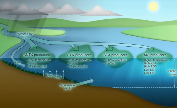
Conclusions
Studies investigating the ecological and physiological roles of cyanotoxins indicate that there are likely to be multiple functions performed by each of these mole-cules. Figure 1 attempts to convey the most commonly observed drivers for production of each cyanotoxin class under natural settings. Much of our understand-ing of cyanotoxins comes from culture studies usunderstand-ing so-called toxic and non-toxic strains. However, based on the several dozen cyanobacterial genomes that have been sequenced to date, it is now apparent that there are significant genomic differences between even seemingly closely related strains. In Microcystis aerugi-nosa, the core genome – defined as those genes found in all strains of M. aeruginosa – is estimated to only be ~2400 genes, or ~47% of the genome (Hum-bert et al., 2013). As such, any studies attempting to compare different strains should be cognizant that there may be considerable differences in their overall genetic makeup, which will influence their ability to adapt to varied environmental conditions (Harke et al., 2016a). At present, M. aeruginosa is the only HAB-forming cyanobacterium for which isogenic mutants (e.g. DmcyB-) have been successfully created, thereby allowing direct assessments of the physiological
function of MC in this strain. As new gene editing tools, such as various CRISPR-Cas systems evolve (Selle and Barrangou, 2015; Yao et al., 2016), genetic manipulation of other genera of cyanobacteria should become accessible.
In the meantime, much can be inferred about the puta-tive functions of cyanotoxins by studying cyanobacterial blooms in situusing a variety of -omics techniques. It is now possible to extract nearly complete cyanobacterial genomes from shotgun sequencing data sets using dif-ferential binning approaches (Albertsen et al., 2013; Brown et al., 2016; Otten et al., 2016). With reference genomes in hand that are specific to the system being investigated, metatranscriptomics can be used to mas-sively survey the system and the resulting gene tran-scripts corresponding to CyanoHAB taxa can be mapped back to their cognate hosts. This methodology performed over a time-series will provide unparalleled insights into cyanobacterial physiology and toxin produc-tion that cannot be obtained using cultured isolates. These methods are starting to gain traction (e.g. Penn et al., 2014; Harke et al., 2016b) and in the coming years we anticipate an explosion of metagenomic, meta-transcriptomic and proteomic data which will usher in a new understanding of the drivers of bloom initiation, col-lapse and toxicity.
References
Albertsen, M., Hugnholtz, P., Skarshewski, A., Nielsen, K.L., Tyson, G.W., and Nielsen, P.H. (2013) Genome sequences of rare, uncultured bacteria obtained by differ-ential coverage binning of multiple genomes.Nat Biotech-nol31:533–538.
Alexova, R., Haynes, P.A., Ferrari, B.C., and Neilan, B.A. (2011a) Comparative protein expression in different strains of the bloom-forming cyanobacterium Microcystis aeruginosa.Mol Cell Proteomics10:M110.003749. Alexova, R., Fujii, M., Birch, D., Cheng, J., Waite, T.D.,
Ferrari, B.C., and Neilan, B.A. (2011b) Iron uptake and toxin synthesis in the bloom-forming Microcystis aerugi-nosa under iron limitation. Environ Microbiol 13: 1064–1077.
Bacsi, I., Vasas, G., Suranyi, G., M-Hamvas, M., Mathe, C., Toth, E., et al. (2006) Alteration of cylindrospermopsin production in sulfate- or phosphate-starved cyanobac-teriumAphanizomenon ovalisporum.FEMS Microbiol Lett
259:303–310.
Banker, R., Carmeli, S., Werman, M., Teltsch, B., Porat, R., and Sukenik, A. (2001) Uracil moiety is required for toxic-ity of the cyanobacterial hepatotoxin cylindrospermopsin.
J Toxicol Environ Health Part A62:281–288.
Borges, H.L.F., Branco, L.H.Z., Martins, M.D., Lima, C.S., Barbosa, P.T., Lira, G.A.S.T., et al. (2015) Cyanotoxin production and phylogeny of benthic cyanobacterial strains isolated from the northeast of Brazil. Harmful Algae43:46–57.
Bormans, M., Lengronne, M., Brient, L., and Duval, C. (2013) Cylindrospermopsin accumulation and release by the benthic cyanobacteria Oscillatoria sp. PCC 6506 under different light conditions and growth phases. Bull Environ Contam Toxicol92:243–247.
Brentano, D.M., Giehl, E.L.H., and Petrucio, M.M. (2016) Abiotic variables affect STX concentration in a meso-oli-gotrophic subtropical coastal lake dominated by Cylin-drospermopsis raciborskii(Cyanophyceae).Harmful Algae
56:22–28.
Briand, E., Yepr emian, C., Humbert, J.F., and Quiblier, C. (2008) Competition between microcystin- and non-micro-cystin-producing Planktothrix agardhii (cyanobacteria) strains under different environmental conditions. Environ Microbiol10:3337–3348.
Brown, N.M., Mueller, R.S., Shepardson, J.W., Landry, Z.C., Morre, J.T., Maier, C.S.,et al.(2016) Structural and func-tional analysis of thefinished genome of the recently iso-lated toxicAnabaenasp. WA102.BMC Genom17:457. Casero, M.C., Ballot, A., Agha, R., Quesada, A., and Cires,
S. (2014) Characterization of saxitoxin production and release and phylogeny of sxt genes in paralytic shellfish poisoning toxin-producing Aphanizomenon gracile.
Harmful Algae37:28–37.
Catterall, W.A. (1980) Neurotoxins that act on voltage-sensi-tive sodium channels in excitable membranes.Annu Rev Pharmacol Toxicol20:15–43.
Christiansen, G., Fastner, J., Erhard, M., B€orner, T., and Dittmann, E. (2003) Microcystin biosynthesis in Plank-tothrix: genes, evolution, and manipulation. J Bacteriol
185:564–572.
Cires, S., and Ballot, A. (2016) A review of the phylogeny, ecology and toxin production of bloom-forming Aphani-zomenon spp. and related species within the Nostocales (cyanobacteria).Harmful Algae54:21–43.
Davis, T.W., Berry, D.L., Boyer, G.L., and Gobler, C.J. (2009) The effects of temperature and nutrients on the growth and dynamics of toxic and non-toxic strains of
Microcystis during cyanobacteria blooms. Harmful Algae
8:715–725.
Devlin, J.P., Edwards, O.E., Gorham, P.R., Hunter, N.R., Pike, R.K., and Stavric, B. (1977) Anatoxin-a, a toxic alka-loid from Anabaena flos-aquae NRC-44h. Can J Chem
55:1367–1371.
Dyble, J., Tester, P., and Litaker, R.W. (2006) Effects of light intensity on cylindrospermopsin production in the cyanobacterial HAB species Cylindrospermopsis raci-borskii.African J Mar Sci28:309–312.
Falconer, I.R., and Humpage, A.R. (1996) Tumour promo-tion by cyanobacterial toxins.Phycologia35(6S):74–79. Fetscher, A.E., Howard, M.D.A., Stancheva, R., Kudela,
R.M., Stein, E.D., Sutula, M.A., et al. (2015) Wadeable streams as widespread sources of benthic cyanotoxins in California, USA.Harmful Algae49:105–116.
Gagnon, A., and Pick, F.R. (2012) Effect of nitrogen on cellu-lar production and release of the neurotoxin anatoxin-a in a nitrogen-fixing cyanobacterium.Front Microbiol3:211. Gallon, J.R. (1992) Tansley Review No. 44/Reconciling the
incompatible: N2 fixation and O2. New Phytol 122: 571– 609.
Harke, M.J., and Gobler, C.J. (2013) Global transcriptional responses of the toxic cyanobacterium,Microcystis aerug-inosa, to nitrogen stress, phosphorus stress, and growth on organic matter.PLoS ONE8:e69834.
Harke, M.J., Steffen, M.M., Gobler, C.J., Otten, T.G., Wilhelm, S.W., Wood, S.A., and Paerl, H.W. (2016a) A review of the global ecology, genomics, and biogeography of the toxic cyanobacterium,Microcystisspp.Harmful Algae54:4–20. Harke, M.J., Davis, T.W., Watson, S.B., and Gobler, C.J.
(2016b) Nutrient-controlled niche differentiation of Wes-tern Lake Erie cyanobacterial populations revealed via metatranscriptomic surveys. Environ Sci Technol 50: 604–615.
Healy, F.P. (1982) Phosphate. InThe Biology of Cyanobac-teria. Carr, N.G., and Whitton, B.A. (eds). Oxford: Black-well Scientific Publications, pp. 105–124.
Heath, M.W., Wood, S.A., Barbieri, R.F., Young, R.G., and Ryan, K.G. (2014) Effects of nitrogen and phosphorus on anatoxin-a, homoanatoxin-a, dihydroanatoxin-a and dihy-drohomoanatoxin-a production byPhormidium autumnale.
Toxicon92:179–185.
Hoeger, S.J., Schmid, D., Blom, J.F., Ernst, B., and Dietrich, D.R. (2007) Analytical and functional characterization of microcystins [Asp3]MC-RR and [Asp3, Dhb7]MC-RR: con-sequences for risk assessment?Environ Sci Technol 41: 2609–2616.
Humbert, J.F., Barbe, V., Gugger, M., Calteau, A., Coursin, T., Lajus, A.,et al. (2013) A tribute to disorder in the gen-ome of the bloom-forming freshwater cyanobacterium
Microcystis aeruginosa.PLOS One8:e70747.
cytotoxicity: role of cytochrome P-450 and oxidative stress.J Toxicol Environ Health Part A68:739–753. Jahnichen, S., Ihle, T., Petzoldt, T., and Benndorf, J. (2007)
Impact of inorganic carbon availability on microcystin pro-duction byMicrocystis aeruginosa PCC 7806.Appl Envi-ron Microbiol73:6994–7002.
Jiang, Y., Xiao, P., Yu, G., Sano, T., Pan, Q., and Li, R. (2012) Molecular basis and phylogenetic implications of deoxycylindrospermopsin biosynthesis in the cyanobac-terium Raphidiopsis curvata. Appl Environ Microbiol 78: 2256–2263.
Jiang, Y., Song, G., Pan, Q., Yang, Y., and Li, R. (2015) Identification of genes for anatoxin-a biosynthesis in Cus-pidothrix issatschenkoi.Harmful Algae46:43–48. Kaebernick, M., and Neilan, B.A. (2001) Ecological and
molecular investigations of cyanotoxin production.FEMS Microbiol Ecol35:1–9.
Kehr, J.C., Zilliges, Y., Springer, A., Disney, M.D., Ratner, D.D., Bouchier, C.,et al. (2006) A mannan binding lectin is involved in cell-cell attachment in a toxic strain of
Microcystis aeruginosa.Mol Microbiol59:893–906. Kellmann, R., and Neilan, B.A. (2007) Biochemical
charac-terization of Paralytic Shellfish Toxin biosynthesis in vitro.
J Phycol43:497–508.
Kellmann, R., Mihali, T.K., and Neilan, B.A. (2008) Identifi -cation of a saxitoxin biosynthesis gene with a history of frequent horizontal gene transfers. J Mol Evol 67: 526– 538.
Loftin, K.A., Graham, J.L., Hilborn, E.D., Lehmann, S.C., Meyer, M.T., Dietze, J.E., and Griffith, C.B. (2016) Cyan-otoxins in inland lakes of the United States: occurrence and potential recreational health risks in the EPA National Lakes Assessment 2007.Harmful Algae56:77–90. Lukac, M., and Aegerter, R. (1993) Influence of trace metals
on growth and toxin production ofMicrocystis aeruginosa.
Toxicon31:293–305.
Mann, S., Cohen, M., Chapuis-Hugon, F., Pichon, V., Maz-mouz, R., Mejean, A., and Ploux, O. (2012) Synthesis, configuration assignment, and simultaneous quantification by liquid chromatography coupled to tandem mass spec-trometry, of dihydroanatoxin-a and dihydrohomoanatoxin-a together with the pdihydrohomoanatoxin-arent toxins, in dihydrohomoanatoxin-axenic cydihydrohomoanatoxin-anobdihydrohomoanatoxin-acteridihydrohomoanatoxin-al strains and in environmental samples.Toxicon60:1404– 1414.
Mazmouz, R., Chapuis-Hugon, F., Mann, S., Pichon, V., Mejean, A., and Ploux, O. (2010) Biosynthesis of cylin-drospermopsin and 7-epicylincylin-drospermopsin in Oscillato-ria sp. Strain PCC 6506: identification of the cyr gene cluster and toxin analysis. Appl Environ Microbiol 76: 4943–4949.
Mejean, A., Mann, S., Maldiney, T., Vassiliadis, G., Lequin, O., and Ploux, O. (2009) Evidence that biosynthesis of the neurotoxic alkaloids anatoxin-a and homo-anatoxin-a in the cyanobacteriumOscillatoriaPCC 6506 occurs on a modular polyketide synthase initiated by L-proline. J Am Chem Soc131:7512–7513.
Mejean, A., Paci, G., Gautier, V., and Ploux, O. (2014) Biosynthesis of anatoxin-a and analogues (anatoxins) in cyanobacteria.Toxicon91:15–22.
Mihali, T.K., Kellmann, R., Muenchhoff, J., Barrow, K.D., and Neilan, B.A. (2008) Characterization of the gene
cluster responsible for cylindrospermopsin biosynthesis.
Appl Environ Microbiol74:716–722.
Negri, A.P., and Jones, G.J. (1995) Bioaccumulation of par-alytic shellfish poisoning (PSP) toxins from the cyanobac-terium Anabaena circinalis by the freshwater mussel
Alathyria condola.Toxicon33:667–678.
Neilan, B.A., Pearson, L.A., Muenchhoff, J., Moffitt, M.C., and Dittmann, E. (2013) Environmental conditions that influence toxin biosynthesis in cyanobacteria. Environ Microbiol15:1239–1253.
Nishiwaki-Matsushima, R., Ohta, T., Nishiwaki, S., Suga-numa, M., Kohyama, K., Ishikawa, T.,et al. (1992) Liver tumor promotion by the cyanobacterial cyclic peptide toxin microcystin-LR.J Cancer Res Clin Oncol118:420–424. Norris, R.L., Eaglesham, G.K., Pierens, G., Shaw, G.R.,
Smith, M.J., Chiswell, R.K.,et al. (1999) Deoxycylindros-permopsin, an analog of cylindrospermopsin from Cylin-drospermopsis raciborskii.Environ Toxicol14:163–165. Oh, H.M., Lee, S.J., Jang, M.H., and Yoon, B.D. (2000)
Micro-cystin production byMicrocystis aeruginosain a phospho-rus-limited chemostat.Appl Environ Microbiol66:176–179. Ongley, S.E., Pengelly, J.J.L., and Neilan, B.A. (2016) Ele-vated Na+and pH influence the production and transport of saxitoxin in the cyanobacteria Anabaena circinalis
AWQC131C and Cylindrospermopsis raciborskii T3.
Microbiol18:427–438.
Orr, P.T., and Jones, G.J. (1998) Relationship between microcystin production and cell division rates in nitrogen-limited Microcystis aeruginosa cultures. Limnol Oceanogr
43:1604–1614.
Otten, T.G., Graham, J.L., Harris, T.D., and Dreher, T.W. (2016) Elucidation of taste-and-odor producing bacteria and toxigenic cyanobacteria by shotgun metagenomics in a Midwestern drinking water supply reservoir. Appl Envi-ron Microbiol82:1–11.
Paerl, H.W. (1982) Chapter 17. Interactions with bacteria. In
The Biology of Cyanobacteria. Carr, N.G., and Whitton, B.A. (eds). Oxford: Blackwell Scientific Publications, pp. 441–461.
Paerl, H.W. (2014) Mitigating harmful cyanobacterial blooms in a human- and climatically-impacted world.Life 4:988– 1012. doi:10.3390/life40x000x.
Paerl, H.W., and Millie, D.F. (1996) Physiological ecology of toxic cyanobacteria.Phycologia35:160–167.
Penn, K., Wang, J., Fernando, S.C., and Thompson, J.R. (2014) Secondary metabolite gene expression and inter-play of bacterial functions in a tropical freshwater cyanobacterial bloom.ISME J8:1866–1878.
Pomati, F., Rossetti, C., Calamari, D., and Neilan, B.A. (2003) Effects of saxitoxin (STX) and veratridine on bacte-rial Na+ -K+ fluxes: a prokaryote-based STX bioassay.
Appl Environ Microbiol69:7371–7376.
Pomati, F., Rossetti, C., Manarolla, G., Burns, B.P., and Neilan, B.A. (2004) Interactions between intracellular Na+ levels and saxitoxin production in Cylindrospermopsis raciborskiiT3.Microbiology150:455–461.
Potts, M., and Whitton, B.A. (2000) The Biology and Ecol-ogy of Cyanobacteria. Oxford: Blackwell Scientific Publi-cations.
producing Aphanizomenon flos-aquae (Cyanobacte-ria) isolated from two German lakes. Toxicon 47: 156– 162.
Preußel, K., Wessel, G., Fastner, J., and Chorus, I. (2009) Response of cylindrospermopsin production and release inAphanizomenon flos-aquae(Cyanobacteria) to varying light and temperature conditions.Harmful Algae 8: 645– 650.
Preußel, K., Chorus, I., and Fastner, J. (2014) Nitrogen limi-tation promotes accumulation and suppresses releases of cylindrospermopsins in cells of Aphanizomenon sp. Tox-ins6:2932–2947.
Puddick, J., Prinsep, M.R., Wood, S.A., Miles, C.O., Rise, F., Cary, S.C., et al. (2013) Structural characterization of new microcystins containing tryptophan and oxidized tryp-tophan residues.Mar Drugs11:3025–3045.
Qi, Y., Rosso, L., Sedan, D., Giannuzzi, L., Andrinolo, D., and Volmer, D.A. (2015) Seven new microcystin variants discovered from a native Microcystis aeruginosa strain -unambiguous assignment of product ions by tandem mass spectrometry. Rapid Commun Mass Spectrom 29: 220–224.
Rantala, A., Fewer, D.P., Hisbergues, M., Rouhiainen, L., Vaitomaa, J., B€orner, T., and Sivonen, K. (2004) Phyloge-netic evidence for the early evolution of microcystin biosynthesis.Proc Natl Acad Sci USA101:568–573. Rapala, J., and Sivonen, K. (1998) Assessment of
environ-mental conditions that favor hepatotoxic and neurotoxic
Anabaenaspp. strains cultured under light limitation at dif-ferent temperatures.Microb Ecol36:181–192.
Rapala, J., Sivonen, K., Luukkainen, R., and Niemela, S.I.€ (1993) Anatoxin-a concentration in Anabaenaand Apha-nizomenon under different environmental conditions and comparison of growth by toxic and non-toxic Ana-baena-strains – a laboratory study. J Appl Phycol 5: 581–591.
Raven, J.A. (2002) Commentaries on cyanobacterial sym-bioses.Proc R Irish Acad Biol Environ102B:3–6. Reynolds, C.S. (2006) Ecology of Phytoplankton (Ecology,
Biodiversity and Conservation). Cambridge, UK: Cam-bridge University Press.
Rohrlack, T., and Hyenstrand, P. (2007) Fate of intracellular microcystins in the cyanobacterium Microcystis aerugi-nosa (Chroococcales, Cyanophyceae). Phycologia 46: 277–283.
Rouhiainen, L., Vakkilainen, T., Siemer, B.L., Buikema, W., Haselkorn, R., and Sivonen, K. (2004) Genes coding for hepatotoxic heptapeptides (microcystins) in the cyanobac-terium Anabaena Strain 90. Appl Environ Microbiol 70: 686–692.
Rucker, J., St€ €uken, A., Nixdorf, B., Fastner, J., Chorus, I., and Wiedner, C. (2007) Concentrations of particulate and dissolved cylindrospermopsin in 21Aphanizomenon -domi-nated temperate lakes.Toxicon50:800–809.
Runnegar, M.T., Kong, S., and Berndt, N. (1993) Protein phosphatase inhibition in vivo hepatotoxicity of micro-cystins.Am J Physiol265:G224–G230.
Saker, M.L., Eaglesham, G.K. (1999) The accumulation of cylindrospermopsin from the cyanobacterium Cylindros-permopsis raciborskii in tissues of the Redclaw crayfish
Cherax quadricarinatus.Toxicon37:1065–1077.
Saker, M.L., and Griffith, D.J. (2000) Effects of temperature on growth and cylindrospermopsin content of seven iso-lates ofCylindrospermopsis raciborskii (Nostocales, Cya-nophyceae) from water bodies in northern Australia.
Phycologia39:349–354.
Saker, M.L., and Neilan, B.A. (2001) Varied diazotrophies, morphologies, and toxicities of genetically similar isolates of Cylindrospermopsis raciborskiifrom northern Australia.
Appl Environ Microbiol67:1839–1845.
Savela, H., Spoof, L., Per€al€a, N., Preede, M., Lamminmaki,€ U., Nybom, S., et al. (2015) Detection of cyanobacterial
sxtgenes and paralytic shellfish toxins in freshwater lakes and brackish water on Aland Islands, Finland. Harmful Algae46:1–10.
Schatz, D., Keren, Y., Vardi, A., Sukenik, A., Carmeli, S., B€orner, T., et al. (2007) Towards clarification of the bio-logical role of microcystins, a family of cyanobacterial tox-ins.Environ Microbiol9:965–970.
Schopf, J.W. (2000) The fossil record: tracing the roots of the cyanobacterial lineage. In The Ecology of Cyanobac-teria. Whitton, B.A., and Potts, M. (eds). Dordrecht: Kluwer Academic Publishers, pp. 13–35.
Selle, K., and Barrangou, R. (2015) Harnessing CRISPR-Cas systems for bacterial genome editing.Trends Micro-biol23:225–232.
Shalev-Malul, G., Lieman-Hurwitz, J., Viner-Mozzini, Y., Sukenik, A., Gaathon, A., Lebendiker, M., and Kaplan, A. (2008) An AbrB-like protein might be involved in the regu-lation of cylindrospermopsin production by Aphani-zomenon ovalisporum.Environ Microbiol10:988–999. Shimizu, K., Sano, T., Kubota, R., Kobayashi, N., Tahara,
M., Obama, T., et al. (2014) Effects of the amino acid constituents of microcystin variants on cytotoxicity to pri-mary cultured rat hepatocytes.Toxins6:168–179. Sivonen, K. (1990) Effects of light, temperature, nitrate,
orthophosphate and bacteria on the growth of and hepa-totoxin production by Oscillatoria agardhii strains. Appl Environ Microbiol56:2658–2666.
Soto-Liebe, K., Mendez, M.A., Fuenzalida, L., Krock, B., Cembella, A., and Vasquez, M. (2012) PSP toxin release from the cyanobacteriumRaphidiopsis brookiiD9 (Nosto-cales) can be induced by sodium and potassium ions.
Toxicon60:1324–1334.
Stucken, K., John, U., Soto-Liebe, K., and Vasquez, M. (2014) Impact of nitrogen sources on gene expression and toxin production in the diazotrophCylindrospermopsis raciborskiiCS-505 and non-diazotrophRaphidiopsis broo-kiiD9.Toxins6:1896–1915.
St€uken, A., and Jakobsen, K.S. (2010) The cylindrosper-mopsin gene cluster of Aphanizomenon sp. strain 10E6: organisation and recombination. Microbiology156: 2438– 2451.
Tillett, D., Dittmann, E., Erhard, M., van D€ohren, H., B€orner, T., and Neilan, B.A. (2000) Structural organization of microcystin biosynthesis in Microcystis aeruginosa
PCC7806: an integrated peptide-polyketide synthetase system.Chem Biol7:753–764.
Utkilen, H., and Gjolme, N. (1995) Iron-stimulated toxin pro-duction inMicrocystis aeruginosa.Appl Environ Microbiol
61:797–800.
Van de Waal, D.B., Verspagen, J.M., Finke, J.F., Vourna-zou, V., Immers, A.K., Kardinaal, W.E.,et al. (2011) Rev-ersal in competitive dominance of a toxic versus non-toxic cyanobacterium in response to rising CO2. ISME J 5: 1438–1450.
Vezie, C., Rapala, J., Vaitomaa, J., Seitsonen, J., and Sivo-nen, K. (2002) Effect of nitrogen and phosphorus on growth of toxic and non-toxic Microcystis strains and on intracellular microcystin concentrations. Microb Ecol 43: 443–454.
Whitton, B.A. (2012) Ecology of Cyanobacteria II: Their Diversity in Space and Time. Dordrecht, The Netherlands: Springer.
Wiese, M., D’Agostino, P.M.D., Mihali, T.K., Moffitt, M.C., and Neilan, B.A. (2010) Neurotoxic alkaloids: saxitoxin and its analogs.Mar Drugs8:2185–2211.
Wilhelm, S.W., and Trick, C.G. (1994) Iron-limited growth of cyanobacteria: multiple siderophore production is a com-mon response.Limnol Oceanogr39:1979–1984.
Willis, A., Adams, M.P., Chuang, A.W., Orr, P.T., O’Brien, K.R., and Burford, M.A. (2015) Constitutive toxin produc-tion under various nitrogen and phosphorus regimes of three ecotypes ofCylindrospermopsis raciborskii((Woł os-zynska) Seenayya et Subba Raju). Harmful Algae 47: 27–34.
Wimmer, K.M., Strangman, W.K., and Wright, J.L.C. (2014) 7-Deoxy-desulfo-cylindrospermopsin and 7-deoxy-desulfo-12-acetylcylindrospermopsin: two new cylindrospermopsin analogs isolated from a Thai strain ofCylindrospermopsis raciborskii.Harmful Algae37:203–206.
Wood, S.A., Smith, F.M.J., Heath, M.W., Palfroy, T., Gaw, S., Young, R.G., and Ryan, K.G. (2012) Within-mat vari-ability in anatoxin-a and homoanatoxin-a production among benthicPhormidium (Cyanobacteria) strains. Tox-ins4:900–912.
Yang, Z., Kong, F., Shi, X., Yu, Y., and Zhang, M. (2015) Effects of UV-B radiation on microcystin production of a toxic strain ofMicrocystis aeruginosaand its competitive-ness against a non-toxic strain. J Hazard Mater 283: 447–453.
Yao, L., Cengic, I., Anfelt, J., and Hudson, E.P. (2016) Mul-tiple gene repression in cyanobacteria using CRISPRi.
ACS Synth Biol5:207–212.
Zilliges, Y., Kehr, J.C., Mikkat, S., Bouchier, C., de Marsac, N.T., B€orner, T., and Dittmann, E. (2008) An extracellular glycoprotein is implicated in cell-cell contacts in the toxic cyanobacterium Microcystis aeruginosa PCC 7806. J Bacteriol190:2871–2879.
Zilliges, Y., Kehr, J.C., Meissner, S., Ishida, K., Mikkat, S., Hagemann, M., et al. (2011) The cyanobacterial hepato-toxin microcystin binds to proteins and increases the fi t-ness of Microcystis under oxidative stress conditions.