Genome-Wide Association Study for Incident
Myocardial Infarction and Coronary Heart
Disease in Prospective Cohort Studies: The
CHARGE Consortium
Abbas Dehghan1☯*, Joshua C. Bis2☯, Charles C. White3☯, Albert Vernon Smith4,5☯, Alanna C. Morrison6☯, L. Adrienne Cupples3,7, Stella Trompet8,9, Daniel I. Chasman10,
Thomas Lumley11,12, Uwe Völker13,14, Brendan M. Buckley15, Jingzhong Ding16, Majken K. Jensen17,18, Aaron R. Folsom19, Stephen B. Kritchevsky20, Cynthia J. Girman21,22,
Ian Ford23, Marcus Dörr14,24, Veikko Salomaa25, André G. Uitterlinden1,26,
Gudny Eiriksdottir4, Ramachandran S. Vasan7,27,28,29,30, Nora Franceschini21, Cara
L. Carty31, Jarmo Virtamo25, Serkalem Demissie3, Philippe Amouyel32,
Dominique Arveiler33, Susan R. Heckbert2,34,35, Jean Ferrières36, Pierre Ducimetière37,
Nicholas L. Smith34,38, Ying A. Wang3,39, David S. Siscovick40, Kenneth M. Rice11, Per-Gunnar Wiklund41, Kent D. Taylor42,43, Alun Evans44, Frank Kee44, Jerome I. Rotter42,43,
Juha Karvanen25,45, Kari Kuulasmaa25, Gerardo Heiss21, Peter Kraft46, Lenore J. Launer47, Albert Hofman1, Marcello R. P. Markus14,48, Lynda M. Rose10, Kaisa Silander25,49,
Peter Wagner25,49, Emelia J. Benjamin7,27,30, Kurt Lohman50, David J. Stott51, Fernando Rivadeneira1,26,52, Tamara B. Harris47, Daniel Levy7, Yongmei Liu50, Eric
B. Rimm17,18, J. Wouter Jukema8,53,54, Henry Völzke14,48, Paul M. Ridker10, Stefan Blankenberg55‡, Oscar H. Franco1‡, Vilmundur Gudnason4,5‡, Bruce M. Psaty2,34,35,56‡, Eric Boerwinkle6,57‡, Christopher J. O'Donnell7,58,59‡*
1Department of Epidemiology, Erasmus University Medical Center, Rotterdam, The Netherlands,
2Cardiovascular Health Research Unit, Department of Medicine, University of Washington, Seattle, Washington, United States of America,3Department of Biostatistics, Boston University School of Public Health, Boston, MA, United States of America,4Icelandic Heart Association, Kopavogur, Iceland,
5University of Iceland, Reykjavik, Iceland,6Human Genetics Center, and Division of Epidemiology, Human Genetics, and Environmental Sciences, University of Texas Health Science Center at Houston, Houston, TX, United States of America,7Boston University’s and National Heart Lung and Blood Institute’s Framingham Heart Study, Framingham, MA, United States of America,8Department of Cardiology, Leiden University Medical Center, Leiden, The Netherlands,9Department of Gerontology and Geriatrics, Leiden University Medical Center, Leiden, The Netherlands,10 Division of Preventive Medicine, Brigham and Women's Hospital and Harvard Medical School, Boston, Massachusetts, United States of America,11Department of Biostatistics, University of Washington, Seattle, WA, United States of America,12 Department of Statistics, University of Auckland, Auckland, New Zealand,13 Interfaculty Institute for Genetics and Functional Genomics, University Medicine Greifswald, Greifswald, Germany,14 DZHK (German Center for Cardiovascular Research), partner site, Greifswald, Germany,15 Department of Pharmacology and Therapeutics, University College, Cork, Ireland,16 Department of Internal Medicine, Division of Geriatrics, Wake Forest University, Winston-Salem, North Carolina, United States of America,17 Department of Nutrition, Harvard School of Public Health, Boston, MA, United States of America,18Channing Division of Network Medicine, Harvard Medical School, Boston, MA, United States of America,19Division of Epidemiology & Community Health, School of Public Health, University of Minnesota, Minneapolis, United States of America,20 Sticht Center on Aging, Wake Forest School of Medicine, Winston-Salem, NC, United States of America,21 Department of Epidemiology, Gillings School of Global Public Health, University of North Carolina, Chapel Hill, NC, United States of America,22 Department of Epidemiology, Merck Research Laboratories, Merck Sharp & Dohme Corp., Whitehouse Station, NJ, United States of America,23Robertson Centre for Biostatistics, University of Glasgow, Glasgow, United Kingdom,24 Department of Internal Medicine B, University Medicine Greifswald, Greifswald, Germany,25 National Institute for Health and Welfare, Helsinki, Finland,26Department of Internal Medicine, Erasmus University Medical Center, Rotterdam, The Netherlands,27Department of Epidemiology, Boston University School of Public Health, Boston, MA, United States of America,28Department of Medicine, Boston University School of Medicine, OPEN ACCESS
Citation:Dehghan A, Bis JC, White CC, Smith AV, Morrison AC, Cupples LA, et al. (2016) Genome-Wide Association Study for Incident Myocardial Infarction and Coronary Heart Disease in Prospective Cohort Studies: The CHARGE Consortium. PLoS ONE 11(3): e0144997. doi:10.1371/journal. pone.0144997
Editor:Marie-Pierre Dubé, Universite de Montreal, CANADA
Received:April 29, 2015
Accepted:November 25, 2015
Published:March 7, 2016
Copyright:This is an open access article, free of all copyright, and may be freely reproduced, distributed, transmitted, modified, built upon, or otherwise used by anyone for any lawful purpose. The work is made available under theCreative Commons CC0public domain dedication.
Data Availability Statement:Data are available from dbGaP with Study Accession: phs000930.v1.p1.
Boston, MA, United States of America,29Department of Preventive Medicine, Boston University School of Medicine, Boston, MA, United States of America,30 Section of Cardiovascular Medicine, Department of Medicine, Boston University School of Medicine, Boston, MA, United States of America,31 Public Health Sciences, Fred Hutchinson Cancer Research Center, Seattle, WA, United States of America,32Department of Epidemiology and Public Health, Pasteur Institute of Lille, Lille, France,33Department of Epidemiology and Public Health, EA 3430, University of Strasbourg, Strasbourg, France,34 Department of Epidemiology, University of Washington, Seattle, WA, United States of America,35Group Health Research Institute, Group Health Cooperative, Seattle, United States of America,36 Departments of Cardiology and Epidemiology, Toulouse University Hospital, Toulouse, France,37 National Institute of Health and Medical Research (U258), Paris, France,38Seattle Epidemiologic Research and Information Center of the Department of Veterans Affairs Office of Research and Development, Seattle, WA, United States of America,39Novartis Institutes for Biomedical Research, 250 Massachusetts Avenue, Cambridge, MA, United States of America,
40The New York Academy of Medicine, New York, NY, United States of America,41 Department of Medicine, UmeåUniversity Hospital, Umeå, Sweden,42Institute for Translational Genomics and Population Sciences, Los Angeles Biomedical Research Institute, Torrance, CA, United States of America,
43Department of Pediatrics, Harbor-UCLA Medical Center, Torrance, CA, United States of America,
44UKCRC Centre of Excellence for Public Health Research (Northern Ireland), Queen’s University of Belfast, Belfast, United Kingdom,45Department of Mathematics and Statistics, University of Jyväskylä, Jyväskylä, Finland,46 Department of Epidemiology, Harvard School of Public Health, Boston, MA, United States of America,47 Laboratory of Epidemiology, Demography, and Biometry, National Institute on Aging, National Institutes of Health, Bethesda, MD, United States of America,48 Institute for Community Medicine, University Medicine Greifswald, Greifswald, Germany,49 Institute for Molecular Medicine FIMM, University of Helsinki, Helsinki, Finland,50 Department of Epidemiology & Prevention, Public Health Sciences, Wake Forest School of Medicine, Winston-Salem, NC, 27157, United States of America,51 Institute of
Cardiovascular and Medical Sciences, Faculty of Medicine, University of Glasgow, Glasgow, United Kingdom,52Netherlands Genomics Initiative (NGI)-sponsored Netherlands Consortium for Healthy Aging (NCHA), Leiden, The Netherlands,53Durrer Center for Cardiogenetic Research, Amsterdam, The Netherlands,54Interuniversity Cardiology Institute of the Netherlands, Utrecht, The Netherlands,
55Department of General and Interventional Cardiology, University Heart Center Hamburg-Eppendorf, Hamburg, Germany,56 Department of Health Services, University of Washington, Seattle, WA, United States of America,57 Department of Medicine, Baylor College of Medicine, Houston, Texas, United States of America,58 Division of Intramural Research, National Heart, Lung and Blood Institute, Bethesda, MD, United States of America,59 Cardiology Section, Department of Medicine, Boston Veteran’s Administration Healthcare, Boston, MA, United States of America
☯These authors contributed equally to this work.
‡These authors also contributed equally to this work.
*[email protected]. (AD);[email protected](CJO)
Abstract
Background
Data are limited on genome-wide association studies (GWAS) for incident coronary heart disease (CHD). Moreover, it is not known whether genetic variants identified to date also associate with risk of CHD in a prospective setting.
Methods
We performed a two-stage GWAS analysis of incident myocardial infarction (MI) and CHD in a total of 64,297 individuals (including 3898 MI cases, 5465 CHD cases). SNPs that passed an arbitrary threshold of 5×10⁻⁶in Stage I were taken to Stage II for further discovery. Further-more, in an analysis of prognosis, we studied whether known SNPs from former GWAS were associated with total mortality in individuals who experienced MI during follow-up.
Results
In Stage I 15 loci passed the threshold of 5×10⁻⁶; 8 loci for MI and 8 loci for CHD, for which one locus overlapped and none were reported in previous GWAS meta-analyses. We took salary for author Ying A. Wang, but did not have any
additional role in the study design, data collection and analysis, decision to publish, or preparation of the manuscript. The specific role of this author is articulated in the‘author contributions’section.
60 SNPs representing these 15 loci to Stage II of discovery. Four SNPs nearQKIshowed nominally significant association with MI (p-value<8.8×10⁻³) and three exceeded the genome-wide significance threshold when Stage I and Stage II results were combined (top SNP rs6941513: p = 6.2×10⁻⁹). Despite excellent power, the 9p21 locus SNP (rs1333049) was only modestly associated with MI (HR = 1.09, p-value = 0.02) and marginally with CHD (HR = 1.06, p-value = 0.08). Among an inception cohort of those who experienced MI during follow-up, the risk allele of rs1333049 was associated with a decreased risk of subsequent mortality (HR = 0.90, p-value = 3.2×10⁻³).
Conclusions
QKIrepresents a novel locus that may serve as a predictor of incident CHD in prospective studies. The association of the 9p21 locus both with increased risk of first myocardial infarc-tion and longer survival after MI highlights the importance of study design in investigating genetic determinants of complex disorders.
Introduction
There is strong and consistent evidence that coronary heart disease (CHD) is highly heritable and is influenced by a wide range of genetic factors [1,2]. Recently genome-wide association studies (GWAS) identified common genetic variants involved in cardiovascular disease and its risk factors [3]. The loci reported by the latest and largest GWAS altogether explain around 10% of CHD heritability [4].
To date, GWAS for CHD have been conducted mostly in cross-sectional case-control setting, and this design, which uses prevalent cases, typically oversamples those with long post-event sur-vival times. Although such a design often makes it possible to collect information from a large number of patients, this approach may incorrectly identify factors that are associated with a high or low case-fatality rate. For instance, a factor associated with a low case-fatality will be enriched among surviving cases and may appear to increase the risk of disease when prevalent cases are compared with controls. This bias is known as incidence-prevalence (Neyman) bias [5,6]. One major advantage of studying incident cases rather than prevalent cases is that incident cases properly represent the fatal cases and persons with only brief post-event survival. To date the strong and reliable evidence for identifying and assessing factors such as LDL-cholesterol and systolic blood pressure that predict future clinical disease are provided by well-designed popula-tion-based, prospective cohort studies that collect large number of incident cases [7].
Here we aimed to study genetic variants that affect the incidence of myocardial infarction (MI) and CHD in prospective, population-based cohorts and whether the genetic variants identified to date are also associated with risk of CHD in a prospective setting. Moreover, we investigated whether the known genetic variants are associated with total-mortality after MI. To this end we used the data from the Cohorts for Heart and Aging Research in Genome Epi-demiology (CHARGE) Consortium [8] and collaborating prospective studies.
Methods
Study Population
Susceptibility Reykjavik Study (AGES) [9]; the Atherosclerosis Risk in Communities (ARIC) Study [10]; the Cardiovascular Health Study (CHS) [11]; the Framingham Heart Study (FHS) [12]; and the Rotterdam Study (RS) [13,14]. Stage II comprised individuals from: The Health, Aging, and Body Composition (Health ABC) Study; The Health Professionals Follow-Up Study (HPFS); The Nurses’Health Study (NHS); PROSPER/PHASE Study; the Study of Health in Pomerania (SHIP); The Women’s Genome Health Study (WGHS); the MOnica Risk, Genet-ics, Archiving and Monograph (MORGAM) Study comprising the Alpha-Tocopherol, Beta-Carotene (ATBC) Study; The FINRISK Study; The PRIME Study (including the PRIME cohorts of Belfast, Lille, Strasbourg and Toulouse); The Northern Sweden Study. Participants in Stages I and II were of European ancestry. Participants with a history of MI or CHD at base-line were excluded. All studies had protocols approved by local institutional review boards. Participants provided written informed consent and gave permission to use their DNA for research purposes. The Supplementary Document provides details about the design and char-acteristics for these studies.
Case Definitions for MI and CHD
The definitions of incident MI were consistent among the participating studies, including both fatal and non-fatal MI. CHD included fatal or non-fatal MI, and in most studies fatal CHD or sudden death. The definition of MI and CHD for each cohort study is summarized inS1 Table
andS2 Table.
Statistical Analysis
The date of entry to the analysis was the date of cohort entry (AGES, ARIC, CHS, RS) or DNA collection (FHS). Within each study, Cox proportional hazards regression models were used to test the association between each SNP and time to incident MI or CHD, while adjusting for sex and baseline age. FHS adjusted for familial correlation by clustering on pedigree. Analyses in CHS and ARIC were adjusted for study site and in FHS, for generation and additionally for ancestry using principal components [15]. The censor date was the time of MI or CHD diagno-sis, the time of death, last date of contact, or at the end of follow-up, whichever came first. For each SNP, additive genetic models were used to estimate the regression coefficient for the haz-ard ratio (HR) for allele dosage and its respective standhaz-ard error. For each analysis, a genomic control coefficient (λ) was calculated, which estimated the extent of underlying population structure. Further information on the analysis methods can be found inS3 TableandS4 Table.
Information regarding the genotyping and imputation as well as genotype quality control are found inS5 TableandS6 Table. SNPs with a minor allele frequency of less than 1%, impu-tation quality less than 0.3 or very large regression coefficients (absolute value larger than 5) were excluded from meta-analysis. Results from individual studies were meta-analyzed for a total of 2,543,842 autosomal SNPs based on Phase 2 HapMap. A fixed effects inverse variance weighted meta-analysis approach was implemented in METAL [16] to combine the regression coefficients and their standard errors, producing a summary regression coefficient and stan-dard error from which a p-value was computed. An arbitrary significance threshold for follow-up in Stage II was set at 5.0×10⁻⁶. When more than one SNP clustered at a locus, we carried forward four SNPs with smallest p-values in the associated locus for further investigation in Stage II.
meta-analysis, we used inverse-variance weighted fixed effects meta-analysis to evaluate the Stage II results. We applied a Bonferroni correction for 60 SNPs and set 8.3×10⁻⁴as the signifi-cance threshold. Finally, results from all studies in Stage I and II were combined using inverse-variance weighted fixed effects meta-analysis.
We further studied each of the 46 SNPs reported by the CARDIoGRAMplusC4D Consor-tium [4], for association with incident events in our meta-analysis of longitudinal cohort stud-ies. Moreover, the SNPs were combined into a weighted genetic risk score using beta estimates from the CARDIoGRAMplusC4D Consortium report [4]. The association of each SNP, as well as the score from the combination of all 46 SNPs, was examined with incident MI and CHD using the results of Stage I meta-analysis.
We applied a Cox proportional hazards model adjusted for age and sex to examine the asso-ciation of the known SNPs with mortality after MI. Five studies including AGES, ARIC, CHS, FHS and the Rotterdam Study provided data for this analysis and in total 2953 individuals were followed after incidence of MI of which 1828 died. The median follow up time ranged from 2.3 years in AGES to 4.7 years in FHS. The baseline characteristics of the study popula-tions for this analysis are presented inS7 Table. Since this analysis was meant to explore poten-tial reasons for weak association or lack of association with incident MI and CHD, we limited the analysis to three SNPs with more than 80% power in Stage I to study its estimated associa-tions with incident MI and CHD.
Results
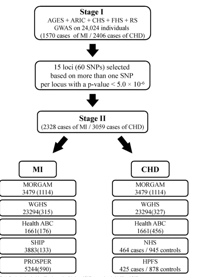
Fig 1describes Stage I and Stage II of the study. The Stage I panel included five prospective cohort studies comprising a total of 24,024 participants who were free of MI and CHD at base-line. The average age ± standard deviation ranged from 54.1±5.6 in ARIC to 74.6±5.5 in AGES. More than half of the participants (54.5%) were women. The basic characteristics of the partici-pating studies are shown inTable 1. A total of 1570 incident MI events (6.5%) and 2406 inci-dent CHD events (10.0%) occurred over an average of 8.2 years and 8.1 years of follow-up for MI and CHD, respectively. The average age at the time of MI ranged from 65.2 years in ARIC to 80.8 years in CHS.
Theλcoefficient within each cohort was small (1.03), suggesting negligible genomic infla-tion. We combined the results of associations for all SNPs across the five cohorts.S1A Figand
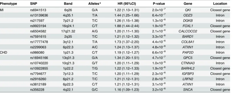
S1B Figpresents the Q-Q plots of combined p-values against the expected p-value distribution for MI and CHD, respectively. The evidence for population admixture was small, both for MI (λ= 1.017) and CHD (λ= 1.022).S2A FigandS2B Figillustrates the p-values of the meta-anal-ysis for each of the SNPs across the 22 autosomal chromosomes for MI and CHD, respectively. In Stage I, 27 SNPs in 8 loci reached our arbitrary threshold of 5×10⁻⁶for MI and 29 SNPs in 8 loci reached this threshold for CHD (Table 2). The most significant association with MI was seen for rs6941513 located on chromosome 6 upstream ofQKI(Hazard Ratio = 1.22 [95% Confidence Interval: 1.13, 1.31], p-value = 2.0×10⁻⁷). For CHD, rs986080, a SNP located on chromosome 1 between two genes (SNX7andPAP2D) showed the strongest association (HR = 1.19 [95%CI: 1.12, 1.27], p-value = 6.6×10⁻⁸).
Fig 1. Study design for identification and validation of SNPs associated with MI and CHD.
Table 1. Baseline characteristics of participants included in incident MI/CHD analysis stratified by cohort.
Characteristic AGES ARIC CHS FHS RS
Participants, n 3219 7406 3291 4134 5974
Age, years 76.4 (5.5) 54.1 (5.57) 72.3 (5.4) 64.5 (12.8) 69.4 (9.1)
Women, % 58.0 54.7 39.1 56.7 59.4
Hypertension1, % 80.6 25.7 52.8 45.3 33.4
Diabetes2, % 11.5 7.7 11.8 10.2 10.6
Current smoker3, % 12.7 24.8 11.3 14.0 22.4
Total cholesterol, mg/dL 217 (45) 214 (41) 213 (39) 203 (40) 255 (47)
HDL cholesterol, mg/dL 61 (17) 51 (17) 55 (16) 52 (17) 52 (14)
Triglyceride, mg/dL 107 (59) 135 (91) 140 (76) 144 (127) NA
Body mass index, kg/m2 27.1 (4.4) 27.0 (4.9) 26.3 (4.5) 27.7 (5.2) 26.3 (3.7)
Incident MI, N cases 86 486 537 165 296
Mean MI follow-up time 2.7 9.2 12.0 5.5 10.1
Incident CHD, N cases 209 575 660 201 761
Mean CHD follow-up time 2.6 9.1 12.0 5.5 9.9
Incident MI Age, years 79.1 (5.5) 65.17 (6.9) 80.8 (6.2) 75.2 (12.2) 80.6 (10.1)
Numbers in table are Mean (SD) or percentage. AGES = Age, Gene/Environment Study; ARIC = Atherosclerosis Risk in Communities Study; CHS = Cardiovascular Health Study; FHS = Framingham Heart Study; HDL = high density lipoprotein; RS = The Rotterdam Study
1 Hypertension was defined as blood pressure140/90 mmHg or on anti-hypertensive medication
2 Diabetes was defined as fasting blood glucose>125 mg/dL, a random blood glucose of>200 mg/dL, or use of insulin or oral hypoglycemic agents (Rotterdam: diabetes definition: Using anti-diabetic medication or random glucose or oral glucose test more than 200 mg/dl)
3 Current cigarette smoking was defined as self-reported cigarette smoking of at least 1 cigarette per day for a year at any attended exam
doi:10.1371/journal.pone.0144997.t001
Table 2. Description and association of SNPs of the top loci associated with incident MI and CHD in Stage I.
Phenotype SNP Band Alleles* HR (95%CI) P-value Gene Location
MI rs6941513 6q26 G/A 1.22 (1.13–1.31) 2.0×10⁻⁷ QKI Closest gene
rs13139636 4q35.1 T/A 1.44 (1.25–1.66) 6.4×10⁻⁷
ODZ3 Intron
rs217597 7p21.2 T/C 1.26 (1.15–1.38) 1.3×10⁻⁶ DGKB Intron
rs9923194 16q24.1 C/T 1.88 (1.44–2.44) 1.9×10⁻⁶ FOXL1 Closest gene
rs6504582 17q21.32 A/G 1.20 (1.11–1.30) 2.1×10⁻⁶ CALCOCO2 Closest gene
rs7591615 2q35 T/C 1.21 (1.12–1.32) 3.3×10⁻⁶
BARD1 Intron
rs17777478 3q12.1 T/A 1.73 (1.37–2.20) 4.4×10⁻⁶ COL8A1 Intron
rs2299063 6p22.3 A/C 1.24 (1.13–1.37) 4.8×10⁻⁶ ATXN1 Intron
CHD rs986080 1p21.3 C/T 1.19 (1.12–1.27) 6.6×10⁻⁸ PAP2D Intron
rs16945166 13q31.3 G/A 1.34 (1.20–1.51) 4.7×10⁻⁷
GPC5 Closest gene
rs10740220 10q21.3 G/T 1.20 (1.11–1.29) 1.0×10⁻⁶ CTNNA3 Intron
rs10922855 1p22.2 T/G 1.22 (1.12–1.33) 1.9×10⁻⁶ BARHL2 Closest gene
rs7794677 7p12.3 T/C 1.20 (1.11–1.29) 2.3×10⁻⁶ IGFBP3 Closest gene
rs2916260 6p21.2 T/C 1.21 (1.12–1.31) 2.8×10⁻⁶
LRFN2 Intron
rs3812189 6p22.3 C/T 1.21 (1.12–1.31) 3.1×10⁻⁶ ATXN1 Intron
rs356228 4q22.1 G/C 1.16 (1.09–1.23) 3.2×10⁻⁶ SNCA Closest gene
*Coded/non-coded allele
HR = Hazard Ratio; CI = Confidence Interval
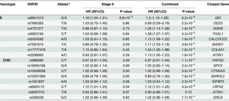
each locus are shown inTable 3. Four SNPs located upstream ofQKIshowed nominal evidence in Stage II for association with MI. The analysis of the combined Stage I and Stage II yielded genome-wide significant associations for three SNPs close toQKI, (rs6941513: HR = 1.21 [95% CI: 1.13, 1.28], p-value = 6.2×10⁻⁹).Fig 2presents the linkage disequilibrium (LD) and p-values of regional markers for this locus. We tested for evidence of replication of this association in 8201 African American individuals including 546 incident cases from the PAGE Study [17], however, rs6941513 was not significantly associated with risk of MI in this population (p = 0.49).
We sought evidence for the association of 46 SNPs recently reported in the largest GWAS to date for coronary artery disease [4] with the incidence of MI and CHD (Table 4). Despite excel-lent power, we found only modest evidence for replication of the association with 9p21 locus (CDKN2A/B), the most established finding from previous cross-sectional case-control GWAS. The most replicated SNP at 9p21 locus, rs1333049, was nominally associated with MI (HR: 1.09 [95%CI: 1.01, 1.18], p-value = 0.02) and marginally with CHD (HR = 1.06 [95%CI: 0.99, 1.13], p-value = 0.08). The most significant association with MI was found for rs15563, a SNP inUBE2Z(HR: 1.12 [95%CI: 1.04, 1.20], p-value = 1.9×10⁻³) and the most significant associa-tion with CHD was found for rs10947789, a SNP within theKCNK5locus (HR: 1.13 [95%CI: 1.05, 1.22], p-value = 5.6×10⁻⁴). We found nominally significant associations (p<0.05) with SNPs annotated toCDKN2A/Bfor MI,LIPAfor CHD andCOL4A2,TCF21,PDGFD,KCNK5, VAMP8,MRAS,UBE2ZandTCF21for both MI and CHD (Table 4). A weighted genetic risk score composed of these 46 SNPs was associated with MI (p-value = 1.3×10⁻³) and CHD (p-value = 1.2×10⁻³) in the Stage I meta-analysis.
Among individuals who experienced MI during follow-up, the risk allele of rs1333049 was associated with a significantly decreased risk of mortality (HR: 0.90 [95% CI: 0.84, 0.97], p-value = 5.5×10⁻³) (Table 5). In both SNPs at 9p21 locus the“risk allele”from cross-sectional
Table 3. Description and association of top SNPs with incident MI and CHD in Stage II and their combined results with Stage I.
Phenotype SNPID Alleles* Stage II Combined Closest Gene
HR (95%CI) P-value HR (95%CI) P-value
MI rs6941513 G/A 1.16 (1.04–1.31) 8.8×10⁻³ 1.2 (1.13–1.28) 6.2×10⁻⁹ QKI
rs7692395 T/G 1.03 (0.75–1.40) 0.86 0.69 (0.59–0.79) 2.5×10⁻⁷ ODZ3
rs4721377 T/G 0.98 (0.87–1.10) 0.73 1.26 (1.14–1.38) 1.9×10⁻⁶ DGKB
rs9923194 C/T 1.04 (0.69–1.58) 0.84 1.58 (1.27–1.97) 4.3×10⁻⁵ FOXL1
rs6504582 A/G 1.03 (0.91–1.15) 0.65 1.15 (1.08–1.22) 1.8×10⁻⁵ CALCOCO2
rs7591615 T/C 0.89 (0.78–1.02) 0.09 1.11 (1.03–1.19) 3.8×10⁻³ BARD1
rs17777478 T/A 1.15 (0.80–1.64) 0.45 1.54 (1.26–1.88) 1.6×10⁻⁵ COL8A1
rs2299063 A/C 0.93 (0.81–1.07) 0.29 1.13 (1.05–1.23) 1.6×10⁻³ ATXN1
CHD rs986080 C/T 0.97 (0.91–1.04) 0.39 0.97 (0.91–1.04) 1.1×10⁻³ PAP2D
rs16945166 G/A 1.02 (0.92–1.14) 0.69 1.02 (0.92–1.14) 2.2×10⁻⁴ GPC5
rs10509258 C/T 1.02 (0.96–1.09) 0.50 1.02 (0.96–1.09) 3.0×10⁻⁴ CTNNA3 rs12031583 G/A 0.89 (0.79–1.00) 0.05 0.89 (0.79–1.00) 7.8×10⁻³ BARHL2
rs1551837 A/G 1.03 (0.94–1.12) 0.58 1.03 (0.94–1.12) 2.3×10⁻⁴ IGFBP3
rs6925172 C/T 1.12 (1.01–1.25) 0.04 1.12 (1.01–1.25) 3.3×10⁻⁶ LRFN2
rs9297015 T/A 0.93 (0.86–1.01) 0.07 0.93 (0.86–1.01) 0.12 ATXN1
rs356228 G/C 1.02 (0.96–1.09) 0.50 1.02 (0.96–1.09) 1.7×10⁻⁴ SNCA
*Coded/non-coded allele
Chr. = Chromosome; HR = Hazard Ratio; CI = Confidence Interval
case-control GWAS was associated with longer survival after MI and would have been enriched in surviving prevalent cases.Fig 3illustrates the inverse association of 78 top SNPs at the 9p21 locus as reported by CARDIoGRAMplusC4D Consortium [4] with survival after MI. We also examined the association of rs6941513 with mortality after MI, however, the association was not significant.
Discussion
We performed a GWAS on incident MI and CHD and examined whether the gene variants identified to date are also associated with risk of CHD in a prospective setting. In a two-stage design, involving 37,561 participants with 2,328 cases of incident MI, we identified a novel genome-wide significant locus,QKI, associated with incident MI. This finding requires further replication. The results also highlighted the difference between the genes identified in prospec-tive versus cross-sectional case-control studies. The 9p21 locus was associated with both an increased risk of incident MI and, during follow-up post-MI, a decreased risk of total mortality, indicating that genetic variants may operate differently in an alternative setting.
Fig 2. Regional plots for the association of SNPs with MI in the region ofQKI.
Table 4. Association of the known SNPs for coronary artery disease with incident MI and CHD in Stage I.
SNP Chr. Freq. Alleles* Reported GWAS
GWAS on Incident MI GWAS on Incident CHD Gene
OR P-value Power HR (95% CI) P-value Power HR (95% CI) P-value
rs3217992 9 0.38 A/G 1.16 7.8×10−57 0.98 1.07 (0.99–1.15) 0.10 0.99 1.04 (0.98–1.11) 0.21 CDKN2A/B
rs1333049 9 0.47 C/G 1.23 1.4×10−52 0.99 1.09 (1.01–1.18) 0.02 0.99 1.06 (0.99–1.13) 0.08 CDKN2A/B
rs602633 1 0.77 C/A 1.12 1.5×10⁻²⁵ 0.73 1.08 (0.99–1.17) 0.10 0.89 1.05 (0.97–1.12) 0.21 PSRC1 rs9369640 6 0.65 A/C 1.09 7.5×10⁻²² 0.62 1.05 (0.98–1.13) 0.17 0.78 1.03 (0.97–1.10) 0.27 PHACTR1 rs11556924 7 0.65 C/T 1.09 6.7×10⁻¹⁷ 0.62 1.05 (0.97–1.14) 0.26 0.78 1.06 (0.99–1.14) 0.07 ZC3HC1
rs9982601 21 0.13 T/C 1.13 7.7×10⁻¹⁷ 0.63 0.98 (0.88–1.09) 0.68 0.79 1.01 (0.93–1.10) 0.81 MRPS6
rs6725887 2 0.11 C/T 1.12 1.2×10⁻¹⁵ 0.51 1.06 (0.95–1.18) 0.29 0.67 0.99 (0.90–1.08) 0.78 WDR12 rs1122608 19 0.76 G/T 1.10 6.3×10⁻¹⁴ 0.62 1.07 (0.98–1.16) 0.13 0.77 1.04 (0.97–1.12) 0.23 SMARCA4 rs12190287 6 0.59 C/G 1.07 4.9×10⁻¹³ 0.45 1.13 (1.04–1.23) 4.4×10⁻³ 0.60 1.09 (1.01–1.16) 0.02 TCF21
rs7173743 15 0.58 T/C 1.07 6.7×10⁻¹³ 0.45 1.05 (0.97–1.13) 0.23 0.60 1.04 (0.98–1.10) 0.23 MORF4L1
rs17114036 1 0.91 A/G 1.11 5.8×10⁻¹² 0.39 1.12 (0.97–1.28) 0.11 0.52 1.06 (0.95–1.19) 0.27 PPAP2B rs9515203 13 0.74 T/C 1.08 5.9×10⁻¹² 0.46 1.06 (0.95–1.17) 0.31 0.61 1.07 (0.99–1.17) 0.09 COL4A2 rs2505083 10 0.42 C/T 1.06 1.4×10⁻¹¹ 0.35 1.05 (0.97–1.13) 0.19 0.47 1.03 (0.97–1.10) 0.36 KIAA1462
rs4773144 13 0.42 G/A 1.07 1.4×10⁻¹¹ 0.45 1.09 (1.01–1.18) 0.03 0.60 1.09 (1.02–1.16) 0.01 COL4A2
rs7692387 4 0.81 G/A 1.06 2.7×10⁻¹¹ 0.39 1.00 (0.91–1.09) 0.95 0.52 1.01 (0.93–1.09) 0.83 GUCY1A3 rs974819 11 0.29 A/G 1.07 3.6×10⁻¹¹ 0.39 0.91 (0.83–0.98) 0.02 0.53 0.93 (0.87–1.00) 0.04 PDGFD rs3184504 12 0.40 T/C 1.07 5.4×10⁻¹¹ 0.44 1.03 (0.95–1.11) 0.47 0.60 1.04 (0.98–1.11) 0.22 SH2B3
rs2075650 19 0.14 G/A 1.11 5.9×10⁻¹¹ 0.53 1.03 (0.91–1.16) 0.67 0.68 1.01 (0.91–1.13) 0.79 TOMM40
rs2048327 6 0.35 G/A 1.06 6.9×10⁻¹¹ 0.33 1.00 (0.93–1.08) 0.98 0.45 1.01 (0.95–1.08) 0.65 SLC22A3 rs9319428 13 0.32 A/G 1.05 7.3×10⁻¹¹ 0.32 0.99 (0.91–1.09) 0.91 0.43 1.03 (0.96–1.11) 0.44 FLT1 rs17514846 15 0.44 A/C 1.05 9.3×10⁻¹¹ 0.45 1.00 (0.93–1.08) 0.94 0.60 1.02 (0.96–1.09) 0.52 FURIN
rs1561198 2 0.45 A/G 1.05 1.2×10⁻¹⁰ 0.35 1.09 (1.01–1.17) 0.02 0.48 1.09 (1.02–1.15) 6.9×10⁻³ VAMP8
rs515135 2 0.83 G/A 1.08 2.6×10⁻¹⁰ 0.29 1.09 (0.98–1.20) 0.10 0.40 1.03 (0.95–1.12) 0.52 APOB rs4845625 1 0.47 T/C 1.04 3.6×10⁻¹⁰ 0.36 1.01 (0.94–1.08) 0.86 0.48 1.03 (0.97–1.10) 0.28 IL6R rs2895811 14 0.43 C/T 1.06 4.1×10⁻¹⁰ 0.35 1.03 (0.95–1.11) 0.44 0.48 1.03 (0.96–1.10) 0.39 KIAA1822
rs4252120 6 0.73 T/C 1.06 4.9×10⁻¹⁰ 0.38 0.99 (0.92–1.07) 0.82 0.51 0.99 (0.93–1.06) 0.74 PLG
rs273909 5 0.14 C/T 1.09 9.6×10⁻¹⁰ 0.26 1.04 (0.91–1.17) 0.59 0.35 1.01 (0.91–1.12) 0.84 SLC22A4 rs12936587 17 0.59 G/A 1.06 1.2×10⁻⁹ 0.35 1.00 (0.92–1.08) 0.94 0.47 0.97 (0.91–1.04) 0.40 RAI1 rs2047009 10 0.48 C/A 1.05 1.6×10⁻⁹ 0.27 1.02 (0.94–1.09) 0.68 0.36 1.03 (0.97–1.1) 0.30 CXCL12
rs501120 10 0.83 A/G 1.07 1.8×10⁻⁹ 0.29 1.08 (0.96–1.21) 0.18 0.39 1.00 (0.91–1.09) 0.96 CXCL12
rs9818870 3 0.14 T/C 1.07 2.6×10⁻⁹ 0.26 1.10 (1.00–1.21) 0.05 0.35 1.09 (1.01–1.18) 0.02 MRAS rs264 8 0.86 G/A 1.05 2.9×10⁻⁹ 0.53 0.99 (0.89–1.10) 0.83 0.68 1.05 (0.96–1.14) 0.31 LPL rs2281727 17 0.36 C/T 1.05 7.8×10⁻⁹ 0.25 1.07 (0.99–1.15) 0.09 0.34 1.02 (0.96–1.09) 0.50 SMG6
rs445925 19 0.9 C/T 1.13 8.8×10⁻⁹ 0.54 1.16 (0.97–1.39) 0.10 0.56 1.05 (0.92–1.19) 0.46 APOC1
rs10947789 6 0.76 T/C 1.06 9.8×10⁻⁹ 0.36 1.12 (1.02–1.22) 0.01 0.48 1.13 (1.05–1.22) 5.6×10⁻⁴ KCNK5 rs579459 9 0.21 C/T 1.07 2.7×10⁻⁸ 0.33 1.06 (0.97–1.17) 0.20 0.45 1.03 (0.96–1.12) 0.39 ABO rs2252641 2 0.46 G/A 1.04 5.3×10⁻⁸ 0.36 1.00 (0.92–1.07) 0.91 0.48 0.97 (0.91–1.03) 0.34 ZEB2
rs12413409 10 0.89 G/A 1.10 6.3×10⁻⁸ 0.38 1.05 (0.92–1.20) 0.49 0.52 1.01 (0.90–1.13) 0.91 CNNM2
rs9326246 11 0.10 C/G 1.09 1.5×10⁻⁷ 0.51 0.93 (0.79–1.08) 0.32 0.41 0.93 (0.82–1.06) 0.26 BUD13 rs11203042 10 0.44 T/C 1.04 6.1×10⁻⁶ 0.19 1.05 (0.98–1.13) 0.15 0.25 1.07 (1.01–1.14) 0.03 LIPA rs15563 17 0.52 C/T 1.04 9.4×10⁻⁶ 0.19 1.12 (1.04–1.20) 1.9×10⁻³ 0.25 1.07 (1.01–1.13) 0.03 UBE2Z
rs2246833 10 0.38 T/C 1.06 9.5×10⁻⁶ 0.34 1.07 (1.00–1.16) 0.06 0.46 1.06 (1.00–1.13) 0.05 LIPA
rs11206510 1 0.84 T/C 1.06 1.8×10⁻⁵ 0.22 1.02 (0.92–1.14) 0.67 0.30 1.01 (0.93–1.10) 0.80 PCSK9 rs12205331 6 0.81 C/T 1.04 4.2×10⁻⁵ 0.14 1.03 (0.93–1.13) 0.58 0.17 1.05 (0.97–1.13) 0.24 ANKS1A rs17464857 1 0.87 T/G 1.05 6.1×10⁻⁵ 0.15 1.11 (0.97–1.27) 0.13 0.19 1.09 (0.97–1.22) 0.14 TAF1A
In this two-stage design, we found evidence for MI-associated genetic variants nearbyQKI (KH domain containing, RNA binding). The combined p-value for three out of four genetic variants that were examined in the region exceeded genome-wide significant threshold. Although these data provide evidence for an association between theQKIlocus and incident MI, this finding should be confirmed by further studies since these variants attained conven-tional levels of genome-wide significant p-value only in the combined meta-analysis.
If confirmed, theQKIfinding may represent a novel pathway in developing CHD.QKIis known to be involved in cell cycle regulation, a pathway for which there is emerging evidence for a key role in developing atherosclerotic plaques and cardiovascular disease [18,19]. A functional study has reported thatQKIis a central regulator of vascular smooth muscle cell phenotypic plas-ticity and that intervention inQKIactivity can improve pathogenic fibro-proliferative responses to vascular injury [20]. Moreover, a recent paper shows that the RNA-binding properties of QKI play a critical role in regulating human monocyte to macrophage differentiation [21]. de Bruin and co-workers identified that the conversion of monocytes to both pro- and anti-inflammatory macrophages with GM-CSF or M-CSF, respectively, markedly increased expression of the QKI, which all were readily detected in CD68+ macrophages of fibrous cap atheromata and atheroscle-rotic lesions with intraplaque hemorrahage. Furthermore, reduced expression of QKI in mono-cytes delayed their differentiation into macrophages, perturbed their capacity to become lipid-engorged foam cells, and led to a reduction in monocyte infiltration in atherosclerotic lesions [21]. Altogether we propose that QKI is involved in inflammatory responses to injury and could be a potential thrapeutic target to prevent cardiovascular disease. Further functional investigation is needed to robustly identify mechanisms involved for this locus.
Prior GWAS which included extremely large sample sizes did not reportQKIthough they should have had enough statistical power to detect a locus with such an effect. However rs6941513 was not associated with CAD in the Cardiogram plusC4D GWAS (OR = 1.01, p-value = 0.45). In contrast to former GWAS, we have used a prospective, longitudinal cohort design to examine genetic association with incident cases of MI and CHD. It is possible that the magnitude of the effect with prevalent cases is smaller than with incident cases; thus the locus was not detected by previously published GWAS that primarily use a case-control design.
Although CHD includes MI events by definition, the loci we found for MI and CHD over-lapped only for one locus (ATX1). One reason could be differences in mechanisms involved in the restrictive diagnosis of MI versus the broader diagnosis of CHD. However, unstable effect
Table 4. (Continued)
SNP Chr. Freq. Alleles* Reported GWAS
GWAS on Incident MI GWAS on Incident CHD Gene
OR P-value Power HR (95% CI) P-value Power HR (95% CI) P-value
rs12539895 7 0.19 A/C 1.08 5.3×10⁻⁴ 0.39 1.03 (0.94–1.12) 0.57 0.52 0.99 (0.92–1.07) 0.87 GPR22
Chr. = Chromosome; Freq. = Frequency; OR = Odds Ratio; HR = Hazard Ratio; CI = Confidence Interval
*Coded / Non-coded allele
doi:10.1371/journal.pone.0144997.t004
Table 5. Association of the known SNPs for coronary artery disease with mortality after MI.
SNP Closest Gene Alleles HR(95%CI) P-value
rs1333049 CDKN2A/B C/G 0.90 (0.84–0.97) 5.5×10⁻³
rs3217992 CDKN2A/B A/G 0.94 (0.88–1.01) 0.10
rs602633 PSRC1 C/A 1.01 (0.93–1.09) 0.93
estimates and p-values due to lack of statistical power could have contributed to this observa-tion as well.
Despite excellent statistical power, we identified only a modest signal at the 9p21 locus. This locus, initially identified by GWAS, has been validated by numerous studies in different geo-graphic and ethnic subgroups. However, our study is not the first study to report a weak signal or lack of association at this locus. In fact, prominent differences have been observed between cross-sectional case-control versus longitudinal studies. For instance, in a meta-analysis by Chan et al [22], cross-sectional analyses of angiographically defined cases and controls show a strong per allele association with 9p21 (OR: 1.31, 95% CI: 1.20, 1.43). However, in a meta-anal-ysis of follow-up studies by Patel et al [23], the per allele hazard ratio of the 9p21 variants for fatal and non-fatal adjudicated MI was 1.09 (95% CI: 1.03–1.16). The latter is the same as what we report in this study, though the meta-analysis includes earlier reports from some of our studies. One explanation for this inconsistency is the incidence-prevalence bias. Most GWAS for coronary artery disease to date have consisted of cross-sectional case-control studies, a design that over represents patients who survived their MI or CHD event. Using data from five population based cohort studies we found that the reported risk alleles for this locus are associ-ated with longer survival after MI. This finding that was previously reported as well [23–25] supports the conjecture. Thus, the high prevalence of the risk allele in various types of
cross-Fig 3. The association of top 79 SNPs with coronary artery disease as reported by CardiogramplusC4D for 9p21 locus and their association with total mortality after MI.
sectional analyses may not be due entirely to a high risk of experiencing MI or CHD, but also to an improved chance of survival after MI.
The molecular biology behind the protective effect of the risk alleles at 9p21 is yet unclear, however, there is a growing body of evidence to show that 9p21 locus is only increasing the risk of CHD for the first event and not for the subsequent events. For instance, Patel et al found no association with subsequent CHD events in a recent meta-analysis of 25,163 individuals with established CHD [23]. Thus, it could be concluded that 9p21 locus is contributing to the forma-tion and progression of plaques and not to their instability prior to events; therefore, the associa-tion is merely observed in early stages of the disease. This is in agreement with the report by Palomaki [26] that suggests a diminished effect of 9p21 locus by age, a finding that is confirmed by Patel et al for secondary events. It should be noted that the mean age of participants was more than 70 years old in two and more than 60 years old in four of the participating cohorts. In this context, the older mean age of our population could be another reason why our findings do not replicate known loci such as 9p21.Our study is the largest collection of population-based pro-spective GWAS on incident MI and CHD and includes high quality genotyping and phenotyping data from well-known cohort studies in the field of cardiovascular disease. Moreover, similar case definitions for MI and CHD, comparable quality control for genotyped data, harmonized imputation strategies and collaboratively designed analysis plans are further strengths of our study. Despite these strengths, there are several limitations that merit discussion. First, nearly all studies who contributed to our GWAS are also members of the CARDIoGRAMPlusC4D Con-sortium [27], however, they have used only their prevalent cases in CARDIoGRAMPlusC4D project and therefore there is no overlap between the two GWAS. Second, since our sample size was limited, further susceptibility variants of weaker effects may have been missed in our study. Third, we have tried to use consistent definitions for MI, however, slight differences exist between the definitions for CHD. This might have introduced heterogeneity in our case definition. Finally, our findings may not be directly generalizable to non-European populations.
A potential clinical application of risk alleles identified from GWAS is the prospective pre-diction of cardiovascular disease. To date, the totality of evidence from prospective studies sug-gests that there is only modest, independent prediction of increased cardiovascular disease risk using genetic information with small to modest incremental reclassification for prediction beyond the known clinical CVD risk scores [28]. This lack of success has been attributed to the small percentage of variance explained by known genetic factors. However, our results also sug-gest that genetic risk prediction needs to consider differences in genetic variants that predict the risk of cardiovascular disease in prospective and cross-sectional settings.
In summary, using the largest collection of population- based prospective genome-wide association studies we have identifiedQKIas a potential locus for incident myocardial infarc-tion. Furthermore, we have shown that the genes associated with risk of cardiovascular disease may differ in effect size when studied in a cross-sectional case-control versus cohort settings. The role of 9p21 locus may be complex, increasing the risk of incident MI and decreasing mor-tality among those with CHD. This highlights the importance of examining longitudinal cohort studies in the study of etiology even for genetic factors. These findings may have implications for application of genetic variants in risk estimation for cardiovascular disease, an effort that so far has not provided strong evidence for incremental risk prediction by genetic markers.
Supporting Information
S1 File. Supplementary document: Methods, acknowledgment and funding for the partici-pating studies.
S1 Fig. a. QQ Plot for Discovery GWAS MI. b. QQ Plot for Discovery GWAS CHD. (ZIP)
S2 Fig. a. Log-plot for Discovery GWAS MI. b. Log-plot for Discovery GWAS CHD. (ZIP)
S1 Table. Phenotype description of the studies in stage I.
(DOCX)
S2 Table. Phenotype description of the studies in stage II.
(DOCX)
S3 Table. Analysis Logistics of the studies in stage I.
(DOCX)
S4 Table. Analysis Logistics of the studies in stage II.
(DOCX)
S5 Table. Genotyping/imputation/QC specifics of the studies in stage I.
(DOCX)
S6 Table. Genotyping/imputation/QC specifics of the studies in stage II.
(DOCX)
S7 Table. Basic description of the study population for the survival after MI analysis.
(DOCX)
S8 Table. Basic description of the cohort studies in stage II.
(DOCX)
S9 Table. Basic description of the case-control studies in stage II.
(DOCX)
S10 Table. Association of the SNPs taken to stage II with MI.
(DOCX)
S11 Table. Association of the SNPs taken to stage II with CHD.
(DOCX)
Author Contributions
Conceived and designed the experiments: DIC ARF SBK CJG VS AGU RSV CLC JV SD PA DA SRH JF PD NLS YAW DSS PGW KDT AE FK JIR JK KK GH LJL AH MRPM KS PW EJB TBH DL YL EBR JWJ HV PMR SB OHF VG BMP EB CJO. Performed the experiments: BMB JD MD MRPM DJS FR. Analyzed the data: AD JCB CCW AVS ACM ST TL UV MKJ IF GE NF YAW GH PK LMR KL. Contributed reagents/materials/analysis tools: LAC TL IF CLC JV PA DA KMR PGW AE FK JK KK PK SB. Wrote the paper: AD JCB CCW LAC KK JWJ BMP EB CJO.
References
1. Lloyd-Jones DM, Nam BH, D'Agostino RB Sr., Levy D, Murabito JM, Wang TJ, et al. Parental cardio-vascular disease as a risk factor for cardiocardio-vascular disease in middle-aged adults: a prospective study of parents and offspring. JAMA. 2004; 291(18):2204–11. Epub 2004/05/13. doi:10.1001/jama.291.18. 2204291/18/2204 [pii]. PMID:15138242.
3. O'Donnell CJ, Nabel EG. Genomics of cardiovascular disease. N Engl J Med. 2011; 365(22):2098– 109. Epub 2011/12/02. doi:10.1056/NEJMra1105239PMID:22129254.
4. Consortium CAD, Deloukas P, Kanoni S, Willenborg C, Farrall M, Assimes TL, et al. Large-scale asso-ciation analysis identifies new risk loci for coronary artery disease. Nat Genet. 2013; 45(1):25–33. Epub 2012/12/04. ng.2480 [pii] doi:10.1038/ng.2480PMID:23202125.
5. Hill G, Connelly J, Hebert R, Lindsay J, Millar W. Neyman's bias re-visited. J Clin Epidemiol. 2003; 56 (4):293–6. Epub 2003/05/28. S0895435602005711 [pii]. PMID:12767404.
6. Neyman J. Statistics; servant of all sciences. Science. 1955; 122(3166):401–6. Epub 1955/09/02. PMID:13246647.
7. Wilson PW. Risk scores for prediction of coronary heart disease: an update. Endocrinol Metab Clin North Am. 2009; 38(1):33–44. Epub 2009/02/17. S0889-8529(08)00084-4 [pii] doi:10.1016/j.ecl.2008. 11.001PMID:19217511.
8. Psaty BM, O'Donnell CJ, Gudnason V, Lunetta KL, Folsom AR, Rotter JI, et al. Cohorts for Heart and Aging Research in Genomic Epidemiology (CHARGE) Consortium: Design of prospective meta-analy-ses of genome-wide association studies from 5 cohorts. Circ Cardiovasc Genet. 2009; 2(1):73–80. Epub 2009/12/25. 2/1/73 [pii] doi:10.1161/CIRCGENETICS.108.829747PMID:20031568; PubMed Central PMCID: PMC2875693.
9. Harris TB, Launer LJ, Eiriksdottir G, Kjartansson O, Jonsson PV, Sigurdsson G, et al. Age, Gene/Envi-ronment Susceptibility-Reykjavik Study: multidisciplinary applied phenomics. Am J Epidemiol. 2007; 165(9):1076–87. Epub 2007/03/14. kwk115 [pii] doi:10.1093/aje/kwk115PMID:17351290; PubMed Central PMCID: PMC2723948.
10. The Atherosclerosis Risk in Communities (ARIC) Study: design and objectives. The ARIC investiga-tors. Am J Epidemiol. 1989; 129(4):687–702. Epub 1989/04/01. PMID:2646917.
11. Fried LP, Borhani NO, Enright P, Furberg CD, Gardin JM, Kronmal RA, et al. The Cardiovascular Health Study: design and rationale. Ann Epidemiol. 1991; 1(3):263–76. Epub 1991/02/01. PMID:1669507.
12. Feinleib M, Kannel WB, Garrison RJ, McNamara PM, Castelli WP. The Framingham Offspring Study. Design and preliminary data. Prev Med. 1975; 4(4):518–25. Epub 1975/12/01. PMID:1208363.
13. Hofman A, Grobbee DE, de Jong PT, van den Ouweland FA. Determinants of disease and disability in the elderly: the Rotterdam Elderly Study. Eur J Epidemiol. 1991; 7(4):403–22. PMID:1833235.
14. Hofman A, van Duijn CM, Franco OH, Ikram MA, Janssen HL, Klaver CC, et al. The Rotterdam Study: 2012 objectives and design update. Eur J Epidemiol. 2011; 26(8):657–86. PMID:21877163. doi:10. 1007/s10654-011-9610-5
15. Price AL, Patterson NJ, Plenge RM, Weinblatt ME, Shadick NA, Reich D. Principal components analy-sis corrects for stratification in genome-wide association studies. Nat Genet. 2006; 38(8):904–9. Epub 2006/07/25. ng1847 [pii] doi:10.1038/ng1847PMID:16862161.
16. Willer CJ, Li Y, Abecasis GR. METAL: fast and efficient meta-analysis of genomewide association scans. Bioinformatics. 2010; 26(17):2190–1. Epub 2010/07/10. btq340 [pii] doi:10.1093/bioinformatics/ btq340PMID:20616382; PubMed Central PMCID: PMC2922887.
17. Matise TC, Ambite JL, Buyske S, Carlson CS, Cole SA, Crawford DC, et al. The Next PAGE in under-standing complex traits: design for the analysis of Population Architecture Using Genetics and Epidemi-ology (PAGE) Study. Am J Epidemiol. 2011; 174(7):849–59. Epub 2011/08/13. kwr160 [pii] doi:10. 1093/aje/kwr160PMID:21836165; PubMed Central PMCID: PMC3176830.
18. Helgadottir A, Thorleifsson G, Manolescu A, Gretarsdottir S, Blondal T, Jonasdottir A, et al. A common variant on chromosome 9p21 affects the risk of myocardial infarction. Science. 2007; 316(5830):1491– 3. Epub 2007/05/05. 1142842 [pii] doi:10.1126/science.1142842PMID:17478679.
19. McPherson R, Pertsemlidis A, Kavaslar N, Stewart A, Roberts R, Cox DR, et al. A common allele on chromosome 9 associated with coronary heart disease. Science. 2007; 316(5830):1488–91. Epub 2007/05/05. 1142447 [pii] doi:10.1126/science.1142447PMID:17478681; PubMed Central PMCID: PMC2711874.
20. van der Veer E, de Bruin RG, Kraaijeveld A, de Vries MR, Bot I, Pera T, et al. The RNA-Binding Protein Quaking is a Critical Regulator of Vascular Smooth Muscle Cell Phenotype. Circ Res. 2013. Epub 2013/08/22. CIRCRESAHA.113.301302 [pii] .
21. de Bruin RG, Shiue L, Prins J, de Boer HC, Singh A, Fagg W.S., et al. Quaking post-transcriptionally promotes monocyte differentiation into pro-atherogenic macrophages by controlling pre-mRNA splicing and gene expression. Nat Commun. 2016.
23. Patel RS, Asselbergs FW, Quyyumi AA, Palmer TM, Finan CI, Tragante V, et al. Genetic variants at chromosome 9p21 and risk of first versus subsequent coronary heart disease events: a systematic review and meta-analysis. J Am Coll Cardiol. 2014; 63(21):2234–45. Epub 2014/03/13. S0735-1097 (14)01383-7 [pii] doi:10.1016/j.jacc.2014.01.065PMID:24607648; PubMed Central PMCID: PMC4035794.
24. Szpakowicz A, Kiliszek M, Pepinski W, Waszkiewicz E, Franaszczyk M, Skawronska M, et al. Polymor-phism of 9p21.3 locus is associated with 5-year survival in high-risk patients with myocardial infarction. PLoS One. 2014; 9(8):e104635. Epub 2014/08/12. doi:10.1371/journal.pone.0104635 PONE-D-14-14817 [pii]. PMID:25105296; PubMed Central PMCID: PMC4126747.
25. Gong Y, Beitelshees AL, Cooper-DeHoff RM, Lobmeyer MT, Langaee TY, Wu J, et al. Chromosome 9p21 haplotypes and prognosis in white and black patients with coronary artery disease. Circ Cardio-vasc Genet. 2011; 4(2):169–78. Epub 2011/03/05. CIRCGENETICS.110.959296 [pii] doi:10.1161/ CIRCGENETICS.110.959296PMID:21372283; PubMed Central PMCID: PMC3101633.
26. Palomaki GE, Melillo S, Bradley LA. Association between 9p21 genomic markers and heart disease: a meta-analysis. JAMA. 2010; 303(7):648–56. Epub 2010/02/18. 303/7/648 [pii] doi:10.1001/jama.2010. 118PMID:20159873.
27. Deloukas P, Kanoni S, Willenborg C, Farrall M, Assimes TL, Thompson JR, et al. Large-scale associa-tion analysis identifies new risk loci for coronary artery disease. Nat Genet. 2012. Epub 2012/12/04. ng.2480 [pii] doi:10.1038/ng.2480PMID:23202125.
28. de Vries PS, Kavousi M, Ligthart S, Uitterlinden AG, Hofman A, Franco OH, et al. Incremental predictive value of 152 single nucleotide polymorphisms in the 10-year risk prediction of incident coronary heart disease: the Rotterdam Study. Int J Epidemiol. 2015; 44(2):682–8. Epub 2015/05/09. dyv070 [pii] doi: