Advances in Biomarkers: Going Beyond the
Carcinoembryonic Antigen
Nicole E. Lopez, MD
1Carrie Y. Peterson, MD
21Division of Surgical Oncology, University of North Carolina, Chapel
Hill, North Carolina
2Division of Colorectal Surgery, Medical College of Wisconsin,
Milwaukee, Wisconsin
Clin Colon Rectal Surg 2016;29:196–204.
Address for correspondenceCarrie Y. Peterson, MD, Medical College of Wisconsin, 9200 West Wisconsin Avenue, Division of Colorectal Surgery, Milwaukee, WI 53226 (e-mail: [email protected]).
Advances in biotechnology in the last decade have been tremendous. The technologies used in molecular biology, genetics, and biochemistry have catapulted our understand-ing of cellular processes and have led to several changes in how we practice medicine; and the coming years will be even more eventful. Not only can we now detect genetic mutations and achieve results in a fraction of the time, but we can do this using smaller tissue samples than ever before. Together, these capacities have enabled increasingly personalized therapeu-tic approaches for patients with colorectal cancer (CRC).
In the traditional paradigm of oncologic treatment strate-gies, three options were available for treating cancer: surgery, cytotoxic chemotherapy, and radiation. The choice of ideal treatment was made based upon the organ of origin, the histologic type of malignancy described, and the stage of the disease. In such a paradigm, all nonmetastatic colon cancers have surgery, and all stage III patients receive the same adjuvant chemotherapeutic agents, resulting in heteroge-neous responses: some do well and some recur. We are rapidly closing in on using each patient’s own unique genetic and tumor profile to help understand these discrepancies and inform medical decision-making, what is termed“ personal-ized medicine.”As we learn more about the genetic changes associated with CRC, we are also learning ways in which these mutations can be used to diagnose, prognosticate outcomes, and measure responses to treatments.
The term“biomarker”is somewhat difficult to define. At its essence, it is any biological substrate that can be detected, which is then used to help guide medical decision-making. Current biomarkers in routine clinical use today for CRC include measur-ing serum carcinoembryonic antigen (CEA) and detectmeasur-ing mis-match repair (MMR) gene andKRASmutations in tumor tissues. Our goal is to review the current state of the science on biomarkers as it relates to CRC. We will discuss the use of CEA for monitoring of recurrence, the role of genetic testing in guiding surveillance, and treatment decisions and how genetic mutations may predict response to chemotherapy and out-comes. We will also briefly review how biomarkers can function as liquid biopsies and explore several new substrates that are currently under investigation. Given the pace of advancements in molecular biology, genetics, and amplification technologies, it is highly likely that in our careers we could do things only seen in the movies: from a single drop of blood we could diagnose the presence of CRC, understand precisely the genetic mutations present, how they impact outcomes and respond to treatment, and even follow that profile as it changes over time.
Carcinoembryonic Antigen
The standard serum biomarker in use today for CRC is CEA. It is a superfamily of glycoproteins found on cell membranes that play an important role in cell recognition and adhesion. It Keywords
►
carcinoembryonic
antigen
►
hereditary colon
cancer
►
polyposis
►
Lynch syndrome
Abstract
Using biologically available markers to guide treatment decisions in colorectal cancer
care is becoming increasingly common, though our understanding of these biomarkers
is in its infancy. In this article, we will discuss how this area is rapidly changing, review
important biomarkers being used currently, and explain how the results in
fl
uence
clinical decision-making. We will also brie
fl
y discuss the possibility of a liquid biopsy and
explore several exciting and new options.
Issue Theme Hot Topics in Colorectal Surgery; Guest Editor: Gregory D. Kennedy, MD, PhD
Copyright © 2016 by Thieme Medical Publishers, Inc., 333 Seventh Avenue, New York, NY 10001, USA.
Tel: +1(212) 584-4662.
DOI http://dx.doi.org/
10.1055/s-0036-1584289.
ISSN 1531-0043.
is thought to be intimately involved in the ability of CRC cells to metastasize; colorectal adenocarcinoma produces larger amounts of this protein as a result of alterations in posttran-scriptional regulation.1,2Interestingly, most newly diagnosed patients present with normal levels as it is cleared in the liver before entering the systemic circulation, though in cases of distal cancers alternate circulation patterns through the internal iliac vessels may be associated with high levels.2 Most modern assays for detecting CEA use a monoclonal technique that has a low false-positive rate; however, it can still be elevated in several disease states such as tobacco use, liver disease, and renal dysfunction.2
Unfortunately, CEA has little utility as a screening tool for diagnosing new CRCs due to poor sensitivity and specificity, particularly in early-stage cancer.2Fletcher calculated that if CEA were used to screen patients for CRC, at a sensitivity and specificity for early-stage disease of 40 and 90%, respectively, there would be 250 false-positive tests while missing 60% of cancers in asymptomatic patients.3Other biomarkers, such as CA 19–9 and CA 242, have fared no better and currently no serum test is recommended as a screening tool for diagnosing CRC.4,5
While CEA may be a poor candidate to assist in screening or diagnosis, it has some utility in predicting prognosis and monitoring for recurrence; elevated levels have been shown to correlate with poor outcomes and might be a useful strategy for defining patients at higher risk of recurrence.1 Preoperative CEA levels also serve as a benchmark to follow treatment outcomes—levels that do not fall within 6 weeks of resection are concerning for residual disease, either local or metastatic. Serial measurements after curative resection can also give an early warning of recurrence or metastatic dis-ease; it is the most frequent indicator of recurrence in asymptomatic patients, is more cost-effective than radiology for detecting curable recurrent disease, is highly sensitive for liver metastases, and can improve survival when included as part of an intensive surveillance plan.1,6,7Using CEA to detect recurrence has been shown to provide 5 months lead time before developing other cancer-related symptoms, though this has not been shown to result in improved survival.8 Current National Comprehensive Cancer Network (NCCN) recommendations include measuring CEA levels at diagnosis and every 3 to 6 months for thefirst 2 years, followed by every 6 months for the next 3 years in all patients with stage I to III CRC who would be candidates for further treatment if de-tected.9 An increase of 30% above baseline is generally considered significant enough to warrant further evaluation, though no widely accepted definition is reported in any recommendation.1
For patients with metastatic disease, CEA changes during treatment can indicate response to therapy. While an elevated preoperative CEA is associated with poor outcomes in all patients, a fall in the CEA to normal levels after complete hepatic disease resection is highly predictive of improved survival.10 Furthermore, CEA is a good biomarker when monitoring response to chemotherapy; the predictive value of a rising CEA during chemotherapy is so suggestive of progression that further imaging and testing may not be
needed before changing treatment regimens.4,11Serial CEA measurements every 2 to 3 months while on chemotherapy are beneficial as persistent rises can detect progression even before it may be apparent on imaging; thus, ineffective regimens can be changed quickly, reducing costs and improv-ing quality of life.4
Despite the demonstrated benefits of using CEA as a biomarker for recurrence and response to treatment, there are some significant limitations. Notably, it has low sensitivity and specificity for early stage CRCs, so it is not only a poor screening tool as a noninvasive alternative to colonoscopy, it has limited ability to prognosticate in patients with early-stage disease who have normal levels—a significant propor-tion of those with the disease. It can also be falsely elevated in several common scenarios, such as patients who smoke and have either renal or liver dysfunction; therefore, a high level in an otherwise asymptomatic patient still demands further diagnostic testing. In addition, there is some concern that serial testing after curative treatment is not cost effective in terms of lives saved and improved survival.2While using CEA as a biomarker has some advantages—its easily measured via a serum test and can be repeated frequently with little impact on the patient—it also has some significant drawbacks; therefore, the search for better substrates continues.
Indications for Genetic Testing
Polyposis Syndromes
CRC is a heterogeneous disease. Most CRC occurs sporadically, but approximately 30% of cases demonstrate a familial predis-position, and a positive family history in unaffected persons doubles their risk of developing CRC.12However, only one-third of patients with a family history have identifiable germline mutations. Given their differential risk profiles, and the benefit of initiating early screening protocols in affected persons, the importance of identification of patients with hereditary syn-dromes is well-recognized. Accordingly, well-defined clinical parameters exist for selecting patients who should undergo genetic testing for suspected hereditary syndromes.13,14
Several polyposis syndromes have been linked to specific genetic mutations, thus any patient presenting with more than 10 polyps, hamartomatous polyps, or polyps at a young age (under 30 years), should prompt genetic evaluation. While the involved genes are known for many of these diseases, de novo mutations within a family can occur, so clinical suspicion outside of family history should prompt evaluation and can be enough to establish the diagnosis as the patient may have a mutation not identifiable by conventional methods.15,16The most commonly occurring hereditary syn-drome is familial adenomatous polyposis. There are several mutations that occur in the APC gene, each with a known associated phenotype that gives rise to several presentations including an attenuated form.17,18Another similar polyposis syndrome has been described that involves an autosomal recessive mutation in the MUTYHgene, known as MUTYH-associated polyposis (MAP). Hamartomatous polyposis syn-dromes are much less common and de novo mutations occur as well, so clinical suspicion is paramount (see►Table 1).
Lynch Syndrome
Lynch syndrome (LS), the most common heritable form of CRC, it is an autosomal dominant inherited disease with variable penetrance, and accounts for approximately 3% of all CRCs.19,20It generally results from inheritance of germline mutations in MMR genes, though rarely de novo mutations, which rise at a rate of about 2%, can also cause the syn-drome.21 Patients with LS can be identified using several strategies. Traditional clinical criteria for identifying persons at risk for LS, such as Amsterdam criteria and Bethesda guidelines, set forth parameters primarily based on family history and age at diagnosis. Both have undergone modifi ca-tions in the efforts of improving sensitivity and specificity. Computational models, such as MMRpredict (Edinburgh, Scotland, UK), offer better sensitivity and specificity, but are less convenient to use in everyday practice.22
While these methods can be used to recognize patients at risk for LS, tumor testing confirms the diagnosis. The Center for Disease Control recommendations support the use of universal genetic screening for the presence of common mutations in MMR genes for all patients with newly diag-nosed CRC.23 However, the utilization of resources with universal genetic testing is significant, and this guideline is not collectively endorsed. Alternative recommendations in-clude screening for patients with CRC under the age of 70 years and those over 70 years with a family history concerning for LS.24,25
Strategies to assess MMR deficiency use either immuno-histochemistry (IHC) to identify impaired protein expression of the four MMR genes commonly mutated in LS (MLH1,
MSH2,MSH6,PMS2), or polymerase chain reaction (PCR) to detect microsatellite instability (MSI), which suggests im-properly functioning MMR proteins. Results of IHC and PCR generally agree, however, IHC is slightly less sensitive and specific. Even so, due to the increased availability of IHC, and its cost savings in comparison to PCR, it is often thefirst step in tumor analysis.26,27 Once an MMR protein deficiency is identified, testing with PCR can confirm MSI. This involves testingfive markers, known as the Bethesda panel (BAT 25, BAT 26, D5S346, D2S123, and D17S250), though other com-mercially available panels are widely used.28,29MSI tumors
are regarded as MSI-H (high) if>30% of the markers are mutated, MSI-L (low) if at least one and<30% of the markers are mutated, and microsatellite stable (MSS) if no markers are mutated.30 To determine the etiology of MSI in patients without a clear family history of LS, further analysis of the pattern of protein loss can be useful. In patients with a MLH1 deficiency, testing for BRAF mutations and MLH1 hyperme-thylation should follow, as these are often seen in sporadic CRC with MSI but rarely seen in LS, which is associated with direct mutations inMLH1orMSH2.31–33Alternatively, if loss of MLH2 and/or MSH6 protein occurs, genetic testing should evaluateMSH2,EpCAM, andMSH6.24,34
Until recently, there has been little clinical utility for understanding the genetics of sporadic CRC. Over the last decade, advances in molecular techniques and genomic pro-filing have improved our understanding of cellular processes, allowing for the development and approval of several tar-geted therapies for use in the CRC. This has resulted in an explosion of scientific interest in establishing genetic bio-markers with prognostic and predictive value that can be used to help guide treatment decisions.
Mutation Analysis to Guide Treatment
Targeted Therapy in Metastatic Colorectal Cancer
The development of several“targeted therapies”for use in metastatic CRC has led to an increasing drive to use each patient’s unique tumor mutational profile to help inform treatment choices (see►Table 2). This is the beginning of personalized medicine—neither two persons, nor do two tumor cells harbor the same profile. Tumor mutation profiles can even change over time as new mutations accumulate or respond to treatment. The beginning of this was the under-standing of the impact of KRASand NRAS mutations. The
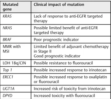
Table 2 Clinical relevance of colorectal cancer genetic mutations
Mutated gene
Clinical impact of mutation
KRAS Lack of response to anti-EGFR targeted therapy
NRAS Possible limited benefit of anti-EGFR targeted therapy
BRAF Poor prognostic indicator MMR with
MSI
Limited benefit of adjuvant chemotherapy in Stage II
Good prognostic indicator
LOH 18q/CIN Possible resistance tofluorouracil
Top 1 Possible increased response to irinotecan
ERCC1 Possible increased response to oxaliplatin orfluorouracil
UGT1A Increased risk of toxicity from irinotecan
DPYD Increased toxicity withfluorouracil
Abbreviation: EGFR, epidermal growth factor receptor; LOH, loss of heterozygosity.
Table 1 Heritable mutations in colorectal cancer
Syndrome Mutated
gene
Inheritance pattern
FAP, attenuated FAP APC Autosomal dominant
MAP MUTYH Autosomal recessive
Peutz–Jeghers STK11, LKB1 Autosomal dominant
Juvenile polyposis SMAD4,
BRMP1A Autosomal dominant
Cowden, Bannayan–
Riley–Ruvalcaba PTEN
Autosomal dominant
Abbreviations: FAP, familial adenomatous polyposis; MAP, MUTYH-as-sociated polyposis.
Source: Table adapted from Syngal et al.14
protein products of these genes are involved in intracellular signaling pathways that promote cell growth and develop-ment via MAP kinase activation. Mutations in these genes result in constitutive activation of the MAPK pathway and are present in 40% of CRCs atKRAS exon 2 (codons 12 and 13).35,36Patients withKRASmutations do not respond to the targeted anti-epidermal growth factor receptor (anti-EGFR) therapy, including cetuximab and panitumumab, which block activation of the pathway in patients with wild type (WT)
KRAS genes. Additionally, and perhaps more importantly, patients with KRAS mutations have been shown to have worse outcomes when given anti-EGFR-targeted chemother-apy, highlighting the importance of assessing KRASstatus before the initiation of these therapies.37,38For those patients with metastatic CRC and no KRAS mutations, anti-EGFR therapy can confer a significant survival benefit.39
Though eliminating patients with known mutations in
KRAS results in improved response rates to EGFR-targeted therapies in combination with chemotherapy, still only ap-proximately 10% ofKRASWT patients respond to cetuximab as monotherapy, suggesting the need for further efforts to im-prove patient selection by better identification of those who will respond to this therapy.38,40–42Accordingly, investigators have become increasingly interested in studies revealing a lack of response among tumors with other RAS family mutations, specifically on theNRASgene, designated“extended RAS”or “expanded RAS” mutations.43–45 Nearly 20% of KRAS WT tumors bear one of these mutations and patients with these mutations may also have limited benefit from anti-EGFR therapies, suggesting an opportunity to further improve pa-tient selection by excluding a significant portion of KRAS WT, who would be unlikely to respond to anti-EGFR therapy.43,45 While testing forKRASmutations in codons 12 and 13 is widely recommended to help guide selection of chemotherapeutic agents, testing for otherKRAS andNRASmutations is more controversial.25,46Still, current NCCN recommendations state that patients with any KRAS or NRAS mutation should not be treated with cetuximab or panitumumab.25
BRAF is another important protein in the MAP kinase pathway. Mutations inBRAFoccur in approximately 15% of CRC, and in contrast toKRAS, evidence concerning the efficacy of anti-EGFR therapy in BRAF mutated CRC is conflicting. Several studies show no benefit of anti-EGFR therapy in patients withBRAFmutations.35,47,48However, pooled data from the CRYSTAL and OPUS trials suggest thatBRAFstatus does not predict a response to anti-EGFR therapy.49Some of the difficulty in arriving at definitive conclusions stems from the relatively low incidence ofBRAF mutations in patients with CRC.35,36The prognostic significance ofBRAFmutations is more certain, as patients with this mutation have consis-tently been shown to have poor outcomes in relation to those with WT BRAF,with reduced overall survival and progres-sion-free survival.44,50,51 Current NCCN recommendations include testing of tumors for BRAF mutations in stage IV disease, though this is of prognostic value only.25 Even so, testing in metastatic patients, particularly those who have failed other chemotherapy regimens, may yield additional benefit to patients in terms of identifying those who might be
eligible to participate in clinical trials withBRAFinhibitors, which are likely to be of more clinical utility.43
Microsatellite Instability in Stage II Colon Cancer
MSI occurs in about 15% of sporadic KRAS WT CRCs. It is associated with right-sided lesions and poorly differentiated, mucinous histology.52,53MSI testing for the purposes of identi-fying patients with LS should be performed according to previ-ously described guidelines. Additionally, the NCCN recommends MSI testing of all tumors in patients with Stage II disease. MSI indicates improved prognosis in comparison to MSS tumors and MSI status can also be used to aid in assessing the likelihood that patients will benefit from adjuvant chemotherapy.25
Despite the predominance of poorly differentiated histolo-gy among patients with MSI tumors, these patients tend to have improved outcomes and a decreased likelihood of metas-tases in comparison to patients with MSS tumors.54 In this respect, MSI is regarded as a strong positive prognostic indicator.55,56 There is evidence to support that patients with MSI tumors and Stage II/III CRC do not have the same improvement in disease-free survival and overall survival with adjuvantfluorouracil (5-FU)-based therapy as compared with their MSS counterparts.52,53Furthermore, the use of 5-FU in MSI patients with Stage II CRC has been associated with adverse outcomes.53 However, these results were found in the setting of regimens lacking oxaliplatin, an agent almost universally used in the adjuvant treatment of CRC today. More recent data from randomized trials (PETACC 3 and QUASAR) have not confirmed thesefindings.57,58Taken together, it is currently recommended that tumors from patients with stage II CRC be tested for MSI. Decisions about whether patients with Stage II CRC and MSI receive 5-FU-based chemotherapy should be individually tailored and based on risk assessment.25The same trend is not seen in stage III CRC, where a clear survival benefit is seen with adjuvant chemotherapy in all patients.59
Chromosomal Instability, Topoisomerase I, and Excision Repair Cross-Complementing Gene 1 Polymorphisms
Chromosomal instability (CIN) describes a state of high rates of gains and losses of chromosomes within tumors. The cause of CIN is unknown, but loss of heterozygosity (LOH) is a hallmark feature and LOH at 18q has generated attention due to its possible association with outcomes.60,61 Additionally, there is some limited evidence to suggest that CIN, in general, may be indicative of resistance to both taxols and 5-FU.62,63 Topoisomerases allow for DNA unwinding, a process that is imperative for DNA replication and upregulated in the CRC.64 The cytotoxic chemotherapeutic agent irinotecan binds and stabilizes the topoisomerase I (Top I)–DNA complex, prevent-ing further DNA replication.65 Accordingly, the UK MRC FOCUS trial showed that Top I overexpression predicts a response to irinotecan and possibly oxaliplatin, though this could not be verified in another study.66,67A similar story is seen for another gene, excision repair cross-complementing
(ERCC1), which encodes the ERCC-1 protein and is responsible for repairing double strand breaks and cross-linking errors in DNA.64ERCC-1 deficiency is common in a CRC and can result
from either genetic mutation or promoter methylation.67 ERCC-1 deficiency has been noted to predict response to oxaliplatin or 5-FU-based chemotherapy, but this could not be confirmed in other retrospective analyses.68,69A deal of additional research evaluating the utility of the above bio-markers is needed before widespread routine clinical use can be recommended.
Predicting Chemotherapy Toxicity
The uridine diphosphate glucuronosyltransferase 1A (UGT1A) gene is located on chromosome 2q37. The gene transcribes several enzymes, among them, UGT1A1 is commonly known for its role in the glucuronidation of bilirubin and a mutation here can result in increased concentrations of the active metabolite of irinotecan, thus increasing tumor responsiveness to this regimen in the presence of the mutation. However, this increased cyto-toxicity also increases the risk of adverse reactions, such as neutropenia and diarrhea. Despite the known risk of toxicity in patients with the mutation, the recommendations do not cur-rently endorse routine testing. Rather, they advise dose reduc-tions in the setting of adverse reacreduc-tions.70Dihydropyrimidine dehydrogenase (DPD) deficiency can also lead to chemotherapy-related toxicity as it is the enzyme responsible for metabolism of 5-FU. Deficiency of this enzyme is present in 3 to 5% of the population and is associated with severe adverse effects in response to 5-FU-based chemotherapy.69,71Deficiency generally rises through mutations in the dihydropyrimidine dehydroge-nase gene (DPYD, mapped to chromosome 1p22) and patients who are homozygous for the mutantDPYDallele can have lethal reactions to 5-FU.72Despite the propensity for patients with DPD deficiency to develop toxicity in response to 5-FU, its association with response to therapy is uncertain. Given decreased adverse effects with newer dosing regimens and the lack of solid evidence to support its ability to predict response to therapy, testing for DPD deficiency is not currently standard.73,74
In addition to DPD, mutations in other enzymes partici-pating in pyrimidine metabolism, such as thymidylate syn-thase and methylenetetrahydrofolate reductase, may predict clinical outcome, response, and toxicity to 5-FU. With regard to biological therapies, investigators have been interested in the utility of PI3K and PTEN to predict response to anti-EGFR therapy.75 Similarly, others are exploring the capacity for soluble vascular endothelial growth factor (VEGF), plasma VEGF, and tumor VEGF to predict responses to treatment with anti-VEGF.74The clinical validity of these markers and more are yet to be defined.
Multigene Assays
Several multigene assays are available for the purposes of risk stratification. Oncotype DX (Genemics Health Inc., Redwood City, CA), ColoPrint (Agendia NV, Amsterdam, Netherlands), and ColDx (Almac, Craigavon, Ireland) each offer genetic panels to aid in predicting recurrence of CRC.25The 12 gene assay, Oncotype DX, was developed using data from NSABP studies and the Cleveland Clinic. Its predictive value, in terms of establishing a recurrence score for patients with Stage II and Stage III CRC, was validated in studies using tissue and data gathered in both the QUASAR and NSABP-07 trials.76–78The
ColoPrint is a similar panel, with 18 genes. In a retrospective cohort study comparing ColoPrint to clinical risk factors out-lined by NCCN guidelines, ColoPrint was more accurate at predicting recurrence of stage II MSS CRC. The study was unable to show a benefit of chemotherapy according to risk stratification, however, this would best be appreciated in a randomized trial that is adequately powered to do so.79Finally, ColDx is a microarray-based assay was validated in its ability to assess recurrence risk in stage II CRC using specimens and data gathered from CALGB 9581.80Despite the capacity of these assays to determine risk of recurrence, no test has displayed the ability to predict a response to chemotherapy, therefore, the true impact of these diagnostics on oncologic outcomes is uncertain, and the utility of these assays in assistance with clinical decision-making has yet to be determined.25
Limitations of Current Biomarkers and
Future Possibilities
As of today, all analyses of tumor characteristics are depen-dent upon invasive biopsy. This includes evaluation of the specific tumor type and histologic features as well as genetic profile. Whether tissue is collected via endoscopic or percu-taneous biopsy, or from a surgical specimen, a reasonably large volume of tissue is needed for interrogation. In most cases, these specimens are collected at the time of diagnosis, but performing the tests evaluating some biomarkers, such as
KRAS, may be delayed until the patient recurs with metastatic disease. This sequencing may pose several problems. First, CRC is a heterogeneous disease with several distinct path-ways to progression, thus each person and even each cell has the potential to harbor a different profile of mutations.81,82 Second, tumor molecular profiles change over time, resulting in different behaviors, such as the ability to metastasize.83 Third, tumor profiles can change with treatment as those cells with targeted features are destroyed by chemotherapy and others without are left behind to develop more genetic alterations.84 A static sample of tissue from a single point in time is not able to reflect these ongoing and dynamic changes. Furthermore, if repeat tissue is needed for analysis, it involves an invasive procedure with inherent complications that can impact patients’care and treatment.
Development of a“liquid biopsy”is an ongoing research topic that is making considerable progress in recent years as amplification technologies are rapidly improving. The goal is to discover a substrate that is sensitive enough to detect the presence of early CRC, but also specific enough to discriminate from other adenocarcinomas, provide molecular profiling of the tumor to direct targeted therapies and assess for re-sponses to treatment, offer prognostic information about the risk of recurrence and response to therapy, and can be drawn repeatedly over the course of treatment with little to no risk. While we have not yet identified the ideal candidate, several targets are showing promise.
Proteins circulating in the serum have long been used as biomarkers, including CEA and CA 19–9. These tests have the benefit being easily reproducible, but are not very sensitive or specific for diagnosing CRC and provide only rough
prognostication.85They also do not offer any guides to treat-ment, but do tend to correlate with disease progression or response.86The potential pool of proteins from which to draw a biomarker is endless, but one candidate, Rab27b, has shown promise. In a recent publication, this GTPase involved in regulating secretory pathways and oncogenesis was shown to have elevated expression in tumor when compared with adjacent normal tissues, and high levels correlated with serum CEA elevations, lymph node metastases, and TNM stage, as well as overall survival rate in a cohort of 116 CRC patients.87 Further work will needed to be done to determine if serum expression of Rab27b correlates or offers improved sensitivity and specificity beyond current biomarkers. Rather than inves-tigate known oncogenic proteinsde novoin each cancer type, another strategy could be a more powerful way to identify candidate proteins. Surinova et al (2015) used a screening method to identify candidate glycoprotein biomarkers from tumor epithelia, then screened selected proteins in the plasma of CRC patients to identify those that reached the circulation. Overall, 88 proteins were identified and 70 fulfilled validation criteria with an independent cohort; further consensus testing identified 5 proteins that consistently predicted disease and performed better than CEA in diagnosing CRC, even when used together with the CEA.88
MicroRNA (miRNA) is another area of active investigation as a source of biomarkers for CRC. miRNA is small, noncoding single strands of RNA that are thought to play a role in posttranscriptional regulation of gene expression and are dysregulated in CRC.89Several candidates have been identi-fied, including miRNA-375, miRNA-183, and miRNA-211 that correlate with detection, recurrence, or prognosis.89–91 How-ever, the understanding of these biomarkers and their native functions is poorly understood. In addition, testing for these substances is highly variable in commercially available kits, calling into question the reproducibility of using these small molecules in the clinical setting.92Furthermore, proteins and miRNA will never have the potential to provide meaningful genetic information about the tumor and its response to treatment, limiting the applicability of these biomarkers.
Perhaps the area showing most potential is in the detection and analysis of circulating DNA and circulating tumor cells (CTCs). Circulating cell-free DNA (cfDNA) is thought to arise from both normal and tumor cells as a result of normal processes as well as apoptosis and necrosis.93Amplification of cfDNA has been increasingly used to diagnose fetal genetic abnormalities in high-risk women, with positive tests warrant-ing more invasive diagnostic procedures, and these cfDNA screening tests have also identified new incidental malignan-cies in the mother due to their high sensitivity.94 Levels of serum cfDNA been shown to correlate with prognosis in metastatic CRC, and it also has the potential to be a source for detection of various genetic mutations, with a high concor-dance of KRAS mutations when compared with standard techniques.93,95Evaluating the level and pattern of methyla-tion in cfDNA samples has also been shown to be sensitive for detecting CRC, especially for stage IV disease.96,97CTC can also be a highly sensitive and specific biomarker and provide specific genetic information about both the tumor and its
response to treatment. Using enrichment and depletion tech-niques, CTC can be isolated and detected by targeting their cell-surface markers, such as CK20, so that further analysis can be performed.98 The discordance between CTC and primary tumor was low for bothKRASandKRASmutations.99 Further-more, patients with known KRAS mutations in CTC were shown to have poor outcomes when treated with anti-EGFR therapy, the same as if primary tumor testing occurred.100
Conclusion
As we approach an era of personalized medicine, the impact of each individual tumor cell and each person’s unique genetic profile will become increasingly important. Our understanding of the utility of these mutations is only in its infancy. We currently use only CEA as a routine serum biomarker, and it has significant limitations. While we are fairly good at identifying heritable mutations, several persons with a strong family history do not have any mutation we currently test for, suggesting that there are still many more genes with influence over the development of CRC. Our understanding of how tumor genetic mutations can predict response to therapy and oncologic outcomes is poor and improvements in this arena could have a huge impact on our everyday practice and advances here will become pro-gressively more valuable. Furthermore, the discovery of a “liquid biopsy”to diagnose, predict response, and prognosti-cate outcomes would be monumental, not only from a treatment standpoint, but from a quality of life and comfort standpoint of our patients. The future in biomarker under-standing is wide open and there remains much to be learned.
References
1 Duffy MJ, van Dalen A, Haglund C, et al. Clinical utility of biochemical markers in colorectal cancer: European Group on Tumour Markers (EGTM) guidelines. Eur J Cancer 2003;39(6): 718–727
2 Goldstein MJ, Mitchell EP. Carcinoembryonic antigen in the staging and follow-up of patients with colorectal cancer. Cancer Invest 2005;23(4):338–351
3 Fletcher RH. Carcinoembryonic antigen. Ann Intern Med 1986; 104(1):66–73
4 Locker GY, Hamilton S, Harris J, et al; ASCO. ASCO 2006 update of recommendations for the use of tumor markers in gastrointesti-nal cancer. J Clin Oncol 2006;24(33):5313–5327
5 Burt RW, Cannon JA, David DS, et al; National comprehensive cancer network. Colorectal cancer screening. J Natl Compr Canc Netw 2013;11(12):1538–1575
6 Mayer RJ, Garnick MB, Steele GD Jr, Zamcheck N. Carcinoem-bryonic antigen (CEA) as a monitor of chemotherapy in dissemi-nated colorectal cancer. Cancer 1978;42(3, Suppl):1428–1433 7 Rosen M, Chan L, Beart RW Jr, Vukasin P, Anthone G. Follow-up of
colorectal cancer: a meta-analysis. Dis Colon Rectum 1998;41(9): 1116–1126
8 Mäkelä JT, Laitinen SO, Kairaluoma MI. Five-year follow-up after radical surgery for colorectal cancer. Results of a prospective randomized trial. Arch Surg 1995;130(10):1062–1067 9 Benson AB III, Bekaii-Saab T, Chan E, et al; National
Comprehen-sive Cancer Network. Localized colon cancer, version 3.2013: featured updates to the NCCN Guidelines. J Natl Compr Canc Netw 2013;11(5):519–528
10Hohenberger P, Schlag PM, Gerneth T, Herfarth C. Pre- and postoperative carcinoembryonic antigen determinations in he-patic resection for colorectal metastases. Predictive value and implications for adjuvant treatment based on multivariate anal-ysis. Ann Surg 1994;219(2):135–143
11Ward U, Primrose JN, Finan PJ, et al. The use of tumour markers CEA, CA-195 and CA-242 in evaluating the response to chemo-therapy in patients with advanced colorectal cancer. Br J Cancer 1993;67(5):1132–1135
12Johnson CM, Wei C, Ensor JE, et al. Meta-analyses of colorectal cancer risk factors. Cancer Causes Control 2013;24(6): 1207–1222
13Network NCC. NCCN Clinical Practice Guidelines in Oncology: Genetic/Familial High-Risk Assessment: Colorectal Version I. 2015. Available at: https://www.nccn.org/professionals/physi-cian_gls/pdf/colorectal_screening.pdf. Access on May 23, 2016 14Syngal S, Brand RE, Church JM, Giardiello FM, Hampel HL, Burt
RW; American College of Gastroenterology. ACG clinical guide-line: Genetic testing and management of hereditary gastrointes-tinal cancer syndromes. Am J Gastroenterol 2015;110(2): 223–262, quiz 263
15Lagarde A, Rouleau E, Ferrari A, et al. Germline APC mutation spectrum derived from 863 genomic variations identified through a 15-year medical genetics service to French patients with FAP. J Med Genet 2010;47(10):721–722
16Aretz S, Vasen HF, Olschwang S. Clinical Utility Gene Card for: Familial adenomatous polyposis (FAP) and attenuated FAP (AFAP)
—update 2014. Eur J Hum Genet 2015;23(6):
17Aretz S, Uhlhaas S, Caspari R, et al. Frequency and parental origin of de novo APC mutations in familial adenomatous polyposis. Eur J Hum Genet 2004;12(1):52–58
18Rustin RB, Jagelman DG, McGannon E, Fazio VW, Lavery IC, Weakley FL. Spontaneous mutation in familial adenomatous polyposis. Dis Colon Rectum 1990;33(1):52–55
19Barnetson RA, Tenesa A, Farrington SM, et al. Identification and survival of carriers of mutations in DNA mismatch-repair genes in colon cancer. N Engl J Med 2006;354(26):2751–2763
20Hampel H, Frankel WL, Martin E, et al. Screening for the Lynch syndrome (hereditary nonpolyposis colorectal cancer). N Engl J Med 2005;352(18):1851–1860
21Win AK, Jenkins MA, Buchanan DD, et al. Determining the frequency of de novo germline mutations in DNA mismatch repair genes. J Med Genet 2011;48(8):530–534
22Green RC, Parfrey PS, Woods MO, Younghusband HB. Prediction of Lynch syndrome in consecutive patients with colorectal can-cer. J Natl Cancer Inst 2009;101(5):331–340
23Evaluation of Genomic Applications in Practice and Prevention (EGAPP) Working Group. Recommendations from the EGAPP Working Group: genetic testing strategies in newly diagnosed individuals with colorectal cancer aimed at reducing morbidity and mortality from Lynch syndrome in relatives. Genet Med 2009;11(1):35–41
24Giardiello FM, Allen JI, Axilbund JE, et al. Guidelines on genetic evaluation and management of Lynch syndrome: a consensus statement by the US Multi-society Task Force on colorectal cancer. Am J Gastroenterol 2014;109(8):1159–1179
25Network NCC. NCCN Clinical Practice Guidelines in Oncology: Colon Cancer Version 3. 2015. Available at: https://www.nccn. org/professionals/physician_gls/pdf/colon.pdf. Access on May 23, 2016
26Bartley AN, Luthra R, Saraiya DS, Urbauer DL, Broaddus RR. Identification of cancer patients with Lynch syndrome: clini-cally significant discordances and problems in tissue-based mismatch repair testing. Cancer Prev Res (Phila) 2012;5(2): 320–327
27Ladabaum U, Wang G, Terdiman J, et al. Strategies to identify the Lynch syndrome among patients with colorectal cancer: a cost-effectiveness analysis. Ann Intern Med 2011;155(2):69–79
28Bacher JW, Flanagan LA, Smalley RL, et al. Development of a
fluorescent multiplex assay for detection of MSI-High tumors. Dis Markers 2004;20(4–5):237–250
29Boland CR, Thibodeau SN, Hamilton SR, et al. A National Cancer Institute Workshop on Microsatellite Instability for cancer detec-tion and familial predisposidetec-tion: development of internadetec-tional criteria for the determination of microsatellite instability in colorectal cancer. Cancer Res 1998;58(22):5248–5257
30Boland CR, Goel A. Microsatellite instability in colorectal cancer. Gastroenterology 2010;138(6):2073–2087.e3
31Affolter K, Samowitz W, Tripp S, Bronner MP. BRAF V600E mutation detection by immunohistochemistry in colorectal car-cinoma. Genes Chromosomes Cancer 2013;52(8):748–752 32Toyota M, Ahuja N, Ohe-Toyota M, Herman JG, Baylin SB, Issa JP.
CpG island methylator phenotype in colorectal cancer. Proc Natl Acad Sci U S A 1999;96(15):8681–8686
33Weisenberger DJ, Siegmund KD, Campan M, et al. CpG island methylator phenotype underlies sporadic microsatellite instabil-ity and is tightly associated with BRAF mutation in colorectal cancer. Nat Genet 2006;38(7):787–793
34Hegde M, Ferber M, Mao R, Samowitz W, Ganguly A; Working Group of the American College of Medical Genetics and Genomics (ACMG) Laboratory Quality Assurance Committee. ACMG techni-cal standards and guidelines for genetic testing for inherited colorectal cancer (Lynch syndrome, familial adenomatous poly-posis, and MYH-associated polyposis). Genet Med 2014;16(1): 101–116
35Maughan TS, Adams RA, Smith CG, et al; MRC COIN Trial Inves-tigators. Addition of cetuximab to oxaliplatin-based first-line combination chemotherapy for treatment of advanced colorectal cancer: results of the randomised phase 3 MRC COIN trial. Lancet 2011;377(9783):2103–2114
36Tveit KM, Guren T, Glimelius B, et al. Phase III trial of cetuximab with continuous or intermittent fluorouracil, leucovorin, and oxaliplatin (Nordic FLOX) versus FLOX alone infirst-line treat-ment of metastatic colorectal cancer: the NORDIC-VII study. J Clin Oncol 2012;30(15):1755–1762
37Tol J, Koopman M, Cats A, et al. Chemotherapy, bevacizumab, and cetuximab in metastatic colorectal cancer. N Engl J Med 2009; 360(6):563–572
38Van Cutsem E, Köhne CH, Hitre E, et al. Cetuximab and chemo-therapy as initial treatment for metastatic colorectal cancer. N Engl J Med 2009;360(14):1408–1417
39Karapetis CS, Khambata-Ford S, Jonker DJ, et al. K-ras mutations and benefit from cetuximab in advanced colorectal cancer. N Engl J Med 2008;359(17):1757–1765
40Bokemeyer C, Bondarenko I, Makhson A, et al. Fluorouracil, leucovorin, and oxaliplatin with and without cetuximab in the
first-line treatment of metastatic colorectal cancer. J Clin Oncol 2009;27(5):663–671
41Jonker DJ, O’Callaghan CJ, Karapetis CS, et al. Cetuximab for the treatment of colorectal cancer. N Engl J Med 2007;357(20): 2040–2048
42Poulin-Costello M, Azoulay L, Van Cutsem E, Peeters M, Siena S, Wolf M. An analysis of the treatment effect of panitumumab on overall survival from a phase 3, randomized, controlled, multi-center trial (20020408) in patients with chemotherapy refractory metastatic colorectal cancer. Target Oncol 2013;8(2):127–136 43Atreya CE, Corcoran RB, Kopetz S. Expanded RAS: refining the
patient population. J Clin Oncol 2015;33(7):682–685
44Douillard JY, Oliner KS, Siena S, et al. Panitumumab-FOLFOX4 treatment and RAS mutations in colorectal cancer. N Engl J Med 2013;369(11):1023–1034
45Sorich MJ, Wiese MD, Rowland A, Kichenadasse G, McKinnon RA, Karapetis CS. Extended RAS mutations and anti-EGFR monoclo-nal antibody survival benefit in metastatic colorectal cancer: a meta-analysis of randomized, controlled trials. Ann Oncol 2015; 26(1):13–21
46 Evaluation of Genomic Applications in Practice and Prevention (EGAPP) Working Group. Recommendations from the EGAPP Working Group: can testing of tumor tissue for mutations in EGFR pathway downstream effector genes in patients with metastatic colorectal cancer improve health outcomes by guiding decisions regarding anti-EGFR therapy? Genet Med 2013;15(7): 517–527
47 De Roock W, Claes B, Bernasconi D, et al. Effects of KRAS, BRAF, NRAS, and PIK3CA mutations on the efficacy of cetuximab plus chemotherapy in chemotherapy-refractory metastatic colorectal cancer: a retrospective consortium analysis. Lancet Oncol 2010; 11(8):753–762
48 Di Nicolantonio F, Martini M, Molinari F, et al. Wild-type BRAF is required for response to panitumumab or cetuximab in metastatic colorectal cancer. J Clin Oncol 2008;26(35): 5705–5712
49 Bokemeyer C, Van Cutsem E, Rougier P, et al. Addition of cetux-imab to chemotherapy asfirst-line treatment for KRAS wild-type metastatic colorectal cancer: pooled analysis of the CRYSTAL and OPUS randomised clinical trials. Eur J Cancer 2012;48(10): 1466–1475
50 Tol J, Nagtegaal ID, Punt CJ. BRAF mutation in metastatic colorec-tal cancer. N Engl J Med 2009;361(1):98–99
51 Bokemeyer C, Bondarenko I, Hartmann JT, et al. Efficacy according to biomarker status of cetuximab plus FOLFOX-4 asfirst-line treatment for metastatic colorectal cancer: the OPUS study. Ann Oncol 2011;22(7):1535–1546
52 Ribic CM, Sargent DJ, Moore MJ, et al. Tumor microsatellite-instability status as a predictor of benefit fromfluorouracil-based adjuvant chemotherapy for colon cancer. N Engl J Med 2003; 349(3):247–257
53 Sargent DJ, Marsoni S, Monges G, et al. Defective mismatch repair as a predictive marker for lack of efficacy offluorouracil-based adjuvant therapy in colon cancer. J Clin Oncol 2010;28(20): 3219–3226
54 Gryfe R, Kim H, Hsieh ET, et al. Tumor microsatellite instability and clinical outcome in young patients with colorectal cancer. N Engl J Med 2000;342(2):69–77
55 Merok MA, Ahlquist T, Røyrvik EC, et al. Microsatellite instability has a positive prognostic impact on stage II colorectal cancer after complete resection: results from a large, consecutive Norwegian series. Ann Oncol 2013;24(5):1274–1282
56 Popat S, Hubner R, Houlston RS. Systematic review of microsat-ellite instability and colorectal cancer prognosis. J Clin Oncol 2005;23(3):609–618
57 Hutchins G, Southward K, Handley K, et al. Value of mismatch repair, KRAS, and BRAF mutations in predicting recurrence and benefits from chemotherapy in colorectal cancer. J Clin Oncol 2011;29(10):1261–1270
58 Tejpar FBS, Delorenzi M, Fiocca R, et al. Microsatellite instability (MSI) in stage II and III colon cancer treated with LV or 5FU-LV and irinotecan (PETACC 3-EORTC 40993-SAKK 60/00 trial). J Clin Oncol 2009;27(15S):4001
59 André T, Boni C, Navarro M, et al. Improved overall survival with oxaliplatin,fluorouracil, and leucovorin as adjuvant treatment in stage II or III colon cancer in the MOSAIC trial. J Clin Oncol 2009; 27(19):3109–3116
60 Watanabe T, Wu TT, Catalano PJ, et al. Molecular predictors of survival after adjuvant chemotherapy for colon cancer. N Engl J Med 2001;344(16):1196–1206
61 Ogino S, Nosho K, Irahara N, et al. Prognostic significance and molecular associations of 18q loss of heterozygosity: a cohort study of microsatellite stable colorectal cancers. J Clin Oncol 2009;27(27):4591–4598
62 Swanton C, Tomlinson I, Downward J. Chromosomal instability, colorectal cancer and taxane resistance. Cell Cycle 2006;5(8): 818–823
63 Lee AJ, Endesfelder D, Rowan AJ, et al. Chromosomal instability confers intrinsic multidrug resistance. Cancer Res 2011;71(5): 1858–1870
64 Kuraoka I, Kobertz WR, Ariza RR, Biggerstaff M, Essigmann JM, Wood RD. Repair of an interstrand DNA cross-link initiated by ERCC1-XPF repair/recombination nuclease. J Biol Chem 2000; 275(34):26632–26636
65 Yu J, Miller R, Zhang W, et al. Copy-number analysis of topoisom-erase and thymidylate synthase genes in frozen and FFPE DNAs of colorectal cancers. Pharmacogenomics 2008;9(10):1459–1466 66 Koopman NKM, Richman S, Seymour M, et al. The correlation
between Topoisomerase-I (Topo1) expression and outcome of treatment with capecitabine and irinotecan in advanced colorec-tal cancer (ACC) patients (pts) treated in the CAIRO study of the Dutch Colorectal Cancer Group (DCCG). Eur J Cancer Suppl 2009; 7(2):321–322
67 Facista A, Nguyen H, Lewis C, et al. Deficient expression of DNA repair enzymes in early progression to sporadic colon cancer. Genome Integr 2012;3(1):3
68 Koopman M, Venderbosch S, van Tinteren H, et al. Predictive and prognostic markers for the outcome of chemotherapy in ad-vanced colorectal cancer, a retrospective analysis of the phase III randomised CAIRO study. Eur J Cancer 2009;45(11): 1999–2006
69 Lu Z, Zhang R, Diasio RB. Dihydropyrimidine dehydrogenase activity in human peripheral blood mononuclear cells and liver: population characteristics, newly identified deficient patients, and clinical implication in 5-fluorouracil chemotherapy. Cancer Res 1993;53(22):5433–5438
70 Evaluation of Genomic Applications in Practice and Prevention (EGAPP) Working Group. Recommendations from the EGAPP Working Group: can UGT1A1 genotyping reduce morbidity and mortality in patients with metastatic colorectal cancer treated with irinotecan? Genet Med 2009;11(1):15–20
71 Etienne MC, Lagrange JL, Dassonville O, et al. Population study of dihydropyrimidine dehydrogenase in cancer patients. J Clin Oncol 1994;12(11):2248–2253
72 Omura K. Clinical implications of dihydropyrimidine dehydroge-nase (DPD) activity in 5-FU-based chemotherapy: mutations in the DPD gene, and DPD inhibitoryfluoropyrimidines. Int J Clin Oncol 2003;8(3):132–138
73 Walther A, Johnstone E, Swanton C, Midgley R, Tomlinson I, Kerr D. Genetic prognostic and predictive markers in colorectal cancer. Nat Rev Cancer 2009;9(7):489–499
74 Schmoll HJ, Van Cutsem E, Stein A, et al. ESMO Consensus Guidelines for management of patients with colon and rectal cancer. a personalized approach to clinical decision making. Ann Oncol 2012;23(10):2479–2516
75 Sood A, McClain D, Maitra R, et al. PTEN gene expression and mutations in the PIK3CA gene as predictors of clinical benefit to anti-epidermal growth factor receptor antibody therapy in pa-tients with KRAS wild-type metastatic colorectal cancer. Clin Colorectal Cancer 2012;11(2):143–150
76 Gray RG, Quirke P, Handley K, et al. Validation study of a quantitative multigene reverse transcriptase-polymerase chain reaction assay for assessment of recurrence risk in patients with stage II colon cancer. J Clin Oncol 2011;29(35):4611–4619 77 O’Connell MJ, Lavery I, Yothers G, et al. Relationship between
tumor gene expression and recurrence in four independent studies of patients with stage II/III colon cancer treated with surgery alone or surgery plus adjuvantfluorouracil plus leuco-vorin. J Clin Oncol 2010;28(25):3937–3944
78 Yothers G, O’Connell MJ, Lee M, et al. Validation of the 12-gene colon cancer recurrence score in NSABP C-07 as a predictor of recurrence in patients with stage II and III colon cancer treated withfluorouracil and leucovorin (FU/LV) and FU/LV plus oxali-platin. J Clin Oncol 2013;31(36):4512–4519
79Kopetz S, Tabernero J, Rosenberg R, et al. Genomic classifier ColoPrint predicts recurrence in stage II colorectal cancer pa-tients more accurately than clinical factors. Oncologist 2015; 20(2):127–133
80Niedzwiecki DFW, Venook AP, Ye X, et al. Association between ColDx assay result and recurrence-free interval in stage II colon cancer patients on CALGB (Alliance) 9581. J Clin Oncol 2014;32 (Suppl 3):abstract number 455
81Bacolod MD, Barany F. Molecular profiling of colon tumors: the search for clinically relevant biomarkers of progression, progno-sis, therapeutics, and predisposition. Ann Surg Oncol 2011; 18(13):3694–3700
82Cancer Genome Atlas N; Cancer Genome Atlas Network. Com-prehensive molecular characterization of human colon and rectal cancer. Nature 2012;487(7407):330–337
83Zalata KR, Elshal MF, Foda AA, Shoma A. Genetic dissimilarity between primary colorectal carcinomas and their lymph node metastases: ploidy, p53, bcl-2, and c-myc expression—a pilot study. Tumour Biol 2015;36(8):6579–6584
84Siravegna G, Mussolin B, Buscarino M, et al. Clonal evolution and resistance to EGFR blockade in the blood of colorectal cancer patients. Nat Med 2015;21(7):795–801
85Giessen-Jung C, Nagel D, Glas M, et al. Preoperative serum markers for individual patient prognosis in stage I-III colon cancer. Tumour Biol 2015;36(10):7897–7906
86Stintzing S, Stremitzer S, Sebio A, Lenz HJ. Predictive and prog-nostic markers in the treatment of metastatic colorectal cancer (mCRC): personalized medicine at work. Hematol Oncol Clin North Am 2015;29(1):43–60
87Bao J, Ni Y, Qin H, et al. Rab27b is a potential predictor for metastasis and prognosis in colorectal cancer. Gastroenterol Res Pract 2014;2014:913106
88Surinova S, Choi M, Tao S, et al. Prediction of colorectal cancer diagnosis based on circulating plasma proteins. EMBO Mol Med 2015;7(9):1166–1178
89Xu L, Li M, Wang M, Yan D, Feng G, An G. The expression of microRNA-375 in plasma and tissue is matched in human colorectal cancer. BMC Cancer 2014;14:714
90Yuan D, Li K, Zhu K, Yan R, Dang C. Plasma miR-183 predicts recurrence and prognosis in patients with colorectal cancer. Cancer Biol Ther 2015;16(2):268–275
91Sümbül AT, Göğebakan B, Bayram S, Batmacı CY, Öztuzcu S. MicroRNA 211 expression is upregulated and associated with poor prognosis in colorectal cancer: a case-control study. Tumour Biol 2015;36(12):9703–9709
92Brunet-Vega A, Pericay C, Quílez ME, Ramírez-Lázaro MJ, Calvet X, Lario S. Variability in microRNA recovery from plasma: Compari-son offive commercial kits. Anal Biochem 2015;488:28–35 93Spindler KL, Pallisgaard N, Andersen RF, Brandslund I, Jakobsen A.
Circulating free DNA as biomarker and source for mutation detection in metastatic colorectal cancer. PLoS ONE 2015; 10(4):e0108247
94Bianchi DW, Chudova D, Sehnert AJ, et al. Noninvasive Prenatal Testing and Incidental Detection of Occult Maternal Malignan-cies. JAMA 2015;314(2):162–169
95Mouliere F, El Messaoudi S, Pang D, Dritschilo A, Thierry AR. Multi-marker analysis of circulating cell-free DNA toward personalized medicine for colorectal cancer. Mol Oncol 2014;8(5):927–941 96Lin CC, Lin JK, Lin TC, et al. The prognostic role of microsatellite
instability, codon-specific KRAS, and BRAF mutations in colon cancer. J Surg Oncol 2014;110(4):451–457
97Lin PC, Lin JK, Lin CH, et al. Clinical Relevance of Plasma DNA Methylation in Colorectal Cancer Patients Identified by Using a Genome-Wide High-Resolution Array. Ann Surg Oncol 2015;22 (Suppl 3):S1419–S1427
98Molnar B, Sipos F, Galamb O, Tulassay Z. Molecular detection of circulating cancer cells. Role in diagnosis, prognosis and follow-up of colon cancer patients. Dig Dis 2003;21(4):320–325 99Lyberopoulou A, Aravantinos G, Efstathopoulos EP, et al.
Muta-tional analysis of circulating tumor cells from colorectal cancer patients and correlation with primary tumor tissue. PLoS ONE 2015;10(4):e0123902
100 Yen LC, Yeh YS, Chen CW, et al. Detection of KRAS oncogene in peripheral blood as a predictor of the response to cetuximab plus chemotherapy in patients with metastatic colorectal cancer. Clin Cancer Res 2009;15(13):4508–4513