A UNIQUE MEMORY B CELL SUBSET
CORRELATES WITH ADVERSE OUTOMES IN
HUMAN SLE
Matilda Wray Nicholas
A dissertation submitted to the faculty of the University of North Carolina at Chapel Hill in partial fulfillment of the requirements for the degree of Doctor of Philosophy in the
Department of Microbiology and Immunology.
Chapel Hill 2007
Approved by:
© 2007
ABSTRACT
MATILDA WRAY NICHOLAS: A Unique Memory B Cell Subset Correlates with Adverse Outcomes in Human SLE
(Under the direction of Stephen H. Clarke)
Systemic lupus erythematosus (SLE) is a devastating systemic autoimmune disease
marked by the production of antinuclear autoantibodies whose etiology has both genetic and
environmental components. We and others have shown that CD19, a positive regulator of B
cell receptor (BCR) signaling, is ~20% decreased on peripheral blood (PB) naïve B cells in
>95% of SLE patients (Pts). We have also identified an expanded subpopulation of IgG+
memory B cells in 25-35% of SLE Pts that display a 2-4 fold increase in CD19 expression
(CD19hi). SLE Pts with an expanded CD19hi population (CD19hi Pts) have a unique pattern of autoantibody production and increased adverse clinical outcomes, particularly end stage
renal disease and neurological complications. CD19hi B cells have an activated phenotype, and sequencing analysis shows they are somatically hypermutated and antigen selected. Our
data indicate they are most likely in G1phase of the cell cycle and are in the early stages of
differentiation to plasma cells.
CD19hi cells also have a ~3 fold increase in basal levels of phosphorylated Syk (pSyk) and ERK1/2 (pERK1/2), suggesting that they have been recently activated. Although
CXCR3 expression is >14-fold elevated in CD19hi cells, and they chemotax effectively towards a CXCR3 ligand, suggesting they are homing to sites of inflammation.
Importantly, CD19hi B cells are enriched for autoreactivity compared to CD19lo B cells from the same patient, and a 2-fold increase in this enrichment is associated with a 100-fold
increase in the serum autoantibody titer, suggesting these cells are precursors to autoantibody
producing plasma cells. Finally, CD19hi Pts are short-term or non-responders to rituximab treatment, indicating a need for a new therapy modality for these Pts.
Taken together, these results suggest that dysregulation of CD19 on B cells plays a
To Mom and Dad, for your endless love, support, and generosity;
to Dr. Otter Von Boondoggle, for the many insightful therapy sessions;
ACKNOWLEDGEMENTS
I am indebted to many for the success of this work. I thank my advisor, Dr. Steve
Clarke, for his insight, ideas, enthusiasm, patience, and support. His mentorship helped me
achieve so many of my goals during this process and taught me how to be a successful
researcher and PI. I will always admire his scientific talent and count him as a good friend.
This work would not be possible without Dr. Donna Culton. She began this project
during her training in Clarke lab, and the majority of the second chapter is the result of her
hard work and intelligence; the third chapter exists only because she laid the groundwork. In
addition to being a highly talented scientist and medical doctor, she is a wonderful person for
whose friendship and advice I am very grateful.
I would also thank two other vital members of the Clarke lab. Suzanne McCray
untangled many complications for me, taught me much about laboratory techniques, baking,
and gardening, and was always a supportive collogue and friend. Kara Conway has been
especially instrumental in the progress of my degree, providing consistent friendship,
support, advice, insight, and much needed laughs. She is a talented scientist and admirable
person, and has talked me down from the ledge many times; without her, I never would have
made it.
Thank you to the UNC Flow Cytometry Facility, in particular Dr. Larry Arnold,
whose generosity with his time and expertise was essential to this work. I am deeply grateful
University SORF facility, who was always willing to re-arrange his schedule or stay late
when I needed it the most.
I also thank my dissertation committee members, Drs. Frelinger, Diaz, Matsushima,
and Vilen. Their guidance and support form the cornerstone of my degree, and they have
taught me much about science, research, and myself. They have always rooted me on, been
patient with my shortcomings, and generous with constructive and insightful suggestions.
Jeff is an admirable leader and student advocate, and his advice has been vital to my
successes and, I am sure, will continue to be. Louis’s unending enthusiasm, optimism, and
success as a physician-scientist are a source of inspiration for me, and I am very grateful for
his continued support and guidance.
I thank the UNC MSTP program, particularly Drs. Eugene Orringer and Gil White,
Kelly Musty, and Liz Garman, for giving me this opportunity, and providing me with a
network of support (and a husband). They, and the students in the program, have become my
family here at UNC and I am grateful for their advice, guidance, camaraderie and laughs.
The UNC Department of Microbiology and Immunology has also provided me with
consistently fantastic support, particularly Dixie Flannery, Sharon Rhone, Lisa Best, Theresa
Duffy, and Mike Loy.
I extend my heartfelt thanks to the clinicians, clinical coordinators, and collaborators
who made this work possible. Dr. Mary Anne Dooley is a key participant in this work,
providing patient samples, clinical data, and insight. Dr. Robert Roubey generously arranged
for the acquisition of many important samples. Brenda Myers, Marcus R. Johnson, Pamela
Sullivan, Sarah Thielman, and particularly Julie Hamera made obtaining patient consent and
Patrick Nachman, and Donna Bunch were responsible for all of the ANCA work in this
dissertation, as well as the providers of enormous support, generosity and knowledge. I am
lucky to have been on their team. In addition, Dr. Chandra Mohan and Quan Li Zhen
developed and employed the proteome array analysis to determine autoantibody specificities
in our patients and healthy controls, Drs. Thomas B. Kepler and Susan Hogan provided
statistical analysis and advice, and Drs. Jennifer Anolik, John Looney, and Inaki Sanz
graciously provided the data describing response to Rituximab.
I am deeply indebted to the many altruistic individuals who generously provided me
with healthy control blood samples and the SLE patients who selflessly participated in this
study for the good of patients everywhere. It has been an honor to meet them, and I only
hope that, one day, I can return the favor.
Finally, I would like to express my unending gratitude to my friends and family. My
wonderful parents, John and Mandy Goodman, have provided me with constant and selfless
emotional, mental, and financial support my entire life. To the extent that I am successful in
my career or in my life, they deserve all of the credit. And last, but most importantly, I thank
my husband Peter, for being someone I can admire and learn from, but also laugh with and
love. I am constantly in awe of him as a student, scientist, and person. His patience,
understanding, support and advice have enabled me to complete this process. He makes my
TABLE OF CONTENTS
LIST OF TABLES ... xii
LIST OF FIGURES... xiii
LIST OF ABBREVIATIONS AND SYMBOLS...xiv
CHAPTER 1: BACKGROUND AND INTRODUCTION ...1
I. B CELLS AND THE IMMUNE SYSTEM ... 2
A. B Cells and the Innate Immune System ... 3
1. Toll-like Receptors... 4
a. B cells and TLRs... 5
2. The Complement Cascade and Complement Receptors... 6
3. B-1 and MZ B cells and Natural Antibody... 8
B. B Cells and the Adaptive Immune System... 9
1. Immunoglobulins ... 10
a. Ig Subtypes... 10
b. Generation of Ig Diversity and Specificity ... 11
2. Central B Cell Development... 12
a. Positive and Negative Selection of B Cells... 13
3. Peripheral Development and B Cell Subsets ... 14
a. Transitional B Cells... 15
b. Mature (Fo) B cells ... 16
c. MZ B cells... 16
a. Signaling Through the BCR... 18
b. The BCR Co-Receptor Complex ... 19
i. CD21 ...20
ii. CD19 ...21
c. Other Molecules Which Regulate B Cells ... 22
5. The B Cell Response... 23
a. B-T Cell Interactions... 24
b. Germinal Centers ... 24
c. Plasma Cells ... 26
d. Memory B Cells... 28
e. Other Effector Functions of B Cells... 30
C. Generation and Maintenance of Self-Tolerance... 31
1. Anergy... 31
2. Other Means of Peripheral Tolerance ... 32
II. AUTOIMMUNE DISEASE ... 33
A. Systemic Lupus Erythematosus ... 34
1. Autoantibodies in SLE... 34
2. Etiology of SLE ... 35
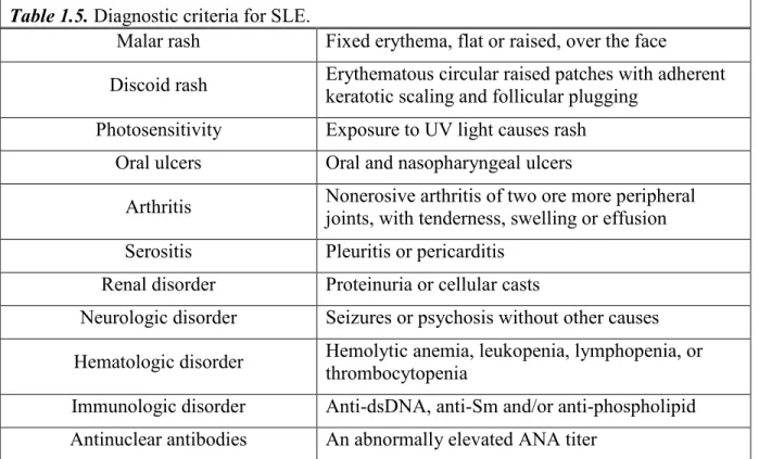
3. Clinical Aspects of SLE... 38
4. Treatment of SLE... 39
B. ANCA-Associated Small Vessel Vasculitis (ANCA-SVV) ... 40
III. SUMMARY OF DISSERTATION... 41
SUGGESTING A SHARED MECHANISM OF B CELL
TOLERANCE LOSS...43
I. ABSTRACT ... 44
II. INTRODUCTION ... 45
III. MATERIALS AND METHODS ... 47
IV. RESULTS... 52
V. DISCUSSION ... 60
VI. CONCLUSION... 65
CHAPTER 3: A UNIQUE SUBSET OF MEMORY B CELLS IS
ENRICHED IN AUTOREACTIVITY AND CORRELATES
WITH ADVERSE OUTCOMES IN SLE ...82
I. ABSTRACT ... 83
II. INTRODUCTION ... 84
III. MATERIALS AND METHODS ... 88
IV. RESULTS... 93
V. DISCUSSION ... 103
CHAPTER 4: GENERAL CONCLUSIONS AND FUTURE
DIRECTIONS ...126
LIST OF TABLES
Table 1.1. Properties of Toll-like receptors. ... 4
Table 1.2. Expression of TLRs on human B cells. ... 6
Table 1.3. Complement receptors, their ligands, function, and expression... 8
Table 1.4. Autoantibodies in human rheumatologic diseases... 35
Table 1.5. Diagnostic criteria for SLE. ... 39
Table 2.1. SLE Patients... 66
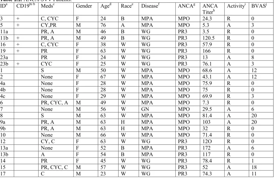
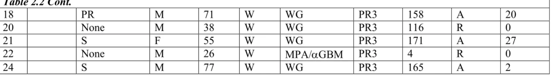
Table 2.2. ANCA-SVV Patients. ... 68
Table 3.1. Clinical outcomes of CD19hi and CD19lo Pts. ... 111
LIST OF FIGURES
Figure 1.1. A brief overview of the classical pathway of complement. ...7
Figure 1.2. A simplified schematic depicting signaling through the BCR. ...22
Figure 1.3. Interactions of transcriptional regulators controlling PC differentiation...27
Figure 2.1. Two B cell populations are detected in SLE and ANCA-SVV Pts based on CD19 expression and size (FSC). ...70
Figure 2.2. CD19hi B cells are present in a subset of SLE or ANCA-SVV Pts...72
Figure 2.3. SLE and ANCA-SVV CD19hi B cells are primarily IgG+ IgD- memory B cells. ...74
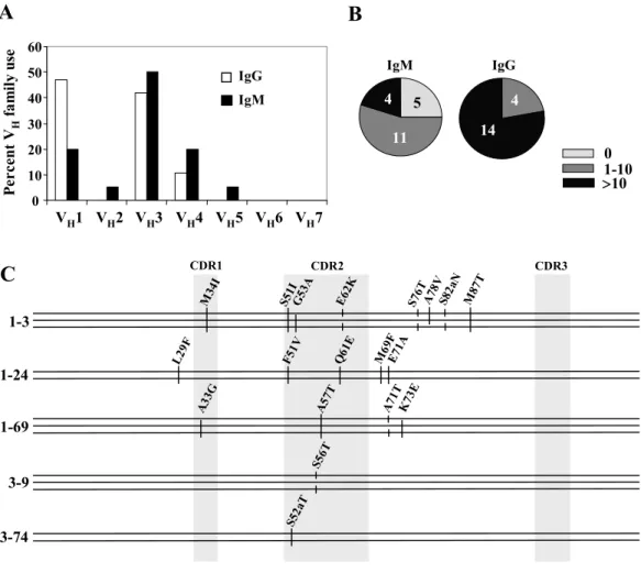
Figure 2.4. CD19hi B cells are somatically mutated and antigen selected...76
Figure 2.5. CD19hi B cells of SLE and ANCA-SVV Pts have upregulated activation markers...77
Figure 2.6. HC memory B cells have higher CD19 expression than naïve B cells. ...78
Figure 2.7. CD19hi Pts have a distinct autoantibody profile...79
Figure 3.1. CD19hi Pts are poor responders to Rituximab treatment...112
Figure 3.2. CD19hi cells are enriched for autoreactivity, and the degree of autoreactivity in these cells correlates exponentially with serum autoantibody...113
Figure 3.3. Basal phosphorylative state of CD19hi cells...114
Figure 3.4. CD19hi cells respond to BCR crosslinking...116
Figure 3.5. CD21 expression is downregulated in CD19hi cells, and sCD21 is decreased in the serum of CD19hi Pts. ...121
Figure 3.6. CD19hi cells appear to be pre-plasma cells. ...122
Figure 3.7. CXCR expression in CD19hi and HC memory cells. ...124
LIST OF ABBREVIATIONS AND SYMBOLS
Ab Antibody
Abs Antibodies
Ag Antigen
AID Activation induced cytidine deaminase ANA Anti-nuclear antibody
ANCA anti-neutrophil cytoplasmic antibody APC Antigen presenting cell
ASC Antibody secreting cell BAFF B cell activating factor
BAFF B cell activating factor of the TNF family BCR B cell receptor
Blimp-1 B lymphocyte induced maturation protein 1 BLyS B lymphocyte stimulator
BM Bone marrow
BSAP B cell specific activating protein Ca2+ Calcium divalent cation
CD19hi cells Memory B cells with increased CD19 expression CD19hi Pts Pts with an expanded CD19hi cell population in the PB CD19lo cells All non-CD19hi B cells (CD19+) in the PB
CD19lo Pts Pts without an expanded CD19hi population CDR3 Complementary determining region 3 CpG Cytosine-phosphate-guanine motif CR Complement receptor
CSR Class switch recombination
CVID Common variable immunodeficiency DC Dendritic cell
dsDNA Double-stranded DNA
ESRD End stage renal disease
Fc Fragment crystallizable (constant) region of an antibody FcR Fc receptor
FDC Follicular dendritic cell Fo Follicular
GC Germinal center
H Heavy
HC Healthy control
IC Immune complex
ICOS Inducable co-stimuator
IFN Interferon
Ig Immunoglobulin IL Interleukin
ITAM Immunoreceptor tyrosine-based activation motif IV Intravenous
JNK c-Jun NH2-terminal kinase
L Light
LPS Lipopolysaccharide MAC Membrane attack complex MAPK Mitogen activated protein kinase MFI Median fluorescence intensity MHC Major histocompatibility complex
MPO Myeloperoxidase
MZ Marginal zone
NFAT Nuclear factor of activated T cells NF- B Nuclear factor B
NSAIDs Non-steroidal anti-inflammatory drugs
p Phosphorylated
PALS Periarterial lymphoid sheath
PAMP Pattern associated recognition motif
PC Plasma cell
PI3K Phosphotidylinositol 3 kinase PKC Protein kinase C
PLC Phospholipase C
PMN Polymorphonuclear leukocyte
PR3 Proteinase-3
Pts Patients
RAG Recombination activating genes RNP Ribonucleoprotein
sCD21 Soluble CD21
SCR Short consensus repeats
SHIP Src homology-2 domain-containing inositol polyphosphate 5’-phosphatase SHM Somatic hypermutation
SHP-1 SH protein-tyrosine phosphatase 1 sIg Surface immunoglobulin SLE Systemic lupus erythematosis SLEDAI SLE disease activity index
Sm Smith antigen
snRNP Small nuclear ribonuclear protein ssRNA Single stranded RNA
SVV Small vessel vasculitis Syk Spleen tyrosine (Y) kinase T regs Regulatory T cells
T1 Transitional 1
T2 Transitional 2
T3 Transitional 3
TCR T cell receptor
TD T-dependent
Th T helper
TI T-independent
TLR Toll-like receptor TNF Tumor necrosis factor
TTP Thrombotic thrombocytopenic purpura UDG Urisil DNA glycolase
V Variable
XBP-1 X-box protein 1 alpha/anti-
beta
epsilon
gamma
I. B CELLS AND THE IMMUNE SYSTEM
The immune system is traditionally divided into two large but overlapping arms: the
innate immune system and the adaptive immune system. Whereas the innate immune system
is primordial, it is none the less effective against many pathogens and is the first line of
defense against invading organisms. However, it is only able to detect unchanging
components common to pathogens, but rare in the host, and does not have the ability to refine
its actions against a specific pathogen nor generate memory against it. The adaptive immune
system, on the other hand, generates exquisite and flexible specificity, allowing it to target
changing aspects of pathogens. It is also able to generate long term memory, protecting the
host against future infections by the same organism. It is, however, slower to respond than
the innate immune system, and, with its nearly limitless ability to adapt to any invader, also
carries the risk of misdirected attack against self, or autoimmunity.
It is only recently that the level to which these two aspects of the immune system
influence each other has been appreciated. We have known for some time that features of the
adaptive system modify and guide the innate system, such as antibodies enhancing the
phagocytosis of pathogens by opsinization, or activating complement in immune complexes
(ICs). We are also beginning to understand how the innate system strongly influences the
adaptive system. Particular complement components, for example, are necessary for an
efficient adaptive response against certain antigens(1-4), and toll-like receptor (TLR) ligands
also control and direct the adaptive response(5-15).
Herein, I will focus on the role of B cells in the normal immune response and in
many other pivotal roles in the immune response. These roles of B cells in health and disease
will be outlined in more detail below.
A. B Cells and the Innate Immune System
Traditionally, the innate immune system consists of specific cells types and soluble
factors. Cell types such as epithelial cells, macrophages, polymorphonuclear leukocytes
(PMNs, or neutrophils), and others are able to participate in the innate immune response.
Epithelial cells can provide early warning of invading pathogens by release of chemokines,
small molecules that direct macrophages, PMNs, and lymphocytes to the site of infection.
These cells in turn are able to phagocytose a wide variety of pathogens and produce toxic
products which indiscriminately kill pathogens. They are also able to secrete cytokines—
small molecules which have specific, receptor-mediated effects on cells of the adaptive and
innate immune systems.
In addition, soluble proteins, particularly complement, are key mediators of innate
immunity and are able to mediate actions on their own (such as directly inducing lysis of
invaders or opsinization) or to enhance the actions of other aspects of the immune system
(such as modifying responses of cell types through complement receptors).
There is not a clear delineation between innate and acquired immunity, however.
Innate immunity is needed for an efficient acquired response, and aspects of the acquired
immune system have innate-like features. Particularly, some subsets of B cells produce what
is known as “natural antibody” which acts as a first line of defense against many pathogens,
1. Toll-like Receptors
Interestingly, many epithelial and immune cells are able to recognize pathogens
through the interaction of common pathogen-associated molecular patterns (PAMPs), such as
lipopolysaccharide (LPS), and pattern recognition receptors (PRRs), such as toll-like
receptors (TLRs)(16, 17). The TLR family contains 10 members, and this group appears to
represent the first evolved type of immune defense. TLRs are highly conserved through many
different species from plants to vertebrates, and recognize conserved lipid, carbohydrate,
nucleic-acid and peptide structures expressed by many pathogens(18, 19). Many of the
subtypes share the common MyD88 signaling pathway and ultimately exert their actions
through the activation of ubiquitous transcription factor NF- B(16, 18, 20). The recognition
patterns and expression location of key TLRs is shown in Table 1.1(21-23). Like
complement components, these ligand-receptor interactions can influence the acquired
immune system(7, 11, 13-15, 24). TLRs, though useful in early defense and immune
regulation, can also become problematic in that many also bind similar antigens derived from
the host (Table 1.1), leading to a break in immunological tolerance(5, 9, 12, 22, 25).
Table 1.1. Properties of Toll-like receptors.
Receptor Location Ligand Exogenous
Source
Endogenous Source
TLR1 Cell Surface Lipoproteins Bacteria n/a
TLR2 Cell Surface Peptidoglycan et al. Gram-positive Bacteria necrotic cells
TLR3 Intracellular dsRNA Viruses necrotic cells
TLR4 Cell Surface LPS Gram-negative
Bacteria n/a
TLR7/8 Intracellular ssRNA Viruses cells, snRNPs dead/dying
a. B cells and TLRs
B cells are among the many cells in the body expressing TLRs and responding to
stimulation by TLR ligands. The effects of TLR stimulation upon B cells are typically highly
stimulatory, although they depend upon B cell differentiative state, presence of concurrent
signaling through other receptors, and the specific ligand and receptor involved(21, 22).
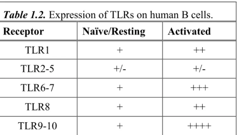
In human B cells, somewhat variable TLRs expression has been reported(26-30).
Most report very low expression of LPS binding TLR4 on these cells(28, 29), however, an
alternate LPS receptor, RP105, may substitute in this case(31). By evaluating mRNA
transcripts, TLRs1, 7, 9, and 10 are expressed in both peripheral blood (PB) and tonsillar B
cells(28, 29). In tonsillar B cells, little variation in expression between naïve, germinal
center (GC), and memory B cells was found in one study(28), but another showed
constitutive expression only in memory B cells(26, 27). Another showed that human naïve,
PB B cells respond robustly to ligands for TLR1/2, 7, and 9, suggesting presence and
functional significance of these TLRs on these cells, although indirect effects via stimulation
of dendritic cells (DC) cannot be ruled out in this study(6). In addition, human memory B
cells can be induced to secrete Ab upon stimulation with the TLR9 ligand CpG(32). A
summary of current knowledge of TLR expression in human B cells can be found in Table
1.2(30).
Much more is known of the function of TLRs in murine B cells, although differences
between these B cells and human B cells are already known. Unlike human B cells, murine
B cells express TLR4(10), and stimulation of these cells with LPS results in significant
proliferation. Of particular import is the recent finding that concurrent stimulation of a B cell
differentiation to an antibody secreting cell (ASC), and can break tolerance in autoreactive B
cells(5, 9, 12, 22, 25). This may be particularly important for B cells which recognize
nuclear components, since these antigens are often associated with various forms of DNA or
RNA that can stimulate TLRs.
Table 1.2. Expression of TLRs on human B cells.
Receptor Naïve/Resting Activated
TLR1 + ++ TLR2-5 +/- +/-
TLR6-7 + +++ TLR8 + ++
TLR9-10 + ++++
+++, strong expression; ++, moderate expression; +, low but detectable expression; +/- low or functionally controversial expression
2. The Complement Cascade and Complement Receptors
Another integral component of the innate immunity is the complement system. The
complement system is composed of numerous small plasma proteins which have the ability
to bind other proteins and molecules with certain properties, such as mannose or polyanionic
surface structures of bacteria or apoptotic cells(1, 17, 33, 34). These proteins are
proteolytically cleaved in complex cascades of activation, the details of which are beyond the
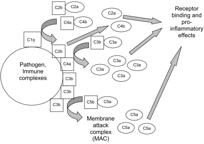
scope of this dissertation. Briefly, there are three pathways for complement activation: the
classical pathway, the mannose-binding lectin pathway, and the alternative pathway. The
classical pathway relies on ICs formed with IgG or IgM (IgM is particularly efficient at
pathogens and apoptotic cells. C1 binds to the invariant regions of the antibodies; bound C1
cleaves C4 and C2, forming C3 convertase. C3 convertase cleaves C3, which continues the
cascade, forms soluble C3a fragments, which have immunological activity, and coats the
surface of the pathogen with C3b. This induces the cleavage of C5 which initiates formation
of the membrane attack complex (MAC), which can directly lyse certain pathogens (Fig.
1.1)(1, 17, 33, 34).
Figure 1.1. A brief overview of the classical pathway of complement.
C3a, C4b, and C5a and some of their smaller products all act as mediators of
inflammation. Complement receptors (CRs) exist for many of these fragments which
enhance responsiveness of the cells which express them(17, 35). Complement receptors 3
(CR3, or CD11b), and CR4 (CD11c) are expressed on macrophages and DCs, among others,
and stimulate phagocytosis as well as regulating the cell response(17, 35, 36). CR2 (CD21)
is a positive regulator of B cell receptor (BCR) signaling, and will be discussed in more
detail in later sections. Its close relative, CR1 (CD35), has similar function and expression
pattern, although it differs between mouse and man. A brief summary of complement
receptors, their ligands, and expression can be found in Table 1.3(17, 35).
Table 1.3. Complement receptors, their ligands, function, and expression.
Receptor Ligands Functions Expression
CR1
(CD35) C3b, C4b, iC3b
Stimulates phagocytosis, transport of ICs
Erythrocytes, macrophages, monocytes, PMNs, B cells, FDCs
CR2 (CD21)
C3d, iC3b, C3dg, EBV, et
al
Part of BCR co-receptor
complex B cells and FDCs
CR3
(CD11b) iC3b Stimulates phagocytosis
Macrophages, monocytes, PMNs, FDCs CR4
(CD11b) iC3b Stimulates phagocytosis
Macrophages, monocytes, PMNs, FDCs
3. B-1 and MZ B cells and Natural Antibody
As mentioned above, some subsets of B cells straddle the divide between the innate
and acquired subparts of the immune system. These subsets are responsible for T cell
independent (TI) production of “natural antibody”, which is typically low-affinity,
cross-reactive, and IgM. Natural antibody represents the bulk of IgM in circulation, and is a key
player in protection from encapsulated bacteria, septic infections, and septic shock(37-42).
B-1 cells may branch off early from the B cell development pathway(43) and are
germline Ab genes(43-45). Interestingly, autoreactive specificities appear to be positively
selected into this subset(46-48). They also differ from B-2 cells (non-B-1 cells, see below),
especially Fo B cells, in their activation and differentiation properties(47, 48).
MZ B cells share some of the activation properties with B-1 B cells, but belong to the
B-2 B cell subset and will be discussed in more detail below.
B. B Cells and the Adaptive Immune System
The three primary cell types of the adaptive response are T lymphocytes (T cells), B
lymphocytes (B cells), and antigen presenting cells (APCs). T cells, so called because they
develop and mature in the thymus, have numerous subtypes which serve diverse functions.
CD4+ helper T cells (Th cells) serve to regulate other cell types, and can drive the immune
response towards either an inflammatory (Type 1, or Th1) or humoral (Type 2, Th2) biased
response, depending on the cytokines they produce(49). CD8+ effector T cells combat
intracellular pathogens by killing infected host cells(49). Finally, T regulatory cells (or T
regs) appear to serve a vital role in maintaining tolerance to self in the periphery(50). The
role of Th2 CD4+ T cells in B cell activation will be discussed in slightly more detail below.
Some non-lymphatic cells, such as dendritic cells (DCs) and stromal cells, have vital
roles in the regulation of B and T cells. Numerous subtypes of DCs exist, and all have
pivotal roles in activation, modulation, and control of the immune response(51-53), and
several subtypes directly influence B cells(51, 54, 55). Follicular dendritic cells (FDCs),
actually a type of stromal cell, are present in germinal centers. These cells can influence B
cell activation and fate through trapping of ICs and antigen and production of cytokines, and
response(56-59). Similarly, T cell maturation and selection in the thymus is guided by specialized thymic
stromal cells.
B cells develop in the bone marrow and serve multiple functions in the immune
system. As producers of Ab, they play roles in both the adaptive and innate immune
systems(60). They also act as antigen presenting cells, activating T cells(61-65), and there is
accumulating evidence they may act in more subtle, regulatory fashions, such as by
production of pivotal cytokines(66-69).
1. Immunoglobulins
Antibodies produced by B cells are diverse in both their recognition of antigen and
their effector function. An antibody, or immunoglobulin (Ig), is a protein consisting of two
identical heavy chains (H) and two identical light chains (L). Both the H and L chains
contain constant and variable regions. The variable regions determine antigen specificity,
while the constant regions confer structural stability. The constant region of the H chain (Fc)
also determines effector function and class of Ig.
a. Ig Subtypes
There are five main classes of Ig: IgM, IgD, IgA, IgE, and IgG(70). IgM exists as a
pentimer and is always the first Ig class generated in any given B cell. This class makes up
the bulk of the TI and natural antibody responses, and is particularly efficient at forming ICs
and activating complement. IgD is still poorly understood, appears to only exist as a surface
molecule, and is not secreted. IgA exists primarily as a dimer and is secreted for protection
of mucosal surfaces. IgE is rarely present in its free form but instead coats the surface of
IgG dominates the secondary and long-term antibody responses to T-dependent antigens.
Several subclasses of IgG exist, each with its own mix of effector functions. The milieu of B
cell activation determines which, if any, class switch will occur when exposed to antigen and
co-stimulation.
DNA splicing prevents multiple Ig classes from being expressed in a single cell at the
same time. A notable exception is the co-expression of IgM and IgD which occurs
commonly and for unknown purpose. Once class switch from IgM occurs, the IgM and IgD
alleles are deleted from the genomic DNA and a single Ig class is expressed for the
remainder of the B cell’s lifespan. This change is made via the process of class switch
recombination (CSR), which relies at least in part on activate-induced cytidine deaminase
(AID)(71, 72).
As stated above, the effector functions of the various Ig classes are determined by the
Fc region of the H chain. Three primary mechanisms are involved: formation of multimers
and/or active transport via Fc-specific transporters into otherwise inaccessible areas (for
example, IgA into the mucous), activation of complement by regions in the Fc (especially by
IgM and subclasses of IgG), and association with receptors specific for the Fc regions of
different subclasses, or Fc receptors. Some Fc receptors are inhibitory and some are
stimulatory, and have profound regulatory ability on cell types such as T and B cells,
dendritic cells, macrophages, eosinophils and basophils.
b. Generation of Ig Diversity and Specificity
Diversity of antigen recognition, generated by the variable regions of the H and L
chains, is generated in four ways; three during development and one after activation(73-76).
chain has only V and J gene segments. There are multiple copies of the genes for H and L
chains and therefore multiple copies of each variable region which can be recombined by
recombination activating genes 1 and 2 (RAG1 and 2)(77-79), leading to the first method of
generating variability: combinatorial diversity(73, 76, 80). During this process, nucleotides
can be randomly added or deleted at the junctions between these regions. This is called
junctional diversity, and can change the reading frame of the remainder of the H or L gene
and therefore generates a non-productive rearrangement in 2 of every 3 attempts(76, 80).
The third process which generates diversity in the Ig repertoire is the fact that both
the H and L chain have variable regions which combine to form the binding site of the
antibody. Thus, a recombined H chain could have very different specificities depending on
the L chain with which it associates(76).
The fourth method for diversity generation, somatic hypermutation (SHM)(81-83),
occurs in the GC and requires T cell help. SHM, dependent largely upon on AID(72, 81-83),
induces extensive mutation in the variable regions of Ig, allowing creation and selection of B
cells with higher specificity for a given antigen. This is accomplished in part via
deamination of dC residues in the variable regions of Ig genes, followed by excision with
uracil-DNA glycosylase (UDG) or replacement(72, 83). The GC reaction will be discussed
in more detail below.
2. Central B Cell Development
The BCR is what allows B cells to identify and respond to foreign antigen. It consists
of the same two H and L chains as the antibody produced by the B cell, along with two
signaling components, Ig and Ig . The expression of this receptor guides the development
B cells begin as common lymphoid progenitors in the bone marrow and differentiate
into pro-B cells, which upregulate the B cell lineage surface marker B220 and activate RAG1
and 2 to begin DJ rearrangement of their H chain under the control of the transcription
factors E2A, EBF, and Pax-5(87-91). Pax-5 in particular is vital for pre-B cell
differentiation, absolute commitment to a B cell lineage and suppression of inappropriate
genes(89, 92, 93). Pre-B cells upregulate CD19, a BCR co-receptor, and proceed with V-DJ
rearrangement. Once the H chain is rearranged, it is expressed with a surrogate Ig L chain to
form the pre-BCR, marking the beginning of the large pre-B cell stage(74, 85). This stage
determines if combinatorial and junctional diversification resulted in a functional H chain. If
the rearranged H chain is not functional, the cell can attempt rearrangement a second time
with the alternate H chain allele(74, 85). If the second rearrangement does not produce
functional H chain, the cell undergoes apoptosis and dies (positive selection, outlined below).
If successful, the large B cell undergoes clonal expansion and proceeds to the small
pre-B cell stage, where it rearranges its L chain gene. When the pre-BCR containing rearranged H
and L chains is finally expressed on the cell surface, the cell is considered an immature B
cell, and must survive another round of positive selection(74, 85, 86).
If a functional BCR is expressed at this stage, the immature B cell leaves the bone
marrow and immigrates to the spleen. There, it proceeds through several transitional stages
before becoming either a MZ or mature (Fo) B cell (discussed below). Mature B cells which
have not encountered their cognate antigen are called naïve B cells.
Developing B cells are subjected to both negative and positive selection in the bone
marrow(94, 95). If the newly generated pre-BCR or BCR is incapable of signaling, it is
eliminated; this constitutes positive selection, since only productive rearrangements are
selected to continue. This mechanism occurs at the pre-B cell and immature B cell stages of
development, and appears to require basal BCR signaling or possibly a very weak or
particular interaction with self(75, 85, 95).
Whereas positive selection requires a sufficient pre-BCR or BCR signal, negative
selection aims to prevent autoreactivity by eliminating immature B cells which recognize self
before they leave the bone marrow. Since only self antigens should be present in the milieu
of the bone marrow, if the BCR of an immature B cell transmits too strong of a signal, it is
triggered to undergo receptor editing, which is a secondary rearrangement of its light
chain(96). If this fails to eliminate autoreactivity, the cell undergoes apoptosis. This
mechanism of regulation is known as clonal deletion(85, 95). Immature B cells that survive
both negative and positive selection leave the bone marrow and migrate to the spleen to
complete their differentiation(97).
3. Peripheral Development and B Cell Subsets
Newly generated immature B cells leave the bone marrow and immigrate to the
spleen. The spleen is broadly divided in to red pulp, where the blood is filtered and iron
recycled, and the white pulp, where lymphocytes reside(98). The white pulp is organized in
sheaths around branching arterial vessels, with T cells residing largely in the periarteriolar
lymphoid sheath (PALS), directly surrounding the arterioles. Adjacent to the PALS, B cells
are arranged in follicles, or B cell zones, of the white pulp(98). Surrounding the PALS and
distinct populations of macrophages, DCs, and MZ B cells(98). Interestingly, MZ B cells are
thought to be vital in the maintenance of the MZ and the other cell types which reside
there(99, 100).
a. Transitional B Cells
Immature B cells immigrating from the bone marrow are directed to the follicle by
the expression of chemokine receptors, particularly CXCR5, whose ligand, CXCL13, is
expressed by follicular stromal cells(98, 101-103). They then proceed through several stages
wherein they are considered transitional B cells. Up until recently, three subsets of
transitional cells (T1, T2 and T3) were recognized; however, recent data suggests cells
previously defined as the T3 subset may in fact be anergic B cells and not transitional cells at
all(104).
T1 and T2 B cells are defined by specific sets of surface markers, half life, location,
and response to BCR crosslinking. As a mechanism for elimination of anti-self specificities,
T1 B cells are eliminated by apoptosis upon antigen encounter. T2 cells, on the other hand,
proliferate and mature when cognate antigen is encountered(75, 105, 106). This additional
phase of negative selection is a vital checkpoint in the elimination of autoreactive
specificities which have escaped central tolerance mechanisms(107-109).
The stage at which MZ B cells branch off and immigrate to the MZ is still unclear(74,
75). Although still under some debate in the field, numerous data suggest that the affinity
displayed by the BCR determines its selection into specific subsets. Stronger signaling
through the BCR, including that which results from weak reactivity to self, may positively
select cells into the MZ subset, while only basal BCR signaling is required for selection of
b. Mature (Fo) B cells
T2 B cells primarily mature into Fo B cells. Fo B cells are responsible for mounting a
high-affinity, long-lived antibody response, and also generate the memory response against a
specific pathogen. Fo B cells exist in the follicle and also recirculate in the blood, lymph,
and lymphoid tissues(112), and have a half-life of 2-3 months(105, 113). When Fo B cells
encounter antigen, they proliferate, upregulate activation markers such as CD80, CD86, and
CD40, and process and present their cognate antigen on the major histocompatibility
complex (MHC) II, priming them for T cell help. When appropriate T cell help is supplied,
the GC reaction is begun. This process will be discussed in more detail below.
c. MZ B cells
MZ B cells are so called because they reside in the marginal zone of the spleen,
which is positioned between the red and white pulp(98). They are therefore some of the first
cells to come into contact with circulating antigen and ICs(37, 114), and as such are perfectly
positioned to be early immune responders. Indeed, MZ B cells are thought to produce the
bulk of the early, TI response(32, 114, 115). The TI response is important in defending
against blood-borne infection and such pathogens as encapsulated bacteria(37) and in the
clearance of LPS, resulting in protection from endotoxic shock(40). In contrast to that of Fo
B cells, the MZ B cell response is typically both short-lived and rapid; recent analysis
indicates that they are molecularly poised to secrete antibody almost immediately after
stimulation(116).
In addition to the TI response, MZ cells play a role in the response to T-cell
processing and presentation of antigen and stimulation of T cells(37, 118, 119), and in their
response to BCR crosslinking and T cell co-stimulation(118, 120, 121).
As mentioned above, multiple lines of evidence suggest that MZ B cells are positively
selected by recognition of self antigen. This may be advantageous in that it may enable
cross-reactivity with common pathogen-derived antigens, or aid in the clearance of apoptotic
cells which could otherwise act as self-antigen (discussed below).
4. The B Cell Receptor
Unlike the T cell receptor (TCR), which respond to processed, digested pieces of
antigen presented by MHC complexes, B cells are able to respond to soluble antigen via the
BCR. Recently, however, it was shown that membrane-bound antigen was more effective in
activating B cells(122), emphasizing a possible role for DCs in B cell activation, as will be
discussed below.
The first step of activation of a Fo B cell is crosslinking of the BCR by antigen (Ag).
The degree of crosslinking directly relates to the strength of the intracellular signal generated.
The intensity of crosslinking is dependent upon properties of the Ag. The BCR or Ab/Ag
interaction can be measured in two ways: affinity and avidity. Affinity is the strength of
interaction between a single Ag epitope and a single variable region of the Ab; the higher the
affinity, the stronger the binding of the variable region to the epitope. Avidity is a more
complicated measure of the total strength of interaction between an Ab and the whole Ag or
Ag complex at all available epitopes present, taking into account multiple antigen binding
regions. Therefore, an Ag or Ag complex (such as an IC) with multiple epitopes would have
higher avidity than one with a single epitope, even though the affinity of the Ab for that
Other properties which can affect efficiency of BCR crosslinking by Ag include
presence of TLR ligands (described above) or complement fragments (described below) in
the Ag complex. Signaling through the BCR can also differ depending on a variety of
circumstances, including differentiative state, subset, previous Ag encounter, and presence of
co-stimulation(85, 86, 123). Pertinent signaling will be described in the following sections.
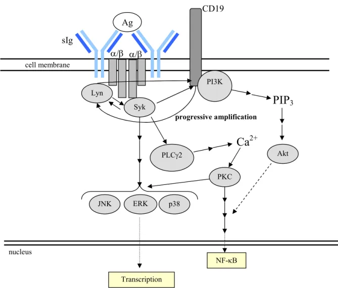
a. Signaling Through the BCR
As mentioned above, the BCR is a membrane-bound version of the Ig currently being
expressed in a given B cell. It is able to signal through its association with the
transmembrane proteins Ig and Ig , which contain immunoreceptor tyrosine-based
activation motifs (ITAMs) in their cytoplasmic tails. Crosslinking of the BCR induces the
formation of the BCR signalosome, a multi-protein complex containing cytosolic and
membrane-bound signaling intermediates(124, 125). The signalosome initiates a
phosphorylation cascade which perpetuates and branches, culminating in the regulation of
key genes.
The cascade begins with the phosphorylation of two tyrosines within the ITAM motif
by Src-family kinases, particularly Lyn(85, 86, 125). This phosphorylation allows
association with and phosphorylation of other tyrosine kinases which contain SH2 domains,
including spleen tyrosine(Y) kinase (Syk)(86, 125, 126). Syk is a key mediator of BCR
signaling, as syk-/- B cells are greatly impaired at both the pro- to pre-B cell and immature to
mature transitions, where positive selection occurs(127-129). Through its regulation of other
intermediates, Syk is essential for many downstream effects of BCR signaling, including
activation of the phosphatidylinositol 3-kinase (PI3K) and phospholipase C 2 (PLC 2)
the Ca2+ flux. The PI3K pathway is needed for efficient activation of the Ser/Thr kinase Akt, which blocks apoptosis in the stimulated cell(85).
Other key signaling molecules downstream of Syk in the BCR signaling pathway
include members of the mitogen-activated protein kinase (MAPK) family, such as
extracellular signal-regulated kinase (ERK), c-Jun NH2-terminal kinase (JNK), and p38
MAPK. These molecules are key regulators of cell proliferation, survival, and
differentiation. In addition, BCR signaling results in activation of transcription factors,
including nuclear factor of activated T cells (NFAT) and the ubiquitously expressed
NF-B(85). A very simplified schematic, including the key member of the co-receptor complex,
CD19, can be found below (Fig. 1.2).
b. The BCR Co-Receptor Complex
The BCR co-receptor complex consists of CD19, a transmembrane glycoprotein with
signaling motifs in its cytoplasmic tail(131-133); CD81 (TAPA-1), which has a role in signal
transduction and cell adhesion(133, 134); Leu-13, which associates with CD81 in B cells and
other cell types(133); and CD21 (CR2), a complement receptor(34, 135). Together, these
molecules enhance BCR signaling through several mechanisms, the most important of which
is thought to involve engagement of CD21 by antigen bound to complement
components(130, 132, 133). Complement-bound antigen, allowing efficient BCR and
co-receptor complex interaction, amplifies the signal through the BCR and elicits a 200-10,000
fold more efficient immune response than antigen alone(1, 4, 132, 133). It also results in
increased antigen processing and presentation than with BCR crosslinking alone(136). At
the finding that crosslinking the BCR/co-receptor complex activates the stimulatory BCR
signaling pathway without concurrent activation of the inhibitory pathway(137).
i. CD21
CD21 is a transmembrane protein with 15 or 16 short consensus repeats (SCRs) and a
short cytoplasmic tail without known signaling motifs(39, 132, 133). CD35 shares extensive
homology with CD21, and, in mice, they are simply splice variants of the same gene. In
humans, each is encoded by a separate but closely related gene and display somewhat
differential expression(138) as described in Table 1.2.
CD21 is unusual in that it binds several diverse ligands, including EBV, CD23, and
IFN- , in addition to the complement fragments iC3b, C3d,g and C3d for which it is
named(39, 135). In mice, only binding to complement fragments has been demonstrated. All
of these ligands bind in the SCR1 and 2 domains and induce a cellular response(135),
indicating that binding of these ligands induces signaling. It is thought that CD19 acts as the
signaling component for CD21, which has traditionally been thought to lack significant
signaling motifs of its own(2, 39). However, its transmembrane domain and cytoplasmic tail
have recently been found to interact with intermediates which effect antigen internalization
and processing(139). CD21 is expressed on B lymphocytes and FDCs in mice, and on these
cells and erythrocytes in humans.
Co-engagement of CD21 with the BCR also increases the internalization of
antigen(140) and expression of stable antigen-MHC II complexes on the cell surface for
presentation of antigen to T cells(3). Interestingly, complement-linked antigen is targeted to
different intracellular compartments and therefore may be processed differently than antigen
cells(41, 144). CD21 also traps ICs on MZ B cells, which then transport and transfer them
onto FDCs(145).
ii. CD19
CD19 is an Ig superfamily transmembrane glycoprotein expressed in B cells and
FDCs(131-133). The 9 tyrosine residues in its cytoplasmic domain are highly conserved in
human, mouse, and guinea-pig, suggesting a critical role in signaling(146). Indeed, these
residues are phosphorylated upon crosslinking and interact with PI3K and Lyn among
others(132, 147, 148). CD19 has been shown to be key in the efficient PI3K-dependent
activation of Akt(149). Additionally, CD19 can augment the phosphorylation of the
Src-family kinases, including Lyn, via a process known as progressive amplification, resulting in
increased phosphorylation of Syk(131, 150). Signaling through CD19 also increases
activation of MEK1/2, which is a kinase for ERK1/2(151). Although CD21 is thought to be
required to bring CD19 into the BCR signaling complex, it is clear that CD19 has signaling
roles even in the absence of CD21, given that the phenotype of CD19 deficient mice is
considerably more profound than that of CD21 deficient mice(132, 133, 146). It may be that
CD19 can somehow associate with the BCR in the absence of CD21; alternatively, some
have proposed there may be an as of yet unidentified ligand for CD19(132, 133).
Given these findings, it is not surprising that even small increases or decreases in
CD19 expression can have dramatic effects on B cell development, function, and notably
tolerance in mouse and man(152-154). This will be discussed in more detail below in the
Figure 1.2. A simplified schematic depicting signaling through the BCR.
c. Other Molecules Which Regulate B Cells
Numerous additional surface receptors modulate B cell response. The most important
are those provided by T cell help, as described below. In addition, B cell activation can be
enhanced by the putative Ca2+ channel CD20(155, 156), numerous cytokines, particularly IL-21 and BLyS(157-161), and by TLR ligands, as described above.
B cell activation and differentiation can be also be inhibited by negative regulators,
such as the IgG receptor Fc RIIb, CD5, and CD22(162-165). Clearly, B cells are never Ag
sIg
/ /
Lyn
Syk
CD19
progressive amplification
PI3K
Akt
PIP
3Ca
2+PLC 2
PKC
NF- B ERK
JNK p38
cell membrane
nucleus
simply “turned on” or “turned off”, but their response depends on the sum of many
competing and overlapping signals.
5. The B Cell Response
B cells can play different roles in the immune response depending on factors such as
their subset, activation state, the properties of antigen, the involvement of other cell types,
and many others. As the details of the B-1 and MZ B cell response are beyond the scope of
this dissertation, I will focus on the typical response mounted by mature B-2 B cells.
The response begins with engagement of the BCR by antigen. Fo B cells are highly
motile, scanning the lymph and blood for soluble antigen, and interacting with FDCs in
search surface-displayed antigen(103). One mechanism by which FDCs may acquire antigen
is the capture of ICs containing antigen by FcRs and/or complement receptors(38, 166).
Interestingly, MZ B cells can capture ICs, bring them to FDCs, and deposit them on their
surface(145). In addition, compelling new data shows that FDCs can engulf, retain and
present intact (non-degraded) antigen to B cells on their surface, eliciting strong BCR
signaling(122, 167, 168).
Once the cognate antigen is encountered, signaling through the BCR “primes” the B
cell to respond to T cell help(60, 81, 103). The antigen is internalized, processed, and
presented on MHCII for recognition by antigen-specific Th cells. In addition, the B cell
proliferates and upregulates the essential co-stimulatory molecules CD40, CD80, and CD86,
whose roles will be discussed below. Importantly, upregulation of the chemokine receptor
CCR7 allows B cells to locate to the B-T cell boundary in search of T cell help(165). If a
a. B-T Cell Interactions
When an activated B cell encounters an activated Th cell whose TCR recognizes the
processed antigen it is presenting, an immunological synapse is formed between the TCR and
the antigen-MHCII complex(60, 81, 169, 170). This interaction is the “first signal” for
activation of the T cell; the B cell’s “first signal” was recognition of antigen. For efficient
activation, it must be followed by the binding of CD40 on B cells with its ligand, CD40L
(CD154), on T cells, without which GC formation and its downstream effects are
blocked(171). This interaction, together with co-stimulation through the B7 molecules CD80
and CD86 on B cells by CD28 and CTLA-4 on Th cells, acts as the key “second signal”
necessary for efficient B and T cell activation(60, 81, 172).
Multiple other cell-bound and secreted factors are also key in the stimulation of B
cells by Th cells, particularly the inducible co-stimulator (ICOS) and its ligand ICOS-L, as
well as IL-4, IL-21, and B cell activating factor belonging to the TNF family (BAFF), also
known as B lymphocyte stimulator (BLyS)(60, 81).
These interactions provide activation and stimulatory signals not just to B cells, but
also to T cells. Among other effects, the formation of these immune synapses stop T cell
migration but not B cell migration, and B cells can be seen “dragging” T cells after synapse
formation in intra vital imaging studies(102).
b. Germinal Centers
The provision of T cell help results in profound clonal expansion of the B cell. Some
of the activated B cells form foci in the T cell zones (extrafollicular foci) and differentiate
signs of SHM(171, 173). The remainder of the B cells proliferate to form a region of
IgD-negative cells within the primary follicle, which, once it acquires a germinal center, is termed
a secondary follicle(60, 81). 7-10 days after initial priming, the secondary follicle polarizes
into two regions: one near the T cell zone consisting of rapidly proliferating B cells, or
centroblasts, and one consisting of quiescent B cells, or centrocytes. These regions are called
the dark and light zones, respectively, and once they form, the secondary follicle is called a
germinal center(60, 81).
As centroblasts divide, they also undergo SHM and CSR(60, 172, 173). They then
proceed as centrocytes into the light zone for selection; competition for relatively limited
supplies of antigen and a default program for cell death account for rigorous selection at this
point(60, 81, 172). Cells which generate lower affinity or lose the function of their BCR die,
whereas cells whose affinity is enhanced through this process are selected to re-enter the
cycle or differentiate into either a long-lived plasma cell (PC) or memory cell(60, 81, 169,
171).
One of the key transcription factors expressed in GC B cells is Bcl-6. In mice lacking
Bcl-6 expression, GC cannot form(174-176). Bcl-6 also acts as a transcriptional repressor of
cyclin dependent kinase inhibitors, allowing the rapid proliferation of centroblasts(177, 178).
It also inhibits B lymphocyte induced maturation protein 1 (Blimp-1), a transcription factor
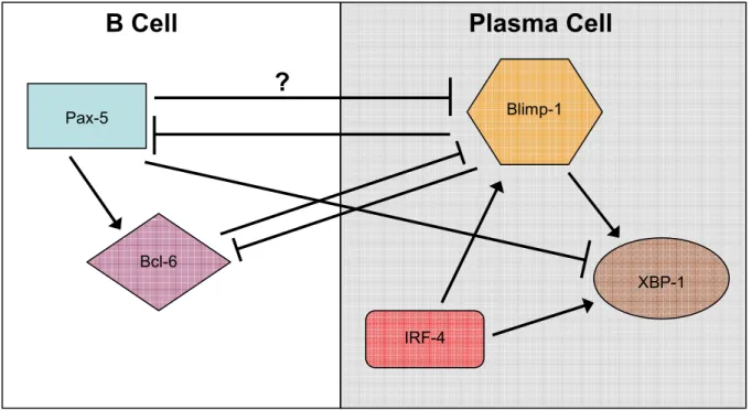
crucial for PC differentiation(174, 177). In addition to Bcl-6, Pax-5 is important in
maintaining the identity of GC B cells, probably in part due to is repression of X-box binding
protein 1 (XBP-1), which is upregulated upon Fo B cell activation and required for PC
extrafollicular response vs. generation of memory, and their role in differentiation to PCs will
be discussed more below.
In addition to the role of Th cells in the GC reaction, many studies have shown that
FDCs play a role in survival and selection of GC B cells by means of antigen held on their
surface(181, 182) and expression of surface-bound and secreted factors, particularly
BLyS(57, 183-187). It was long held that the main function of CR2 on FDCs was capture of
ICs, however, this idea was recently confounded by the finding that expression of CR2 on
FDCs is critical, even in mice lacking any secreted Ab and therefore lacking ICs(188); this
may mean that CR2 has a signaling role in FDCs. The precise role of FDCs in the GC
reaction is still under debate(187).
c. Plasma Cells
Plasma cells are one of two main outcomes of the GC response and represent terminal
differentiation of the B cell lineage. They are long-lived, secrete class-switched, SHM Ab,
and are characterized by the downregulation of B cell specific surface markers such as the
BCR, CD19, CD20, B220, MHCII and others, with concurrent upregulation of syndecan-1,
or CD138(171). Cells committed to this lineage leave the GC as pre-PC, or plasmablasts,
since fully differentiated PC no longer respond to chemotactic signals(171).
While the majority of plasmablasts express CXCR4, which through its interactions
with its ligand CXCL12 direct PCs to the bone marrow (BM), a subset express CXCR3(180,
189-191). The CXCR3 ligands CXCL9 and 10 are expressed at sites of inflammation(192)
and direct a subset of PCs there, where they terminally differentiate and become resident
time, likely decades, depending on their environment(197). The phenomenon of long-lived
PCs residing in extra-lymphatic tissue is prominent in states of autoimmunity(193-195).
As mentioned above, complex transcriptional control is involved in the differentiation
of PCs. In short, Pax-5 is downregulated, leading to a decrease in Bcl-6; the decrease in
expression of these proteins relieves repression of Blimp-1, the master regulator of PC
differentiation(171, 180). Blimp-1 in turn further represses Pax-5, Bcl-6, proliferation, and
other B cell lineage genes, while activating XBP-1(171, 180). Recent studies indicate that
commitment to this lineage also requires expression of the transcription factor IRF-4, which
acts to upregulate Blimp-1 and other PC specific genes(189). A very simplified schematic
depicting the interaction of these proteins can be found in Fig. 1.3(171, 180).
Figure 1.3. Interactions of transcriptional regulators controlling PC differentiation.
Pax-5 Blimp-1
B Cell Plasma
Cell
?
XBP-1 Bcl-6
d. Memory B Cells
The other type of effector B cell generated by the GC response is the memory B cell.
Memory B cells are long lived and express Ig, which has, like that of PCs, undergone SHM
and typically CSR. In humans, they are defined as CD19+CD38- population which also
typically, but not always, expresses CD27(197-200). Class-switched (or IgD-) and IgM
memory cells have been identified, both of which show SHM of their Ig(201, 202) and, in
fact, presence of mutations in the V regions of the BCR has been the “gold standard” of
defining a memory population(203). Antigen-specific memory B cells can be detected for an
impressive 60 years or longer after primary immunization(204).
The role of persistent antigen, in various forms, in the maintenance of this population
is debated(205, 206). It was initially shown that the presence of antigen on the surface of
FDCs was required for the maintenance of memory B cells(205, 207). However, in the
absence of secreted Ig, which precludes IC formation, memory B cells display the same
lifespan and function as those in control mice(208). In another experimental setting, memory
B cells could be manipulated via a genetic switch to change BCR specificity. In this case,
the memory B cells with specificity for an antigen never present in the animal persisted just
as well as memory cells of the originally induced specificity(206). Therefore, the currently
favored hypothesis is that that antigen is not required for maintenance of memory B
cells(197).
Recently, focus has shifted from the study of memory B cells as a homogenous group
to an attempt to delineate them into distinct subsets. Multiple lines of evidence suggest that
memory B cell subsets with distinct functional and more subtle but detectable surface
defined based on surface receptor expression and function(209, 210). Emerging evidence
suggests that this is the case in the human memory B cell pool(197-200), although specific
markers largely remain to be elucidated. One possible marker is Fc receptor homologue H4
(FcRH4), which was found on a subset of largely CD27+ but class-switched and SHM B
cells in human tonsil, although the significance of this finding is still unclear(200). In
addition, it has recently been shown that a small subset of peripheral memory B cells in
humans express CXCR3, are able to chemotax towards the corresponding ligands, and that
expression of this receptor is maintained upon differentiation to a PC(190, 211); these cells
may represent a distinct subset of memory B cells in humans.
Traditionally, cognate antigen and memory Th cells are thought to be required for
activation of quiescent memory B cells(60, 191, 212). However, some evidence exists that
polyclonal stimuli can induce the proliferation and even differentiation of human memory B
cells(213), particularly “bystander” T cell help (soluble cytokines), CpG(26, 213), or
stimulation with CD27 ligand (CD70) and IL-10(214). Overall, memory B cells appear to be
poised to respond rapidly to activating stimuli. In vitro, stimulation through the BCR leads
to increased secretion of Ig by memory B cells compared to naïve B cells, and memory, but
not naïve, B cells are able to differentiate to ASCs upon stimulation with anti-CD40, IL-2
and IL-10 in the absence of antigen(215, 216).
Upon stimulation, memory B cells proliferate, some replenishing the memory cell
pool, and others differentiating into short- or long-lived PCs(60, 191, 197, 209, 216, 217).
They also strongly upregulate B7 molecules and, unlike naïve B cells, are able to effectively
reactivation and proliferation, after which those differentiating to a PC undergo the same
transcriptional shifts as a GC B cell differentiating into a PC.
e. Other Effector Functions of B Cells
Though production of Ab is a central role of B cells, they have other pivotal roles in
immune response and regulation. As mentioned above, B cells can act as APCs, and in some
autoimmune diseases, particularly SLE, arthritis, and non-obese diabetes, appear to be the
required APC for initial T cell activation and break in tolerance (218-220).
B cells also have the capacity for significant production of important cytokines. They
have been shown to produce both Th1 and Th2 cytokines, especially IL-10, IL-6, and
TNF-(221-224). IL-10 and IL-6 have autocrine effects on B cells in addition to their effects on
other cell types, and promote a Th2 response; TNF- is one of the most potent cytokines,
mediating a largely Th1 response and activating CD8+ T cells and macrophages, among
others.
B cells have also been shown to produce IL-2, IL-4 and IFN- , though possibly to a
lesser extent than those listed above(225-228). IL-2 and IFN- mediate Th1 responses, while
IL-4 is considered a Th2 cytokine.
Like Th cells, which can differentiate to Th1 or Th2 types to mediate a Type 1 or
Type 2 response, B cells can become Type 1 or Type 2 B effector cells (Be1 and Be2) which
possess many of the same traits as Th1 or Th2 cells, and can control the development of Th1
and Th2 subtypes from naïve Th cells(229-231). Be1 and Be2 cells produce IL-2 at nearly
equal levels. Be1 cells produce significant quantities of IFN- and IL-12, whereas Be2 cells
IL-6, and IL-10; Be1 cells produce essentially no IL-4, 4-fold less IL-6, and less than half as
much IL-10 comparatively(230).
Most interestingly, Be1 and Be2 cells were able to efficiently generate Th1 or Th2
cells, respectively, from naïve CD4+ T cells, and in vivo responded by producing cytokines
even before Th cells. These data indicate that not only do B cells produce Type 1 and 2
relevant cytokines themselves, but are able to effectively polarize the global response, and
may be among the first cell types to do so(230).
Clearly, B cells play many roles in the innate and acquired immune systems, even
apart from their production of Ab.
C. Generation and Maintenance of Self-Tolerance
Many mechanisms of central and peripheral tolerance via negative and positive
selection have been outlined above, including receptor editing and clonal deletion. There are,
however, additional mechanisms of peripheral tolerance in particular which are crucial to
prevention of autoreactivity. These include anergy, block of differentiation at the pre-PC
stage(232) and suppression by cytokines secreted by DCs and macrophages, which will be
discussed briefly below.
1. Anergy
Anergy is a state of non-responsiveness induced by antigen encounter at an early
differentiative stage or by chronic antigen exposure(233-236). Generally, whereas higher
avidity for self results in receptor editing and clonal deletion, B cells which recognize
Anergic B cells do not proliferate, upregulate activation markers, or secrete Ab in
response to BCR crosslinking(235, 237). This is due, at least in part, to dampened signaling
through the BCR as measured by decreased phosphorylation of substrates and decreased Ca2+ flux(124, 238, 239). Interestingly, although these intracellular responses to BCR
crosslinking are diminished and altered, some are basally elevated compared to naïve B cells,
including intracellular Ca2+ levels(104, 124, 238, 239) and ERK phosphorylation(124, 240). Anergic cells also have a shorter half-life than naïve B cells(241), due in part to their
increased dependence upon BAFF for survival(242).
An emerging mechanism for anergy appears to be activation of Lyn and its
downstream pathways in the absence of Syk activation(233). Among other effects, this
would promote the inhibitory feedback mechanisms involving the phosphatases SHP-1 and
SHIP, blocking the Akt survival pathway and activation of NF- B by Akt and Syk(233).
The precise mechanisms governing anergy remain to be elucidated, and many subtle
subtypes of anergy, or varying states of “non responsiveness” may exist.
2. Other Means of Peripheral Tolerance
Despite all of the above mechanisms to eliminate or silence autoreactive B cells,
some still persist. Numerous additional mechanisms exist to prevent the formation of
high-affinity anti-self Ab and/or memory B cells. Autoreactive B cells experience impaired
selection in the GC and blocks in PC formation(243) or are excluded from the follicle
(“follicular exclusion”)(234). Recently a block was described for autoreactive B cells at the
pre-PC stage, which represents regulation at the latest possible differentiative step(232). Ab
secretion by self-specific mature B cells can also be suppressed by DCs and macrophages
T cells also play a role in preventing B cell autoimmunity; the simple lack of cognate
anti-self T cell help can block humeral autoimmunity. In addition, T regs can suppress B cell
activation and Ab secretion(245).
II. AUTOIMMUNE DISEASE
With the phenomenal ability of B and T cells to generate receptors with nearly
infinite specificity, it is not surprising that many generate receptors which recognize
components of self. The immune system has therefore evolved the complex and
multi-layered mechanisms described above to prevent the immune system from unleashing its
considerable arsenal upon the host. Given the frequency with which autoreactive B and T
cells develop, it is a testimony to these mechanisms that so little autoimmune disease occurs.
In humans, autoimmune disease takes many forms. Often, these diseases are broadly
divided into those primarily mediated by either T cells or B cells. Clearly, as the immune
system is a complex, interdependent system, few if any diseases are mediated by a single cell
type. However, the heterogeneous family of autoimmune disorders mediated by
autoantibody are considered to be chiefly B cell mediated (e.g., SLE), while diseases marked
by the infiltration and destruction of tissue by T cells are considered to be chiefly T cell
mediated (e.g., Type 1 Diabetes). Recently, a pivotal role for B cells, outside of their ability
to produce autoantibody, has been described. It is now clear that in some systems, B cells
acting as APCs are responsible for the initial break of T cell tolerance(246-248).
A summary of autoantibody-mediated diseases can be found in Table 1.4, along with
their common autoantibody targets(249). Interestingly, a majority of these diseases are up to
10 times more common in women than in men, indicating that hormonal environment may