Studying the mechanisms of HP1-mediated heterochromatin formation
and memory of repression
By
Celyn Bregio
Senior Honors Thesis
Department of Chemistry
University of North Carolina at Chapel Hill
April 26th, 2019
Approved:
Studying the mechanisms of HP1-mediated heterochromatin
formation and memory of repression
Celyn Bregio
Center for Integrative Chemical Biology and Drug Discovery, The Division of Chemical Biology and Medicinal Chemistry, UNC Eshelman School of Pharmacy.
Department of Chemistry, University of North Carolina at Chapel Hill,
In fulfillment of a CHEM 692H Research Project under the supervision of Dr. Nate Hathaway.
Committee Members: Paul Kropp (Chair) Brian Hogan Nate Hathaway
TABLE OF CONTENTS
I. Abstract
II. Introduction
III. Materials and Methods
IV. Results
V. Discussion
VI. Conclusions
VII. Acknowledgments
I. ABSTRACT
Heterochromatic pathways play a key role in the regulation of the genetic code. While there
are numerous pathways that achieve the inactivation of specific genes, some have been
evolutionarily conserved due to their effectiveness. One of the best examples of this is concept is
Heterochromatin Protein 1 (HP1), which has been found to be a versatile protein that serves various
roles across the genome. There are 3 different HP1 isoforms known to date: CBX5 (HP1α), CBX1
(HP1), and CBX3 (HP1) that despite their similarities have been reported to behave differently.
In this study, our aim is to determine the interplay between the identity of HP1 isoforms and their
effect on the velocity and memory of gene repression. For this, we used a chromatin in vivo assay
(CiA) system to chemically recruit HP1 to a reporter gene and study its effect. Our experimental
data support the established notion that HP1α is a more efficient repressor than the other isoforms.
Due to the collaborative relationship between biochemical activity, gene repression, and cellular
memory, we believe that investigation of HP1 behavior can be relevant for the development of
therapeutic drugs to treat genetically-linked diseases in which epigenetic misregulation often has
a significant effect.
II. INTRODUCTION
The field of epigenetics revolves around post-transcriptional modifications and their effect
on overall gene expression. Such modifications regulate the status of chromatin, a dynamic
complex that consists of DNA strands wrapped around a tetramer of histone proteins, which in
turn dictates the accessibility of a gene for transcription (Venkatesh & Workman, 2015). The less
compacted and more accessible euchromatin state allows for the interaction of RNA polymerases
adopting a more condensed and inaccessible conformation (Zhang & Reinberg, 2001). These states
are achieved through the manifestation of chromatin modifications, such as methylation,
acetylation, and phosphorylation, which play a key role in regulating transcriptional programs in
cells. These regulatory processes often act in concert with one another, leading to a network of
epigenetic marks that can activate or deactivate genes depending on the context (Tongprasit et al.,
2008). For instance, trimethylation of histone 3 at its lysine 27 tail (H3K27me3) has been reported
to lead to gene repression, yet the same mark at lysine 4 (H3K4me3) has been correlated with
gene activation (Tongprasit et al., 2008).
Accordingly, these subtle but instrumental differences in the nature of a given
post-transcriptional mark are essential for the proper regulation of genes. If improper activation and
inactivation of genes occurs, the consequences can be detrimental. Notably, this concept has
become an important target for the understanding of oncogenesis, given that many types of cancer
have been associated with alterations in epigenetic pathways. For example, malignant follicular
lymphoma contains mutations of a histone methyltransferase in about 90% of the cases (Morin et
al., 2011), DNA hypermethylation has been reported in the silencing of tumor suppressor genes
(Jones, 2012) and studies of heterochromatin protein 1 (HP1) have demonstrated that modulation
of this protein alters the invasive potential of breast cancer cells (Norwood et al., 2006). However,
even though extensive research has been dedicated to studying these epigenetic marks and
pathways, the development of effective therapeutic targets has been challenged by the promiscuity
of the proteins involved in gene regulation.
One of these versatile proteins is HP1: the first non-histone structural component of
heterochromatin to be isolated and that is widely known for its role in the establishment and
mammalian isoforms are known, are encoded by chromobox (CBX) genes. These isoforms are
known as HP1α, HP1, and HP1 and are encoded by CBX5, CBX1, and CBX3, respectively
(Lomberk, Wallrath, & Urrutia, 2006). These proteins possess a characteristic genetic sequence
that has been greatly conserved in many organisms, ranging from Drosophila to mice and humans
(Vermaak, Henikoff, & Malik, 2005), indicating that the specific interactions that arise from the
transcription, and subsequent translation of such sequences into amino acids contribute an
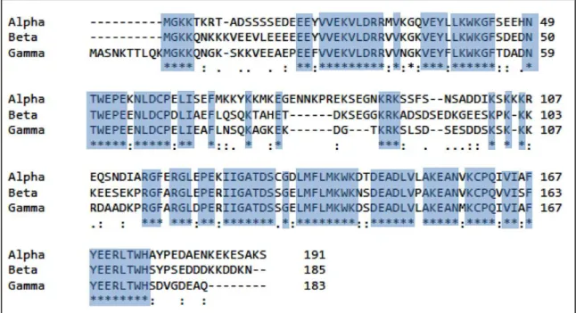
evolutionary advantage. The main organization of the CBX genes comprises an N-terminus
extension (NTE), a chromo-domain (CD), a middle hinge region, a chromoshadow domain (CSD),
and a C-terminus extension (CTE), sequentially (Singh, 2010). The chromodomain is known to
encode the site responsible for HP1 binding to H3K9me2/3, while the chromoshadow is involved
in homo- and/or heterodimerization and interaction with other proteins (Lomberk et al., 2006).
These two areas are very similar among the three isoforms with over 70% identity, while the hinge
region contains the most variable sequences of amino acids and has been shown to highly influence
the localization, interactions, and function of each isoform (Lomberk et al., 2006) (Fig 1).
Interestingly, despite what its name suggests, HP1 is not exclusively present in
heterochromatic regions and its only role is not to silence genes. Studies have made clear that these
proteins have additional nuclear functions, including transcriptional activation and elongation,
chromosome segregation, DNA repair, sister chromatid cohesion, and RNA splicing (Canzio,
Larson, & Narlikar, 2014). As a result, HP1 is dispersed throughout the genome, with different
isoforms concentrating in distinct regions. For example, HP1α and HP1 have been primarily
associated with heterochromatic locations such as telomeres and centromeres, while HP1 has
been largely found in euchromatic regions playing a role in transcriptional elongation (Canzio et
al., 2014). Thus, although the HP1 paralogs possess very similar genetic constructs, they seem to
perform different functions and have specific genomic zones of influence where their subtle
structural differences potentially enhance or diminish their efficiency.
In this project, we aim to study the interplay between the identity of the HP1 isoform and
its effect on the velocity and memory of gene repression. To achieve this, we used the chromatin
in vivo assay (CiA) system as described in Hathaway et al., 2012 to chemically recruit HP1 to a
reporter gene. HP1 paves the way for the formation of heterochromatic domains by luring histone
methyltransferases (HMTs) that insert H3K9 methyl marks, DNA methyltransferases (DNMTs)
that methylate CpG dinucleotides within DNA, and other HP1 proteins that propagate these signals
to neighboring nucleosomes (Fig 1). The CiA system enabled us to selectively repress the reporter
gene and, this way, test the contribution of each HP1 isoform to cellular memory and stability after
III. MATERIALS AND METHODS
Cell culture
Three MEF cell line with a reporter nucEGFP under control of a CMV/EF1α promoter and
adjacent to a 5x array of Gal4 DNA binding site (Vignaux et al., 2019-in review) were grown in
FBS-supplemented growth media as described in (Butler, Chiarella, Jin, & Hathaway, 2018) and
selected with 9 g/mL blasticidin and 6.5 g/mL puromycin to drive csHP1(α, , )-Frb and
FKBP-Gal4 expression.
Lentiviral Infections
293T LentiX cells (Clontech) were transfected using polyethyleneimine with each of the
chromoshadow-Fbr constructs and the Gal4-FKBP fusion proteins.
CIP Heterochromatin Assay
Antibiotic selection was removed from cells the day before experimentation. The chemical
inducer of proximity rapamycin was added to the medium at a 3 nM concentration (3 L of a 10
M stock solution per 10 mL of media).
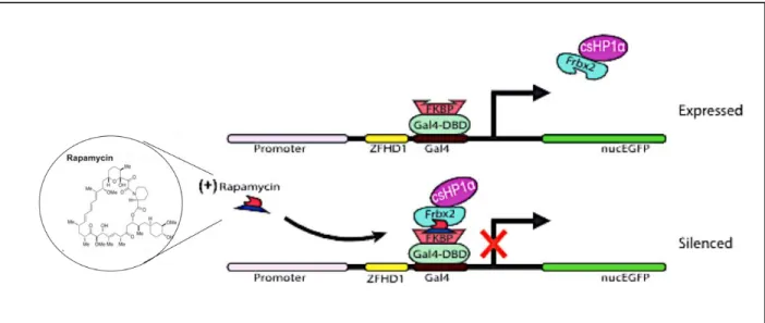
Figure 2. Visual model for HP1-mediate heterochromatin formation mechanism. HP1 recognizes the H3K9me3 site and, after
Removal of CIP and EGFP Re-expression
The removal of rapamycin was achieved by adding FK506 at a 100 nM concentration (10
L of a 100 M stock solution per 10 mL of media) for 48 hours. For the remainder of the time,
the cells were allowed to grow in drug-free media.
Flow Cytometry
Flow cytometry was performed on an Attune Nxt in biological triplicates. The samples
were later analyzed using FlowJo software, where the data was gated to include only live, single,
and non-autofluorescent cells. The number of cells included in the data was normalized to the
sample with the lowest population count through randomized down-sampling.
Statistical Analysis
Statistical analyses were performed using GraphPad Prism 7 software. Replicates from
flow cytometry were subjected to 2-way ANOVA tests, and discoveries were determined using
the Geisser-Greenhouse correction.
IV. RESULTS
In order to compare and contrast the repressive pathways of each HP1 isoform and their
individual effect on heterochromatin formation velocity and memory, 3 different cell lines were
created. Each clonal line was designed to chemically recruit the chromoshadow domain (CSD) of
the specific isoform, giving rise to the lines that in this study will be referred to as HP1α, HP1,
and HP1. To quantify gene expression, we used CRISPR/Cas9 gene editing to insert a nucEGFP
gene driven by a promoter comprised of a CMV enhancer and a core EF1α promoter sequence.
The reporter complex was incorporated outside of the Hbb- gene in the -globin locus in the
We induced heterochromatin formation at this locus by using the CiA system, which uses
a chemical inducer of proximity (CIP) to recruit fusion proteins that reversibly tether the
chromoshadow of HP1 (csHP1) to the site. The binding of the CIP rapamycin to FKBP (FK506
binding protein) will allow for the subsequent binding of a Frb (FKBP-rapamycin binding) domain
with which csHP1 is tagged for recruitment to the CiA locus (Fig 2) (Hathaway et al., 2012). After
CIP addition, the recruited exogenous csHP1 to the site to lure endogenous HP1 proteins, HMTs,
and DNMTs, which act in concert to establish a heterochromatic domain. Conveniently, because
the domain is formed as a result of CIP bridging, this effect can be easily reversed through the
addition of FK506, which effectively displaces rapamycin and csHP1 by binding to FKBP, but not
to Frb (Fig 2). Since csHP1 can be removed, the epigenetic stability and memory of repression
can be studied given that the domain would need to be maintained by the cell’s own machinery.
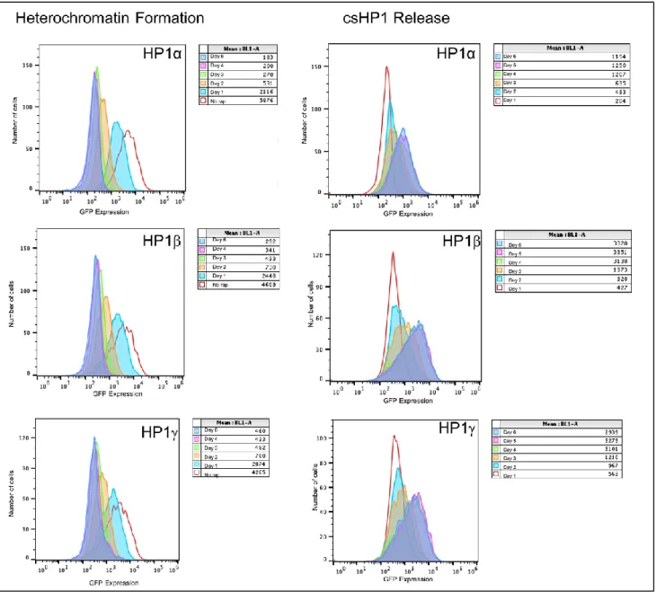
Heterochromatin formation in the three cell lines was monitored by the expression of green
fluorescent protein (GFP), which was quantified through flow cytometry analysis. Expression
levels after CIP addition were measured for 6 consecutive days. Based on the results, all the cell
lines experienced about 50% reduction in expression levels after 1 day of csHP1 recruitment and
almost complete repression after 6 days (Fig 4, 5A, 5B). Although the baseline levels of GFP
expression were different for the 3 cell lines, they all showed similar trends of silencing. These
data show that the structural differences between the isoforms are not enough to cause an impactful
change in heterochromatization velocity.
Following the 6 days of repression, we proceeded to remove the tethered csHP1 complexes
by adding FK506 for 48 hours. We then performed flow cytometry for 6 additional days, which
allowed us to juxtapose the stability of the heterochromatic domain established in each of the cell
lines by evaluating their velocities of re-expression. Interestingly, while HP1 and HP1 reported
similar velocities of GFP expression recovery, HP1α displayed a substantially slower rate of
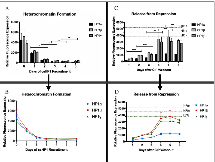
re-expression (Fig. 4, 5C, 5D) By Day 4, the HP1α cell line had recovered about 26% of its baseline
expression level, while HP1 and HP1 recuperated 81% and 67%, respectively (Fig. 2). However,
6 days was not enough for any of the cell lines to completely reach baseline GFP expression values,
Figure 4. Histogram representation of GFP expression after csHP1 recruitment (left) and release (right). In the legends for the
Figure 5. (A and B) Graphical representation of GFP expression as a function of time showing the velocity of heterochromatin
formation for the three isoforms. (C and D) Same as described for A and B, but showing the trend for GFP re-expression. Two-way ANOVA statistical analysis validates a significant change in expression between days.
V. DISCUSSION
This study evaluates the behavior of the 3 known HP1 isoforms after engagement in
heterochromatin formation by analyzing velocity and memory of repression. Our data reveals that
while the 3 isoforms showed similar rates of silencing, they display variability in their rates of
re-expression. HP1α was considerably slower than HP1 and HP1 with only a 27% restoration of
GFP expression after 5 days as opposed to 82% and 73% by the latter two, respectively. It is
important to note that in previous experiments using the same HP1α cell line, expression was
restored after 6 days (Vignaux et al., 2019-in review). Nevertheless, our results can, in theory, be
A C
explained by analyzing the structural differences between the three isoforms and their typical
genomic location-specific roles.
While HP1α is commonly associated with silenced heterochromatic regions, HP1 and
HP1 have been known to play both gene silencing and activating roles (Larson et al., 2018)
Studies have proposed that this variability might be due to their differences in their amino acid
sequence (Fig 1). As mentioned above, the sequence of the HP1 proteins has been highly
conserved, with all 3 possessing an N-terminal extension (NTE), a chromodomain (CD), a hinge
region, a chromoshadow domain (CSD), and a C-terminal extension (CTE) (Fig 1).
Structural biochemical analyses of the isoforms’ CSD suggest that, given their similarities
in sequence and organization, the CSDs of HP1α/β/γ would recognize histone H3 fragment
similarly (Loppnau et al., 2017). This supports our finding that the rate of heterochromatin
formation for all three cell lines was about the same and any difference was indistinguishable. On
the other hand, when thinking about the more variable regions on the HP1 isoforms complexes,
we encounter a possible explanation for our re-expression results. Liu et al. report that although
not necessary for binding, the CTE enhances HP1 affinity to the H3 protein due to its interaction
with the CSD as it dimerizes with other proteins (Loppnau et al., 2017). This concept is further
investigated by Mendez et al., who rationalize that HP1α selectivity is derived from the
cooperation between its CSD and its C-terminal extension (CTE), a mechanism that has resulted
in preferential interactions with different peptides (Mendez et al., 2011). On the other end of the
isoforms’ sequence, we can find the NTE, in which important differences have been found;
namely, that HP1α NTE region contains phosphorylation sites that are absent in HP1 and HP1
and that have been shown to have a key role in the formation of heterochromatin in cells
variability of the HP1 isoforms, this time targeting the highly variable hinge region, reported that
the HP1α hinge region can independently induce heterochromatin (Smothers & Henikoff, 2002).
In all, structural and biochemical studies support our finding that HP1α forms a potentially
tighter heterochromatic domain. However, further experimentation would be needed to accurately
conclude if the delay in the HP1α construct GFP re-expression is due to the cell’s own maintenance
of the repressive complex, to csHP1α establishing “tighter” heterochromatic domain. In order to
determine the reason behind these results, a potential future experiment would chromatin
immunoprecipitation (ChIP). This would aid in the quantification of H3K9me3 enrichment, a
variable that directly correlates to the activity of csHP1 in the cell. This will tell us if repression is
being triggered by HP1 or if, potentially, other repressive domains are being recruited by HP1. We
could also use ChIP to investigate if the introduction of exogenous HP1 isoforms preferentially
leads to the recruitment of the same endogenous HP1 isoform to the site of heterochromatization
and, this way, identify any interactions between the isoforms.
Afterward, long-term silencing would be a great way to determine whether each isoform is
able to engage the cell’s own machinery in the maintenance of the heterochromatin after removal
of HP1. Long term silencing via HP1 recruitment has been shown to engage DNMTs, which
facilitate the methylation of CpG dinucleotides in the DNA template (Hathaway et al., 2012).
Because DNA methylation is a mark that is inherited through mitosis and is, therefore, perpetuated
without the need of csHP1 binding, we will be able to determine if the cell’s molecular machinery
is being involved in the maintenance of stable heterochromatin and if this engagement is affected
by the isoform that starts the process (Amabile et al., 2016). A short-term repression experiment
is not likely to engage the endogenous DNMTs to the point of adding the necessary CpG methyl
identify the differences between short-term and long-term silencing by obtaining bisulfite
sequencing data, through which we could compare the levels of DNA methylation before
repression and after short- and long-term exposure to csHP1.
VI. CONCLUSIONS
In this study, we investigate the interplay between the amino acid sequence of HP1
isoforms and their effect on the velocity and memory of gene repression. To achieve this, we used
a chromatin in vivo assay (CiA) system to chemically recruit csHP1to a reporter gene. We
concluded that while the 3 isoforms are very similar in their amino acid sequence, they have
different effects on the memory of heterochromatin formation, with HP1α potentially forming a
tighter heterochromatic domain. Based on our results, we believe that the study of HP1-mediated
heterochromatin formation can help increase the current understanding of how biochemical
activity impacts cellular memory and genetic regulation. This can be relevant for the development
of therapeutic drugs to treat genetically-linked diseases, such as cancer, in which epigenetic
misregulation often plays a central role.
VII. ACKNOWLEDGMENTS
Special thanks to Mrs. Patricia Vignaux and Dr. Nate Hathaway for their patience,
guidance, and support throughout my time as an undergraduate researcher. I also acknowledge the
Hathaway Lab team for their valuable discussions and eagerness to offer help and assistance.
Additionally, I would like to thank Dr. Gidi Shemer who, through the SMART program, offered
me the opportunity to be involved in research at Carolina. Lastly, I would like to thank my honors
VIII. REFERENCES
Amabile, A., Migliara, A., Capasso, P., Biffi, M., Cittaro, D., Naldini, L., & Lombardo, A.
(2016). Inheritable Silencing of Endogenous Genes by Hit-and-Run Targeted Epigenetic
Editing. Cell, 167(1), 219–232.e14. https://doi.org/10.1016/j.cell.2016.09.006
Butler, K. V., Chiarella, A. M., Jin, J., & Hathaway, N. A. (2018). Targeted Gene Repression
Using Novel Bifunctional Molecules to Harness Endogenous Histone Deacetylation
Activity. ACS Synthetic Biology, 7(1), 38–45. https://doi.org/10.1021/acssynbio.7b00295
Canzio, D., Larson, A., & Narlikar, G. J. (2014). Mechanisms of functional promiscuity by HP1
proteins. Trends in Cell Biology, 24(6), 377–386. https://doi.org/10.1016/j.tcb.2014.01.002
Hathaway, N. A., Bell, O., Hodges, C., Miller, E. L., Neel, D. S., & Crabtree, G. R. (2012).
Dynamics and memory of heterochromatin in living cells. Cell, 149(7), 1447–1460.
https://doi.org/10.1016/j.cell.2012.03.052
Hiragami-Hamada, K., Soeroes, S., Nikolov, M., Wilkins, B., Kreuz, S., Chen, C., … Fischle, W.
(2016). Dynamic and flexible H3K9me3 bridging via HP1β dimerization establishes a
plastic state of condensed chromatin. Nature Communications, 7.
https://doi.org/10.1038/ncomms11310
Jones, P. A. (2012). A decade of exploring the cancer epigenome, 11(10), 726–734.
https://doi.org/10.1038/nrc3130.A
Larson, D. R., Day, C. R., Ren, G., Zhao, K., Rodriguez, J., & Chow, C. C. (2018). Intrinsic
Dynamics of a Human Gene Reveal the Basis of Expression Heterogeneity. Cell, 176(1–2),
213–226.e18. https://doi.org/10.1016/j.cell.2018.11.026
Lomberk, G., Wallrath, L. L., & Urrutia, R. (2006). Protein family review The Heterochromatin
Loppnau, P., Zhang, Y., Li, Y., Liu, Y., Lei, M., Qin, S., … Min, J. (2017). Peptide recognition
by heterochromatin protein 1 (HP1) chromoshadow domains revisited: Plasticity in the
pseudosymmetric histone binding site of human HP1. Journal of Biological Chemistry,
292(14), 5655–5664. https://doi.org/10.1074/jbc.m116.768374
Mendez, D. L., Elgin, S. C. R., Khorasanizadeh, S., Kim, D., Chruszcz, M., Stephens, G. E., &
Minor, W. (2011). The HP1a Disordered C Terminus and Chromo Shadow Domain
Cooperate to Select Target Peptide Partners. ChemBioChem, 12(7), 1084–1096.
https://doi.org/10.1002/cbic.201000598
Norwood, L. E., Moss, T. J., Margaryan, N. V., Cook, S. L., Wright, L., Seftor, E. A., …
Wallrath, L. L. (2006). A requirement for dimerization of HP1Hsα in suppression of breast
cancer invasion. Journal of Biological Chemistry, 281(27), 18668–18676.
https://doi.org/10.1074/jbc.M512454200
Singh, P. B. (2010). HP1 proteins—What is the essential interaction? Russian Journal of
Genetics, 46(10), 1257–1262. https://doi.org/10.1134/s1022795410100297
Smothers, J. F., & Henikoff, S. (2002). The Hinge and Chromo Shadow Domain Impart Distinct
Targeting of HP1-Like Proteins. Molecular and Cellular Biology, 21(7), 2555–2569.
https://doi.org/10.1128/mcb.21.7.2555-2569.2001
Tongprasit, W., Ma, Y., Li, X., Jin, W., Li, S., Terzaghi, W., … Stolc, V. (2008).
High-Resolution Mapping of Epigenetic Modifications of the Rice Genome Uncovers Interplay
between DNA Methylation, Histone Methylation, and Gene Expression. The Plant Cell
Online, 20(2), 259–276. https://doi.org/10.1105/tpc.107.056879
Venkatesh, S., & Workman, J. L. (2015). Histone exchange, chromatin structure and the
https://doi.org/10.1038/nrm3941
Vermaak, D., Henikoff, S., & Malik, H. S. (2005). Positive selection drives the evolution of
rhino, a member of the heterochromatin protein 1 family in drosophila. PLoS Genetics, 1(1),
0096-0108. https://doi.org/10.1371/journal.pgen.0010009
Woolcock, B., Meissner, B., Delaney, A., Rimsza, L., Tamura-Wells, J., Gascoyne, R. D., …
Firme, M. R. (2011). Frequent mutation of histone-modifying genes in non-Hodgkin
lymphoma. Nature, 476(7360), 298–303. https://doi.org/10.1038/nature10351
Zhang, Y., & Reinberg, D. (2001). Transcription regulation by histone methylation: Interplay
between different covalent modifications of the core histone tails. Genes and Development,