Dual Targeting of Cell Wall Precursors by Teixobactin Leads to Cell
Lysis
Tomoyuki Homma,a,fAustin Nuxoll,a,eAutumn Brown Gandt,aPatrick Ebner,bIna Engels,c,dTanja Schneider,c,dFriedrich Götz,b Kim Lewis,aBrian P. Conlona,g
Antimicrobial Discovery Center, Department of Biology, Northeastern University, Boston, Massachusetts, USAa
; Microbial Genetics, University of Tübingen, Tübingen, Germanyb
; Institute of Pharmaceutical Microbiology, University of Bonn, Bonn, Germanyc
; German Centre for Infection Research (DZIF), Partner Site Bonn-Cologne, Bonn, Germanyd
; Biology Department, University of Nebraska at Kearney, Kearney, Nebraska, USAe
; Discovery Research Laboratory for Core Therapeutic Areas, Shionogi & Co., Ltd., Toyonaka, Osaka, Japanf
; Department of Microbiology and Immunology, University of North Carolina at Chapel Hill, North Carolina, USAg
Teixobactin represents the first member of a newly discovered class of antibiotics that act through inhibition of cell wall
synthe-sis. Teixobactin binds multiple bactoprenol-coupled cell wall precursors, inhibiting both peptidoglycan and teichoic acid
syn-thesis. Here, we show that the impressive bactericidal activity of teixobactin is due to the synergistic inhibition of both targets,
resulting in cell wall damage, delocalization of autolysins, and subsequent cell lysis. We also find that teixobactin does not bind
mature peptidoglycan, further increasing its activity at high cell densities and against vancomycin-intermediate
Staphylococcus
aureus
(VISA) isolates with thickened peptidoglycan layers. These findings add to the attractiveness of teixobactin as a potential
therapeutic agent for the treatment of infection caused by antibiotic-resistant Gram-positive pathogens.
A
ntibiotic resistance development is a major threat to human
health. Constant development of novel antibiotics is required
to keep pace with the emergence and spread of antibiotic
resis-tance in bacterial pathogens (
1
). The majority of antibiotics in use
today are derivatives of molecules discovered in the early to
mid-twentieth century. The lack of novel compounds, coupled with the
emergence and spread of antibiotic resistance, has resulted in an
increasingly dangerous situation (
2–4
).
One approach to discover novel antibiotics is to improve our
ability to cultivate microorganisms that produce them.
Tradi-tional culturing methods allow access to an estimated 1% of the
biodiversity in soil. A novel cultivation technique, using an
isola-tion chip or iChip, provides access to an untapped reservoir of
natural product antibiotics, produced by bacteria that had
previ-ously eluded cultivation efforts. One such organism,
Eleftheria
terrae
, a previously uncultivated Gram-negative
betaproteobacte-rium, was found to produce a novel depsipeptide antibiotic, called
teixobactin. Teixobactin inhibits cell wall biosynthesis and
repre-sents a new class of antibiotics (
5
).
The bacterial cell wall contains layers of peptidoglycan, a
cross-linked matrix of linear glycan chains (
6
). Peptidoglycan
cross-linking is the target of

-lactam and glycopeptide antibiotics. In
Gram-positive bacteria, teichoic acid (TA) is also a major
compo-nent of the cell wall. TA includes wall teichoic acid (WTA),
con-nected to peptidoglycan, or lipoteichoic acid (LTA), anchored in
the cytoplasmic membrane. TA plays important roles in bacterial
physiology (
7
,
8
), and teichoic acid biosynthesis is an important
target for antibiotic development (
9
). Recently, it was shown that
inhibition of teichoic acid biosynthesis can restore
susceptibil-ity to methicillin in methicillin-resistant
Staphylococcus aureus
(MRSA) (
10
).
In a previous study, we found that teixobactin binds lipid II, a
precursor of peptidoglycan biosynthesis, and lipid III, a precursor
of teichoic acid biosynthesis (
5
). Interestingly, teixobactin was
ca-pable of superior bactericidal and bacteriolytic activity compared
to other cell wall-acting antibiotics, the

-lactam oxacillin, or the
glycopeptide vancomycin. Also, resistance to teixobactin was not
detected in a number of
in vitro
studies. Here, we show that the
ability of teixobactin to simultaneously inhibit peptidoglycan and
teichoic acid biosynthesis triggers synergistic effects, resulting in
increased cell wall damage, delocalization of autolysins, and
sub-sequent lysis and cell death. Furthermore, we demonstrate that
teixobactin does not bind mature peptidoglycan and hence is
ca-pable of effectively targeting vancomycin-intermediate
S. aureus
(VISA) strains that have increased cell wall density.
MATERIALS AND METHODS
Antimicrobial agents, bacterial strains, and primers.Teixobactin was purified according to the procedure described previously (5). Vancomy-cin and tunicamyVancomy-cin were purchased from Sigma-Aldrich. The bacterial strains and primers are listed in Table S2 in the supplemental material. JE2, HG003, and SA113 were used throughout the study. HG003 and SA113 are closely related laboratory methicillin-susceptible S. aureus
(MSSA) strains. JE2 is a MRSA USA300 isolate. All strains displayed the same MIC to teixobactin. Strains were selected for specific assays due to preexisting characterized mutants in those strain backgrounds.
MIC.The MIC was determined by the broth microdilution method according to CLSI guidelines. Muller-Hinton broth (MHB) was supple-mented with 0.1% Tween 80 to prevent the absorption of compounds to plastic surfaces. Cell concentration was adjusted to about 5⫻105CFU/ ml, and cells were incubated for 20 h at 37°C. The MIC was defined as the lowest concentration of antimicrobial agents that resulted in no visible
Received16 May 2016Returned for modification9 June 2016 Accepted6 August 2016
Accepted manuscript posted online22 August 2016
CitationHomma T, Nuxoll A, Gandt AB, Ebner P, Engels I, Schneider T, Götz F, Lewis K, Conlon BP. 2016. Dual targeting of cell wall precursors by teixobactin leads to cell lysis. Antimicrob Agents Chemother 60:6510 – 6517.
doi:10.1128/AAC.01050-16.
Address correspondence to Brian P. Conlon, [email protected].
Supplemental material for this article may be found at
http://dx.doi.org/10.1128/AAC.01050-16.
hyde with 0.1 M sodium cacodylate buffer (pH 7.2) for 1 h and treated in a graded series of 1% OsO4with 0.1 M sodium cacodylate buffer. Cells were then dehydrated in ethanol and critical-point dried using CO2. The samples were sputter-coated with a 5-nm platinum coating and examined at a 3.0-kV accelerating voltage with a Hitachi S-4800 field emission scan-ning electron microscope.
Time-dependent killing.An overnight culture ofS. aureuswas diluted in MHB and incubated at 37°C and 225 rpm for 3 h. Antibiotics were added at 10⫻MIC, and cultures were incubated at 37°C and 225 rpm. At intervals, 100-l aliquots were removed and centrifuged at 10,000⫻gfor 1 min and resuspended in 100l of sterile phosphate-buffered saline (PBS). Tenfold serially diluted suspensions (10l) were plated on Muller-Hinton agar plates. The plates were incubated at 37°C overnight.
Bacteriolytic assay.The bacteriolytic activity in supernatant of cell cultures was determined by the change of turbidity of the substrate, heat-killed RN4220 cells (11). The cells were incubated at 37°C until reaching mid-exponential phase (OD600of 0.4 to 0.6). Antibiotics were added at 10⫻MIC, and cultures were incubated at 37°C and 225 rpm for 4 h. Filter-sterilized culture supernatant was mixed with heat-killed cells ad-justed to an OD600of 0.5 and incubated at 37°C for 6 h. The turbidity was measured every 30 min. The remaining quantity was expressed as the ratio to initial values.
Zymographic analysis.Murein hydrolase profiles were analyzed by zymogram (12,13). Antimicrobial agents at 10⫻MIC were added to mid-exponential-phase JE2 cells in 15 ml Trypticase soy broth (TSB), and the cells were cultivated for an additional 4 h. Cultures were centrifuged and supernatants and cells were collected separately. The supernatants were concentrated using an Ultra-4 3000 molecular-weight-cutoff filter (Amicon) to 500l. The concentration of protein was determined by the Bio-Rad protein assay method by following the manufacturer’s instruc-tions. The cells were washed with 0.1 M Tris-HCl (pH 6.8) once and treated with sample buffer (2% SDS with-mercaptoethanol) and incu-bated at 65°C for 5 min. After centrifugation, the supernatants were stored at⫺80°C until analysis could be performed. The samples were electro-phoresed in an 8% SDS-PAGE gel containing heat-killed RN4220 cells as a substrate (final OD600, 10). After electrophoresis, the gel was washed with Tris-buffered saline (TBS; 50 mM Tris, 150 mM NaCl, and 3 mM KCl, pH 7.5) buffer containing 1% Hanks buffer (Sigma) and 2.5% Tri-ton-X and incubated in TBS buffer containing 1% Hanks buffer and 10 mM CaCl2at 37°C overnight. The clear zone, which indicated a protein with autolysis activity, appeared as a dark band inFig. 2B(see also Fig. S1A in the supplemental material).
Quantitative real-time PCR.RNA purification was performed as pre-viously reported (14). Antimicrobial agents at 10⫻MIC were added to mid-exponential-phase USA300 cells, and the cells were incubated for 1 h. The cells were collected and treated with RNAprotect bacterial reagent (Qiagen) to ensure RNA integrity. Cells were pelleted, suspended in 50 mM EDTA with 0.6 mg/ml lysostaphin (Sigma), and incubated at 37°C for 2 min to lyse the cell. Total RNA was isolated using an RNeasy minikit (Qiagen) according to the manufacturer’s instructions. To remove DNA, a Turbo DNA-free kit (Ambion) was used. The integrity of purified RNA was confirmed by an RNA 600 Nano kit and bioanalyzer (Agilent). RNA was reverse transcribed into cDNA using high-capacity cDNA reverse transcription kits (Applied Bioscience). Quantitative real-time PCR was
antibody (12) for 1 h. Finally, the membrane was washed and incubated with anti-rabbit IgG secondary antibody. After incubation of the mem-brane with substrate, the blot was analyzed by a gel imaging system (Bio-Rad).
Isolation of peptidoglycan.Isolation of peptidoglycan for competi-tion assay was described previously (15). Exponential-phase HG003 cells were harvested by centrifugation, washed with ice-cold saline, and resus-pended in 2 ml of saline. The cells were boiled for 20 min. After centrifu-gation, the cells were resuspended with saline, mixed with glass beads, and lysed by bead beater (2 rounds, with 20 s at the highest speed and cooling on ice for 5 min). After harvesting the suspensions, the cells were collected by centrifugation, suspended in 1 ml 2% SDS, and boiled for 30 min. After cooling to room temperature, the cells were washed with distilled water at least 5 times. The pellets were dissolved in 0.1 M Tris-HCl (pH 6.8) with trypsin (0.5 mg/ml; Sigma) and incubated at 37°C overnight. Finally, the pellets were washed with distilled water 3 times. This suspension was stored at⫺80°C until used for competition assay.
Isolation and detection of cell wall teichoic acid.Isolation of teichoic acid was conducted as follows (16). Antimicrobial agents (teixobactin and vancomycin [10⫻MIC] as well as tunicamycin [0.025⫻MIC]) were added to mid-exponential-phase HG003 cells in MHB, and the cells were cultivated for an additional 4 h. Cell pellets were collected by centrifuge, washed with 50 mM 2-(N-morpholino)ethanesulfonic acid (MES; pH 6.5), resuspended in 0.5 ml 50 mM MES (pH 6.5) with 4% (wt/vol) SDS, and boiled for 1 h. The cells then were collected by centrifugation and washed with 50 mM MES (pH 6.5) with 4% SDS twice, washed with 50 mM MES (pH 6.5) with 2% (wt/vol) NaCl, and finally washed with 50 mM MES (pH 6.5). The cells were resuspended with 20 mM Tris-HCl (pH 8.0) with trypsin (0.5 mg/ml) and incubated at 37°C overnight. Following this, the pellets were washed with distilled water at least 3 times. Pellets were then suspended with 0.1 ml of 0.1 M NaOH and incubated at room temperature overnight. Supernatants were used for competition assay or detection of WTA by SDS-PAGE. Samples were run in SDS-PAGE gel (16% Tris-Tricine gel; Novex), and the gel was stained by a Pierce silver stain kit (Thermo Fisher) by following the manufacturer’s procedure.
Purification of Atl R1–3and conjugation with Cy3 dye.The three-Atl-repeat domain, R1–3(17,18), was expressed with a N-terminal His6tag in
Escherichia coli M15 using the isopropyl--D-thiogalactopyranoside
min. The cells were washed twice with PBS and incubated with Cy3-R1–3 for 5 min at room temperature. After washing twice with PBS, a 2-l cell suspension was applied to glass slides and distribution of fluorescence was confirmed with a Leica fluorescence microscopy system.
RESULTS
Teixobactin-induced lysis is dependent on the Atl autolysin.
Teixobactin has been shown to have excellent bactericidal activity
against
S. aureus
(
5
). We examined the effect on cell morphology
of teixobactin and vancomycin after 4 h of exposure at 10
⫻
MIC
by electron microscopy (
Fig. 1A
). Exposure to teixobactin results
in collapse of the cell wall, while vancomycin damage to the cell
wall was less severe.

-Lactam-induced lysis is known to be mediated by Atl, the
major cell wall autolysin of
S. aureus
(
17
,
22
). To investigate the
contribution of Atl to the activity of teixobactin, the
antibac-terial activity against an
⌬
atl
mutant from the Nebraska
trans-poson insertion library (available through BEI [
https://www
.beiresources.org/
]) was examined and compared to wild-type
strain JE2. Although the MIC of teixobactin was not affected by
mutation of
atl
(see Table S1 in the supplemental material), the
bactericidal activity was markedly reduced. Teixobactin did not
significantly reduce the viable cell count of the
⌬
atl
mutant after
24 h of exposure, compared to a 3-log reduction in CFU per
mil-liliter in the wild-type strain (
Fig. 1B
). Also, 24 h of teixobactin
exposure did not cause lysis in an
atl
mutant, confirming the lysis
phenotype is Atl dependent (
Fig. 1C
).
Teixobactin causes a decrease in
atl
expression.
Teixobactin’s
bactericidal activity is dependent on Atl; hence, we decided to
examine the lytic capacity of the supernatant of a culture treated
with teixobactin. Atl undergoes maturation by proteolytic
pro-cessing, resulting in generation of two extracellular lytic enzymes,
an amidase and a glucosaminidase (
12
,
14
,
23
). If teixobactin
treatment resulted in increased lytic enzyme production, we
would expect the supernatant to have increased lysis capacity. To
test this, we performed bacteriolytic assays with supernatants of
treated and untreated cultures (
Fig. 2A
). The untreated control
supernatant had marked lytic activity and strongly reduced the
turbidity of a cell suspension over time. On the other hand, the
supernatant of a teixobactin-treated culture did not have any
vis-ible lytic capacity. To further investigate this, we examined the
lysis profile of the extracellular and cell wall-anchored Atl with
zymography (
Fig. 2B
). Both supernatant and cell wall-associated
protein isolated from cells treated with 10
⫻
MIC of teixobactin
for 4 h showed reduced murein hydrolase activity compared to
those from untreated samples (see Fig. S1A in the supplemental
material). Furthermore, real-time PCR revealed that expression of
atl
in teixobactin-treated cells was about 25-fold lower than that in
untreated control cells (
Fig. 2C
). Finally, the reduced Atl levels
were confirmed by Western blot analysis (see Fig. S1B).
Vanco-mycin also reduced the murein hydrolase activity (
Fig. 2A
to
C
),
the time of addition of the antibiotic.
Teixobactin-induced lysis is enabled by inhibition of cell
wall teichoic acid biosynthesis.
The localization of Atl at the
sep-tum to mediate cell division has been proposed to occur via
exclu-sion from the rest of the cell wall by the presence of teichoic acids
(
21
). In the absence of WTA, Atl binding on the cell surface is
delocalized, causing lysis (
21
). Furthermore, WTA are required
for

-lactam resistance in methicillin-resistant
S. aureus
, and the
cells which lack WTA are sensitized to

-lactam-induced cell lysis
(
26–28
). The genes involved in teichoic acid biosynthesis are
called the
tar
genes (for teichoic acid ribitol). TarO (previously
referred to as TagO), an
N
-acetylglucosamine-1-phosphate
trans-ferase, catalyzes the first step in this biosynthetic pathway, and
mutation of
tarO
results in a teichoic acid-deficient strain (
29
).
We examined cell lysis of wild-type and
tarO
mutant strains in the
presence of vancomycin and teixobactin. As previously reported,
teixobactin causes increased lysis of the wild-type strain compared
to vancomycin. Both antibiotics caused lysis of the
tarO
mutant,
demonstrating that lipid II inhibition results in cell lysis, but only
in a teichoic acid null background (
Fig. 3A
and
B
). As teixobactin
blocks lipid II and lipid III, this suggests that the lytic activity of
teixobactin is due to the combined inhibition of both targets. To
FIG 2Teixobactin causes downregulation ofatlexpression. (A) Bacteriolytic assay using supernatant from JE2 incubated with teixobactin (10⫻MIC) or vancomycin (10⫻MIC) for 4 h. (B) Zymography of teixobactin-treated sam-ples on SDS-PAGE gel containing heat-killedS. aureusRN4220 as a substrate. The dark bands indicate the clear zone in the SDS-PAGE gel, which is caused by the lysis of substrate. Left and right lanes were supernatant (Sup) and cell wall-associated samples (CW), respectively. (C) Comparison ofatl transcrip-tion by quantitative real-time PCR. The bars indicate the relative values com-pared to the no-antibiotic-treatment sample. These data represents the means and SD from 3 independent experiments.
further examine a potential synergistic relationship between
inhi-bition of peptidoglycan and teichoic acid biosynthesis, we
exam-ined the bactericidal and lytic activity of vancomycin with
tunica-mycin, a known inhibitor of WTA synthesis against cultures of the
S. aureus
laboratory strain HG003 (
27
) (
Fig. 3C
and
D
). We found
that indeed, the combination of tunicamycin and vancomycin
re-sults in increased killing relative to vancomycin alone, and cell
lysis was increased (
Fig. 3C
and
D
). We next examined the effect of
teixobactin on teichoic acid levels. Teichoic acids were isolated
from the cell wall before and after 4 h of teixobactin treatment.
Results clearly showed that teixobactin treatment results in
mark-edly reduced quantities of teichoic acid in the cell wall (
Fig. 3E
).
In an attempt to identify the signal transduction pathway
lead-ing to
atl
downregulation, we examined the MIC activity of
teixo-bactin, oxacillin, and vancomycin against 12 mutants of
two-com-ponent regulatory systems (see Table S3 in the supplemental
material). These mutants were from the JE2 ordered mutant
li-brary. The
⌬saeS
mutation resulted in a dramatic 16-fold
reduc-tion in MIC to teixobactin but only a modest 2-fold decreased
MIC to vancomycin. We then performed killing experiments and
found that vancomycin displayed bactericidal killing against a
⌬saeS
mutant but not the JE2 parental strain (
Fig. 4A
).
Surpris-ingly, preliminary analysis showed an apparent decreased
abun-dance of WTA in the
⌬saeS
mutant (
Fig. 4B
). SaeRS is known to
regulate virulence determinants, including surface proteins,
tox-ins, and capsule biosynthesis components (
30–32
), and the
saeR
mutant shows high susceptibility to

-lactams in
Staphylococcus
epidermidis
(
33
). This increased susceptibility to cell wall-acting
antibiotics may be due to decreased teichoic acids in the cell wall.
This would further corroborate the role of teichoic acids in
pre-vention of lysis mediated by lipid II inhibition. Again,
teixobac-tin’s ability to inhibit both peptidoglycan and teichoic acid
bio-synthesis explains its ability to kill and lyse
S. aureus
so efficiently.
More work is required to more precisely measure the impact of
saeS
mutation on teichoic acid content of the cell wall and
eluci-date how the SaeRS two-component system could influence WTA
levels in
S. aureus
.
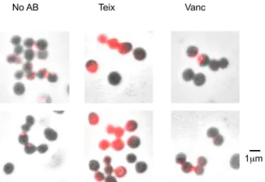
Teixobactin treatment results in delocalization of
autoly-sins.
It has been proposed that teichoic acids control autolysin
binding to peptidoglycan by an exclusion principle, whereby cell
wall teichoic acid interaction with peptidoglycan inhibits
autoly-sin binding (
21
,
34
). Consequently, an absence of teichoic acids at
the septum facilitates appropriate localization of autolysins
dur-ing cell division. It was also shown that mutation of
tarO
results in
delocalization of amidase (
21
). Atl possesses three repeat
se-quences, each about 150 amino acids long (R
1–3), which bind to
peptidoglycan (
19
,
35
). We examined the localization of Atl using
a fluorescently labeled R
1–3repeat domain (Cy3-R
1–3) (
21
). We
found that Cy3-R
1–3delocalization occurs in cells treated with
teixobactin for 30 min, similar to that previously seen in a
tarO
mutant. These experiments were performed against the model
laboratory strain HG003 (
Fig. 5
; see also Fig. S2 in the
supplemen-tal material). This suggested that teixobactin treatment resulted in
delocalization of Atl due to inhibition of WTA biosynthesis of the
cell. This delocalization explains the intense lytic capacity of
teixo-bactin. Vancomycin did not cause delocalization of the amidase
over a similar time period. A longer 4-h treatment with
vancomy-cin did result in delocalization of the fluorescent amidase.
Delo-calization by teixobactin is rapid and does not allow the cell time
to reduce autolysin production and limit Atl-mediated damage.
Collectively, these results suggest that coinhibition of lipid II and
lipid III by teixobactin causes a pronounced weakening of the cell
wall compared to lipid II inhibition alone. This results in increased
delocalization of autolysins, leading to cell lysis and death.
Teixobactin does not bind cell wall peptidoglycan.
We have
previously shown that teixobactin has killing activity superior to
that of vancomycin against dense populations of
S. aureus
(
5
).
This is due in part to teixobactin’s ability to inhibit teichoic acid
production. However, a further important limitation of
vancomy-cin activity, particularly at high cell densities, is the binding of
vancomycin to mature peptidoglycan. Vancomycin has no
anti-bacterial activity when bound to mature peptidoglycan. This
ex-plains the reduced bactericidal activity of vancomycin against
dense populations of cells. Furthermore, this circumstance can
lead to vancomycin-intermediate resistance. Many VISA strains
have thicker cell walls with altered cross-linking (
36
). This leads to
increased binding of vancomycin to mature peptidoglycan at the
D
-Ala-
D-Ala pentapeptide and sequestration of the antibiotic.
Teixobactin does not bind the pentapeptide; hence, it may not
FIG 4Mutation ofsaeSresults in sensitization to vancomycin due to de-creased WTA. (A) Time-dependent killing of JE2⌬saeSmutant by vancomy-cin (10⫻MIC). These data represent the means and SD from 3 independent experiments. (B) The detection of WTA purified from JE2⌬saeSmutant in stationary phase. The gel was stained by a silver stain kit.
bind the mature cell wall peptidoglycan. To test this, we purified
peptidoglycan from
S. aureus
HG003 and performed MIC testing
after a preincubation of vancomycin or teixobactin with purified
peptidoglycan (
Table 1
). Interestingly, the MIC to vancomycin
increased 16-fold after this preincubation due to binding with
peptidoglycan decreasing the concentration of active antibiotic in
the medium. Incubation with teixobactin, on the other hand,
re-sulted in only a modest 2-fold increase in MIC, and this did not
increase with higher concentrations of peptidoglycan (note that
the intrinsic variability in MIC determination is also 2-fold). This
showed that teixobactin does not bind mature peptidoglycan,
which likely contributes to its activity against dense populations of
S. aureus
and its activity against all VISA strains examined.
DISCUSSION
Teixobactin represents a recently discovered class of antibiotics,
and it exhibits a number of unique and desirable characteristics.
These include an apparent absence of resistance development and
an improved lytic capacity compared to the cell wall-acting
anti-biotics oxacillin and vancomycin. In this study, we sought to
fur-ther explore the mechanism of action of teixobactin and how it
yields the intense lytic and bactericidal activity against
S. aureus
,
including VISA isolates. We find that teixobactin exhibits
excel-lent bacterial killing, likely due to the synergistic inhibition of both
peptidoglycan and WTA biosynthesis. In spite of downregulation
of Atl, inhibition of teichoic acid biosynthesis and peptidoglycan
results in significant Atl-mediated lysis and cell death. We also
find that teixobactin does not bind to and hence is not
antago-nized by mature peptidoglycan, explaining activity against VISA
isolates. We find that sub-MIC levels of tunicamycin enhanced the
bactericidal activity of vancomycin without affecting the MIC. At
this concentration (0.4
g/ml), tunicamycin inhibits the
biosyn-thesis of WTA without affecting cell growth. This showed that lack
of WTA enhanced the killing by lipid II inhibition. Bactericidal
activity of teixobactin plus tunicamycin was stronger than that of
vancomycin plus tunicamycin. This may be due to the different
stages of teichoic acid biosynthesis that are inhibited by
tunicamy-cin and teixobactin. In the WTA biosynthesis pathway, the first
two enzymes, TarO and TarA, are not essential under laboratory
conditions, while most of the downstream factors are essential
(
37
). The mechanism of this lethality may be due to accumulation
of toxic intermediates or to depletion of cellular pools of cell wall
precursors (
37
). Teixobactin is thought to inhibit the later steps of
WTA biosynthesis by binding to lipid III outside the cell
mem-brane (
5
). Interestingly, the teichoic acid profile from cells treated
with tunicamycin or teixobactin is markedly different,
presum-ably due to the different stages of biosynthesis inhibited by each
compound.
Teixobactin causes the downregulation of
atl
gene expression.
displays upregulated
atl
transcription (
40
). GraRS is a
well-stud-ied system which regulates the resistance to cationic antimicrobial
peptides. A
⌬
graRS
mutant displays reduced expression of
atl
(
41
). These findings suggest that
atl
transcription is inactivated via
WalKR and/or GraRS systems under cell wall stress conditions.
Here, we find that mutation of
saeS
of the SaeRS two-component
system resulted in increased sensitivity to killing by vancomycin
independent of an effect on
atl
expression. Mutation of
saeS
re-sults in loss of teichoic acid in the cell wall, resulting in sensitivity
to lysis by lipid II inhibition. With this in mind, inhibitors of
two-component systems, particularly WalKR, GraRS, and SaeRS,
have the potential to further sensitize cells to cell wall-acting
an-tibiotics and may result in improved bactericidal activity.
Intriguingly, teixobactin exhibits potent activity against
M.
tu-berculosis
(
5
). Mycobacterium has a unique cell wall structure
composed of arabinogalactan, peptidoglycan, and mycolic acid
(
42
). The mechanism of ethambutol, an antituberculosis agents, is
inhibition of arabinogalactan biosynthesis (
43
). Recently, it was
reported that the inhibitor of WecA, which is the ortholog of TarO
and is involved in arabinogalactan biosynthesis, has good
antitu-bercular activity (
44
). It will be interesting to examine
teixobac-tin’s proposed ability to inhibit arabinogalactan and how the dual
inhibition of peptidoglycan and arabinogalactan synthesis may
result in enhanced bactericidal activity against this important
hu-man pathogen.
ACKNOWLEDGMENTS
We thank Dallas Hughes (NovoBiotic Pharmaceuticals) for providing teixobactin and Jeffrey L. Bose (The University of Kansas) for providing anti-Atl antibody. We also thank William Fowle (Northeastern Univer-sity) for conducting scanning electron microscope experiments.
This work was partially supported by NIH grant R01AI110578 to K.L. and by a Charles A. King fellowship to B.P.C.
The study was designed by F.G., T.S., K.L., and B.P.C. Experiments were designed by T.H., A.N., A.B.G., P.E., and B.P.C. Experiments were conducted by T.H., A.B.G., and P.E. The paper was written by T.H., A.N., and B.P.C.
All authors except for T.H. have no conflicts of interest to declare. T.H. is an employee of Shionogi & Co. Ltd.
FUNDING INFORMATION
This work, including the efforts of Kim Lewis, was funded by HHS | NIH | National Institute of Allergy and Infectious Diseases (NIAID) (R01AI110578). This work, including the efforts of Brian P. Conlon, was funded by Charles A. King Trust.
REFERENCES
1.Boucher HW, Talbot GH, Bradley JS, Edwards JE, Gilbert D, Rice LB, Scheld M, Spellberg B, Bartlett J.2009. Bad bugs, no drugs: no ESKAPE! An update from the Infectious Diseases Society of America. Clin Infect Dis
48:1–12.http://dx.doi.org/10.1086/595011.
charitable funding from 2008 to 2013 for bacteriology and antibiotic re-search in the UK: an observational study. Lancet Infect Dis14:857– 868.
http://dx.doi.org/10.1016/S1473-3099(14)70825-4.
3.Chambers HF, Bartlett JG, Bonomo RA, Chiou C, Cosgrove SE, Cross HR, Daum RS, Downing M, Evans SR, Knisely J, Kreiswirth BN, Lautenbach E, Mickley BS, Patel R, Pettigrew MM, Rodvold KA, Spell-berg B, Fowler VG, Jr.2014. Antibacterial resistance leadership group: open for business. Clin Infect Dis58:1571–1576.http://dx.doi.org/10 .1093/cid/ciu132.
4.Schaberle TF, Hack IM.2014. Overcoming the current deadlock in an-tibiotic research. Trends Microbiol22:165–167.http://dx.doi.org/10.1016 /j.tim.2013.12.007.
5.Ling LL, Schneider T, Peoples AJ, Spoering AL, Engels I, Conlon BP, Mueller A, Schaberle TF, Hughes DE, Epstein S, Jones M, Lazarides L, Steadman VA, Cohen DR, Felix CR, Fetterman KA, Millett WP, Nitti AG, Zullo AM, Chen C, Lewis K.2015. A new antibiotic kills pathogens without detectable resistance. Nature517:455– 459.http://dx.doi.org/10 .1038/nature14098.
6.Vollmer W, Blanot D, de Pedro MA.2008. Peptidoglycan structure and architecture. FEMS Microbiol Rev32:149 –167.http://dx.doi.org/10.1111 /j.1574-6976.2007.00094.x.
7.Neuhaus FC, Baddiley J.2003. A continuum of anionic charge: structures and functions of D-alanyl-teichoic acids in gram-positive bacteria. Micro-biol Mol Biol Rev 67:686 –723. http://dx.doi.org/10.1128/MMBR.67.4 .686-723.2003.
8.Xia G, Kohler T, Peschel A.2010. The wall teichoic acid and lipoteichoic acid polymers ofStaphylococcus aureus. Int J Med Microbiol300:148 –154.
http://dx.doi.org/10.1016/j.ijmm.2009.10.001.
9.Pasquina LW, Santa Maria JP, Walker S.2013. Teichoic acid biosynthe-sis as an antibiotic target. Curr Opin Microbiol16:531–537.http://dx.doi .org/10.1016/j.mib.2013.06.014.
10. Lee SH, Wang H, Labroli M, Koseoglu S, Zuck P, Mayhood T, Gill C, Mann P, Sher X, Ha S, Yang SW, Mandal M, Yang C, Liang L, Tan Z, Tawa P, Hou Y, Kuvelkar R, DeVito K, Wen X, Xiao J, Batchlett M, Balibar CJ, Liu J, Xiao J, Murgolo N, Garlisi CG, Sheth PR, Flattery A, Su J, Tan C, Roemer T.2016. TarO-specific inhibitors of wall teichoic acid biosynthesis restore beta-lactam efficacy against methicillin-resistant staphylococci. Sci Transl Med 8:329ra32. http://dx.doi.org/10.1126 /scitranslmed.aad7364.
11. Mani N, Tobin P, Jayaswal RK.1993. Isolation and characterization of autolysis-defective mutants ofStaphylococcus aureuscreated by Tn917-lacZ mutagenesis. J Bacteriol175:1493–1499.
12. Bose JL, Lehman MK, Fey PD, Bayles KW.2012. Contribution of the
Staphylococcus aureusAtl AM and GL murein hydrolase activities in cell division, autolysis, and biofilm formation. PLoS One7:e42244.http://dx .doi.org/10.1371/journal.pone.0042244.
13. Saising J, Dube L, Ziebandt AK, Voravuthikunchai SP, Nega M, Götz F.
2012. Activity of gallidermin onStaphylococcus aureusandStaphylococcus epidermidisbiofilms. Antimicrob Agents Chemother56:5804 –5810.http: //dx.doi.org/10.1128/AAC.01296-12.
14. Houston P, Rowe SE, Pozzi C, Waters EM, O’Gara JP.2011. Essential role for the major autolysin in the fibronectin-binding protein-mediated
Staphylococcus aureusbiofilm phenotype. Infect Immun79:1153–1165.
http://dx.doi.org/10.1128/IAI.00364-10.
15. Bertsche U, Weidenmaier C, Kuehner D, Yang SJ, Baur S, Wanner S, Francois P, Schrenzel J, Yeaman MR, Bayer AS.2011. Correlation of daptomycin resistance in a clinicalStaphylococcus aureusstrain with increased cell wall teichoic acid production and D-alanylation. Anti-microb Agents Chemother 55:3922–3928. http://dx.doi.org/10.1128 /AAC.01226-10.
16. Meredith TC, Swoboda JG, Walker S.2008. Late-stage polyribitol phos-phate wall teichoic acid biosynthesis inStaphylococcus aureus. J Bacteriol
190:3046 –3056.http://dx.doi.org/10.1128/JB.01880-07.
17. Götz F, Heilmann C, Stehle T.2014. Functional and structural analysis of the major amidase (Atl) inStaphylococcus. Int J Med Microbiol304:156 – 163.http://dx.doi.org/10.1016/j.ijmm.2013.11.006.
18. Zoll S, Schlag M, Shkumatov AV, Rautenberg M, Svergun DI, Götz F, Stehle T.2012. Ligand-binding properties and conformational dynamics of autolysin repeat domains in staphylococcal cell wall recognition. J Bac-teriol194:3789 –3802.http://dx.doi.org/10.1128/JB.00331-12.
19. Biswas R, Voggu L, Simon UK, Hentschel P, Thumm G, Götz F.2006. Activity of the major staphylococcal autolysin Atl. FEMS Microbiol Lett
259:260 –268.http://dx.doi.org/10.1111/j.1574-6968.2006.00281.x.
20. Hirschhausen N, Schlesier T, Schmidt MA, Götz F, Peters G, Heilmann C.2010. A novel staphylococcal internalization mechanism involves the major autolysin Atl and heat shock cognate protein Hsc70 as host cell receptor. Cell Microbiol12:1746 –1764.http://dx.doi.org/10.1111/j.1462 -5822.2010.01506.x.
21. Schlag M, Biswas R, Krismer B, Kohler T, Zoll S, Yu W, Schwarz H, Peschel A, Gotz F.2010. Role of staphylococcal wall teichoic acid in targeting the major autolysin Atl. Mol Microbiol75:864 – 873.http://dx .doi.org/10.1111/j.1365-2958.2009.07007.x.
22. Sugai M, Yamada S, Nakashima S, Komatsuzawa H, Matsumoto A, Oshida T, Suginaka H.1997. Localized perforation of the cell wall by a major autolysin:atlgene products and the onset of penicillin-induced lysis ofStaphylococcus aureus. J Bacteriol179:2958 –2962.
23. Heilmann C, Hussain M, Peters G, Götz F.1997. Evidence for autolysin-mediated primary attachment ofStaphylococcus epidermidisto a polysty-rene surface. Mol Microbiol24:1013–1024.http://dx.doi.org/10.1046/j .1365-2958.1997.4101774.x.
24. Kuroda M, Kuroda H, Oshima T, Takeuchi F, Mori H, Hiramatsu K.
2003. Two-component system VraSR positively modulates the regulation of cell-wall biosynthesis pathway inStaphylococcus aureus. Mol Microbiol
49:807– 821.
25. Sieradzki K, Tomasz A.2006. Inhibition of the autolytic system by vancomycin causes mimicry of vancomycin-intermediate Staphylococ-cus aureus-type resistance, cell concentration dependence of the MIC, and antibiotic tolerance in vancomycin-susceptibleS. aureus. Antimi-crob Agents Chemother50:527–533.http://dx.doi.org/10.1128/AAC .50.2.527-533.2006.
26. Brown S, Xia G, Luhachack LG, Campbell J, Meredith TC, Chen C, Winstel V, Gekeler C, Irazoqui JE, Peschel A, Walker S.2012. Methi-cillin resistance in Staphylococcus aureusrequires glycosylated wall teichoic acids. Proc Natl Acad Sci U S A109:18909 –18914.http://dx.doi .org/10.1073/pnas.1209126109.
27. Campbell J, Singh AK, Santa Maria JP, Jr, Kim Y, Brown S, Swoboda JG, Mylonakis E, Wilkinson BJ, Walker S.2011. Synthetic lethal com-pound combinations reveal a fundamental connection between wall teichoic acid and peptidoglycan biosyntheses inStaphylococcus aureus. ACS Chem Biol6:106 –116.http://dx.doi.org/10.1021/cb100269f. 28. Maki H, Yamaguchi T, Murakami K.1994. Cloning and characterization
of a gene affecting the methicillin resistance level and the autolysis rate in
Staphylococcus aureus. J Bacteriol176:4993–5000.
29. Weidenmaier C, Kokai-Kun JF, Kristian SA, Chanturiya T, Kalbacher H, Gross M, Nicholson G, Neumeister B, Mond JJ, Peschel A.2004. Role of teichoic acids inStaphylococcus aureusnasal colonization, a major risk factor in nosocomial infections. Nat Med10:243–245.http://dx.doi .org/10.1038/nm991.
30. Voyich JM, Vuong C, DeWald M, Nygaard TK, Kocianova S, Griffith S, Jones J, Iverson C, Sturdevant DE, Braughton KR, Whitney AR, Otto M, DeLeo FR.2009. The SaeR/S gene regulatory system is essential for innate immune evasion byStaphylococcus aureus. J Infect Dis199:1698 – 1706.http://dx.doi.org/10.1086/598967.
31. Mrak LN, Zielinska AK, Beenken KE, Mrak IN, Atwood DN, Griffin LM, Lee CY, Smeltzer MS.2012.saeRSandsarAact synergistically to repress protease production and promote biofilm formation in Staphylo-coccus aureus. PLoS One 7:e38453. http://dx.doi.org/10.1371/journal .pone.0038453.
32. Luong TT, Sau K, Roux C, Sau S, Dunman PM, Lee CY.2011. Staph-ylococcus aureusClpC divergently regulates capsule viasaeandcodYin strain Newman but activates capsule viacodYin strain UAMS-1 and in strain Newman with repairedsaeS. J Bacteriol193:686 – 694.http://dx.doi .org/10.1128/JB.00987-10.
33. Lou Q, Ma Y, Qu D.2016. Two-component signal transduction system SaeRS is involved in competence and penicillin susceptibility in Staphylo-coccus epidermidis. J Basic Microbiol 56:358 –368.http://dx.doi.org/10 .1002/jobm.201500488.
34. Frankel MB, Schneewind O.2012. Determinants of murein hydrolase targeting to cross-wall ofStaphylococcus aureuspeptidoglycan. J Biol Chem287:10460 –10471.http://dx.doi.org/10.1074/jbc.M111.336404. 35. Oshida T, Sugai M, Komatsuzawa H, Hong YM, Suginaka H, Tomasz
tani-Ui Y, Takahashi NK, Sawano T, Inoue R, Kaito C, Sekimizu K, Hirakawa H, Kuhara S, Goto S, Yabuzaki J, Kanehisa M, Yamashita A, Oshima K, Furuya K, Yoshino C, Shiba T, Hattori M, Ogasawara N, Hayashi H, Hiramatsu K. 2001. Whole genome sequencing of meticillin-resistantStaphylococcus aureus. Lancet357:1225–1240.http: //dx.doi.org/10.1016/S0140-6736(00)04403-2.
40. Delaune A, Dubrac S, Blanchet C, Poupel O, Mader U, Hiron A, Leduc
mother33:1493–1499.http://dx.doi.org/10.1128/AAC.33.9.1493. 44. Ishizaki Y, Hayashi C, Inoue K, Igarashi M, Takahashi Y, Pujari V,