OBLIQUELY-STRIATED MUSCLE IS NOT JUST FOR SUPER-ELONGATION
Julia Olszewski-Jubelirer
A thesis submitted to the faculty at the University of North Carolina at Chapel Hill in partial fulfillment of the requirements for the degree of Master of Science in the Department of Biology.
Chapel Hill 2015
Approved by: William M. Kier
© 2015
ABSTRACT
Julia Olszewski-Jubelirer: Obliquely-striated muscle is not just for super-elongation (Under the direction of William M. Kier)
Since the discovery of the sliding filament theory, scientists have made strong
TABLE OF CONTENTS
LIST OF TABLES……….…….vi
LIST OF FIGURES………...vii
CHAPTER 1: A REVIEW OF OBLIQUELY-STRIATED MUSCLES’ DIVERSITY AND FUNCTIONS………...………1
Introduction………..1
Early observations and sliding versus shearing………...……2
Length-tension curves………..2
Changing partners hypothesis: evidence and theory………3
Additional length-tension curves of annelid muscle………5
Length-tension curves and operating lengths of molluscan muscle………6
Major alternative structures of obliquely-striated muscle………...………8
Discussion………8
Evidence for super elongation……….…..10
CHAPTER 2: GEOMETRIC MODEL………..21
Introduction………21
Methods………..22
Results………24
Discussion………..24
CHAPTER 3: CONCLUSIONS AND FUTURE DIRECTIONS……….………34 APPENDIX 1: MATLAB PROGRAM FOR PREDICTING THE LENGTH
LIST OF TABLES
LIST OF FIGURES
Figure 1 – Diagram of cross-striated and obliquely-striated muscle……….……15
Figure 2 – Bilaterians with obliquely-striated muscle………...16
Figure 3 – Illustration of changing partners..………..………...17
Figure 4 – All known length-tension curves of obliquely-striated muscle………...…….19
Figure 5 – Obliquely-striated sarcomeres with dimensions considered in the model…………...27
Figure 6 – Diagram of one set of thick and thin myofilaments………..…………...28
Figure 7 – Maximum length/ Lo changes with initial striation angle………29
Figure 8 – Effects of varying parameters on inflection points in the model.………….…………30
CHAPTER 1: A REVIEW OF OBLIQUELY-STRIATED MUSCLE DIVERSITY AND FUNCTION
Introduction
One goal of organismal biology is to relate structures to functions. Biologists who study muscle have been successful in establishing a link between structure and function across a large scale- from the level of whole muscles interacting with tendons (Roberts and Azizi, 2010) down to the proteins that build sarcomeres (Kier, 1991). This understanding is particularly well
developed in cross-striated muscle. The sliding-filament theory coupled with the relatively simple geometry of the sarcomeres allows scientists to make and test predictions about how the lengths of thin and thick myofilaments and their relative overlap affect the amount of force or velocity a muscle produces (Gordon et al., 1966).
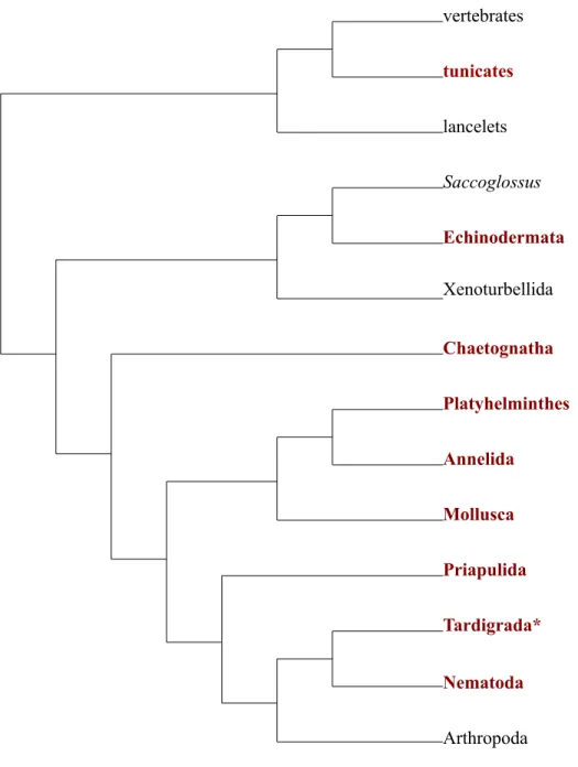
In obliquely-striated muscle, the relationship between structure and function is less well established. Obliquely-striated muscle has Z-bodies that serve as anchors for the thin
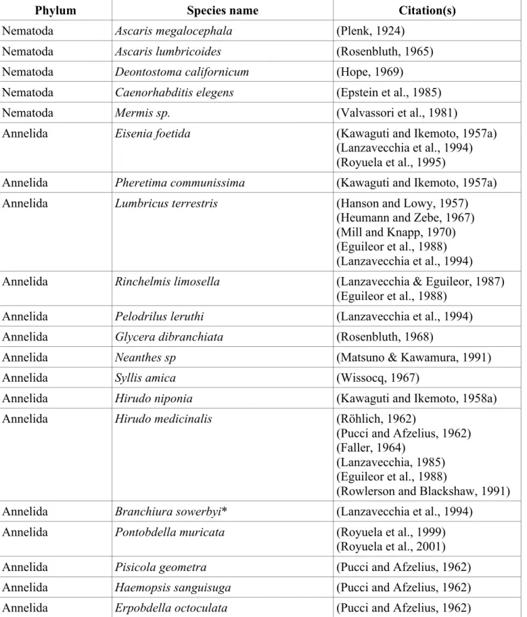
evidence that other obliquely-striated muscles are used to produce forces at longer lengths. Obliquely-striated muscle is present across bilateria (Fig. 1, Table 1) in more phyla than has been recognized in previous reviews (Lanzavecchia et al., 1977) but physiological data are only
available from annelids and mollusks (Fig. 2). Previous reviews have focused on the structure and implications of the geometry for the function of obliquely-striated muscle (Paniagua et al., 1996). The following is a review of the functional data available for obliquely-striated muscles. Early observations and sliding versus shearing
Initially, obliquely-striated muscle was thought to contract via two separate mechanisms: sliding and shearing (Rosenbluth, 1967). In the proposed sliding mechanism, the thick and thin myofilaments interacted in a manner similar to that proposed in the vertebrate sliding-filament theory (Gordon et al., 1966; Rosenbluth, 1967). In addition to sliding, a separate shearing mechanism was proposed which involved a change in the stagger of adjacent thick filaments as the muscle shortened and extended (Rosenbluth, 1967). Later, a geometric model of the muscle suggested that the observed shearing was a passive property of the geometry of the muscle fiber. In this case, the fiber actively contracts with a conventional sliding filament mechanism and the shortening of the long axis of the fiber coupled with the fact that muscle is essentially
isovolumetric means that the angle of striation and the stagger of the myofilaments (shearing) must change during contraction. It is this passive shearing coupled with the active sliding mechanism that was thought to make obliquely-striated muscle more extensible, or capable of producing forces at longer lengths than cross-striated muscle (Lanzavecchia, 1977).
Length-tension curves
document the length-tension relationship. Muscles produce different amounts of tension (tension is the force divided by the cross-sectional area) due to changes in the overlap between thick and thin myofilaments, which depends on muscle length. To understand how force changes with length, scientists stretch a muscle in vitro to a particular length, stimulate the muscle to contract isometrically, and measure the amount of force produced. They then repeat this procedure for a range of lengths. Plots of these forces versus length data are known as length-tension curves. Customarily, length (L) is standardized by the length at which the muscle produces maximum force (Lo) and tension (P) is standardized by the maximum tension (Po) produced by the muscle. Length-tension curves in vertebrate cross-striated muscle consist of an ascending limb in which the force increases as length increases, a plateau at the maximum force, and then a descending limb in which the force decreases with further increase in length.
Changing partners hypothesis: evidence and theory
allowed the muscle to produce relatively high amounts of force until it was stretched to such a length that there was no overlap with a consequent rapid decrease in force with length.
the curves. They also do not plot all three of the curves with LOcoincident, nor do they indicate the relative scaling of the length axis for each muscle type. Their conclusion is that based on the TEM data and the discrepancy between their theoretical and the actual curves, changing partners must be occurring (Lanzavecchia & Arcidiacono, 1981). These studies leave the field in a tenuous position. Because they do not provide enough explanation to evaluate or replicate their results, the validity of their conclusions is uncertain and difficult to test. But scientists who might attempt to repeat their experiments run the risk of devoting large amounts of time to a TEM study that fails to obtain novel insights.
In a follow-up study, Lanzavecchia modified the geometric model he used to differentiate sliding versus shearing mechanisms (Lanzavecchia, 1977) to include the effects of changing partners (Lanzavecchia, 1985). Then, in a manner not made explicit in the study, he used this model to predict morphological characteristics of the sarcomere. He compared these parameters, including distance between filaments, number of filaments, ratio of thick and thin filaments, width of the sarcomere, and filament length, to the observed values in TEMs of the leech, Hirudo medicinalis, once again a different species than the one used by Miller (1975), and reported that his model agreed with the micrographs. He concluded that in order to explain the Miller (1975) length-tension curve, the thick and thin filaments must change partners twice over the range from fully contracted to fully elongated (Lanzavecchia, 1985).
Additional length-tension curves of annelid muscle
There are length-tension curves reported for earthworms (Tashiro and Yamamoto, 1971). They do not appear to be super elongating (Figs. 4A and B).
is capable of super elongation (Gerry and Ellerby, 2011). This study used a portion of the longitudinal muscle of a single body segment rather than the entire dorsal body wall like Miller (1975), and found that the muscle produced active force from about 40-160% of resting length. When serotonin was present the muscle produced greater force at each length, shifting the length-force curve upwards. Additionally, in vivo sonomicrometry of suction feeding, crawling, and swimming suggested that this muscle operates at 75-240% of its resting length (Gerry and Ellerby, 2011). A later study that employed work loop procedures for the same muscle found that without serotonin the muscle acted as a brake, absorbing some of the energy, and that serotonin increased the net work of the muscle by decreasing the negative work due to passive stiffness (Gerry et al., 2012).
Length-tension curves and operating lengths of molluscan muscle
All known length-tension curves (Figs. 4A and B) and all known data on the in vivo
operating lengths suggest that the obliquely-striated muscle found in molluscs is not capable of super elongation.
The obliquely-striated mantle muscles of Alloteuthis subulata (squid) and Sepia
officinalis (cuttlefish) have length-tension and force-velocity curves that are similar to vertebrate cross-striated muscle and do not appear to be super elongating (Milligan et al., 1997).
The obliquely-striated muscle fibers of the arm of the squid Doryteuthis pealeii
(previously known as Loligo pealei) have longer thick filaments (7.41 +/- 0.44 um) than the thick filaments of the cross-striated muscle found in the tentacles (0.81 +/- 0.08 um). As expected from the thick filament lengths, the arm muscle fibers produce higher peak tensions and lower
the length ranges from about 60% to 110% of the resting length of the muscle (Kier and Curtin, 2002).
There are two different types of obliquely-striated circular fibers in the mantle muscle of the squid Doryteuthis pealeii: centrally located, mitochondria-poor (CMP) and superficially located, mitochondria-rich (SMR). CMP fibers have shorter thick filaments (1.78 +/- 0.27 um) and faster shortening velocities. SMR fibers have longer thick filaments (3.12 +/- 0.56 um) and higher peak forces. For both types of muscle, the length-tension curve ranges 90-105% of the resting length (Thompson et al., 2008). Both CMP and SMR increase in thick filament length and decrease in striation angle as the squid develop from paralarvae to adults (Thompson et al. 2010).
Further work on the CMP muscle fibers found length-tension curves with lengths from about 65% to 105% of resting length. Fibers taken from the inner portion of the mantle produced higher forces after being scaled to Po than fibers taken from the outer portion of the mantle. In vivo sonomicrometry experiments on CMP muscle fibers from both locations suggest that the muscles work primarily on the ascending limb of the length-tension curve (Thompson et al., 2014).
The funnel refractor muscle of the squid Doryteuthis pealeii was initially called helical smooth (Hanson and Lowy, 1957), but subsequent work reclassified it as obliquely-striated (Rosenbluth et al., 2010). In vivo sonomicrometry showed that it does not undergo large length changes. Instead, its operating range is around 4% of its resting length. The authors also
Despite there being no evidence for squid muscle producing force over a large range of operating lengths, authors continue to argue that the squid muscle might be capable of this because of the structural similarities between squid and leech muscle (Thompson et al. 2010). However, kinematic data on squid tentacle muscle shows the muscle is capable of extending 70-100% of its resting length, suggesting it might be capable of super elongation (Kier and Leeuwen, 1997).
Major alternative structures of obliquely-striated muscle
In the nematodes Caenorhabditis elegans and Mermis nigrescens, unlike in annelids and molluscs, the longitudinal body wall muscles, which are used to bend the body during movement, possess z-bodies that are directly attached to the muscle cell membrane. The z-body/cell
membrane attachments are proposed to allow the force of contraction to be transmitted laterally to the sides of the muscle cell rather than longitudinally at the ends of the muscle cell (Burr and Gans, 1998). The oblique striation pattern is thus thought to evenly distribute the attachment sites and force transmission, making it more likely that the worms will bend rather than kink. To my knowledge, there are no length-tension curves reported for nematode muscles.
Crinoids, a group of echinoderms, have obliquely-striated muscle with continuous Z-lines, unlike all other known types of obliquely-striated muscle (Carnevali et al., 1986). These muscles are found in the hinged system of skeletal elements of the crinoid arm (Carnevali & Saita, 1985b), which is not a hydrostatic system as seen in annelids, nematodes, and mollusks. To my knowledge, there are no length-tension curves reported for echinoderm muscle.
Discussion
obliquely-striated muscle can be found across bilateria, including in one non-hydrostatic skeleton: the hinged arms of crinoids (Carnevali & Saita, 1985b). The lack of evidence of a broad length-tension curve for molluscan obliquely-striated muscle coupled with structural differences between different types of obliquely-striated muscle suggests that the functional theories proposed for annelid and nematode muscle should not simply be applied to all obliquely-striated muscle.
Additionally, there are thirteen phyla in which obliquely-striated muscle has been described but for which length-tension information is not available. Currently, the theoretical understanding of obliquely-striated muscle is based on functional data from annelids, and ignores the contradictory data gathered from molluscs. Increasing the number of phyla for which
functional data is known could help sort out whether annelids, molluscs, or neither are typical of obliquely-striated muscle as a whole.
Further work involving geometric modeling of the results of sliding, shearing, and changing partners on length-tension curves would provide specific, testable hypotheses that could guide future studies and help elucidate the functional significance of the oblique striation pattern.
would show no evidence of changing partners, given the lack of super-elongation reported in previous studies.
Evidence for super elongation
Only one of six species for which there are physiological data shows evidence of super elongation (Fig. 4). Super elongation should no longer be considered the default function of obliquely-striated muscle. Especially within cephalopods, where all evidence points against super elongation, scientists should assume that the muscles are not capable of super elongation unless proven otherwise.
At the moment changing partners is the only theory offered to explain why leeches are capable of super elongation. This theory was proposed and validated through experimental and theoretical methods before the Gerry (2011) data were available. There is currently no feasible method for testing this theory through imaging, but the Lanzavecchia (1981) anesthetized vs. unanesthetized experiments are worth further exploration.
Table 1. Structural descriptions of obliquely-striated muscle. Asterisks indicate muscles thought to be an intermediary structure between obliquely-striated and smooth muscle.
Phylum Species name Citation(s)
Nematoda Ascaris megalocephala (Plenk, 1924)
Nematoda Ascaris lumbricoides (Rosenbluth, 1965)
Nematoda Deontostoma californicum (Hope, 1969)
Nematoda Caenorhabditis elegens (Epstein et al., 1985)
Nematoda Mermis sp. (Valvassori et al., 1981)
Annelida Eisenia foetida (Kawaguti and Ikemoto, 1957a)
(Lanzavecchia et al., 1994) (Royuela et al., 1995)
Annelida Pheretima communissima (Kawaguti and Ikemoto, 1957a)
Annelida Lumbricus terrestris (Hanson and Lowy, 1957)
(Heumann and Zebe, 1967) (Mill and Knapp, 1970) (Eguileor et al., 1988) (Lanzavecchia et al., 1994) Annelida Rinchelmis limosella (Lanzavecchia & Eguileor, 1987)
(Eguileor et al., 1988)
Annelida Pelodrilus leruthi (Lanzavecchia et al., 1994)
Annelida Glycera dibranchiata (Rosenbluth, 1968)
Annelida Neanthes sp (Matsuno & Kawamura, 1991)
Annelida Syllis amica (Wissocq, 1967)
Annelida Hirudo niponia (Kawaguti and Ikemoto, 1958a)
Annelida Hirudo medicinalis (Röhlich, 1962)
(Pucci and Afzelius, 1962) (Faller, 1964)
(Lanzavecchia, 1985) (Eguileor et al., 1988)
(Rowlerson and Blackshaw, 1991) Annelida Branchiura sowerbyi* (Lanzavecchia et al., 1994)
Annelida Pontobdella muricata (Royuela et al., 1999)
(Royuela et al., 2001)
Annelida Pisicola geometra (Pucci and Afzelius, 1962)
Annelida Haemopsis sanguisuga (Pucci and Afzelius, 1962)
Phylum Species name Citation(s) Annelida Glossiphonia complanata (Pucci and Afzelius, 1962)
Annelida Enchytraeus albidus (Eguileor et al., 1988)
Annelida Prinospio caspersi (Eguileor et al., 1988)
Annelida Magelona papillicornis (Wissocq and Boilly, 1977)
Annelida Tubifex tubifex (Lanzavecchia et al., 1994)
Annelida Limnodrilus udekemianus (Lanzavecchia et al., 1994)
Annelida Monopylephorus sp. (Lanzavecchia et al., 1994)
Annelida Peloscolex sp. (Lanzavecchia et al., 1994)
Annelida Rynchelmis limosella (Lanzavecchia et al., 1994) Annelida Lumbriculus variegatus (Lanzavecchia et al., 1994)
Annelida Bythonomus lemani (Lanzavecchia et al., 1994)
Annelida Kincaidiana sp. (Lanzavecchia et al., 1994)
Annelida Phagodrilus sp. (Lanzavecchia et al., 1994)
Annelida Eiseniella tetraedra (Lanzavecchia et al., 1994)
Mollusca Sepia esculenta (Kawaguti and Ikemoto, 1957b)
Mollusca Doryteuthis pealeii (Kier 1985)
(Rosenbluth et al., 2010)
Mollusca Illex illecebrosus (Kier 1985)
Mollusca Alloteuthis subulata (Bone et al., 1981)
Mollusca Sepia officinalis (Amsellem and Nicaise, 1980)
(Kier 1989)
Mollusca Pecten albicans (Kawaguti and Ikemoto, 1958b)
(Nunzi and Franzini-Armstrong, 1981)
Mollusca Crassostrea virginica (Morrison and Odense, 1974)
Mollusca Artica islandica (Morrison and Odense, 1974)
Mollusca Tridacna crocea (Matsuno & Kuga, 1989)
Mollusca Mitilus crassitesa (Kawaguti and Ikemoto, 1957c)
Mollusca Spondilus cruentus (Kawaguti and Ikemoto, 1959)
Mollusca Crassostrea angulata (Hanson and Lowy, 1961)
Mollusca Fragum onedo (Matsuno, 1988)
Mollusca Sepiella japonica (Matsuno, 1987)
Phylum Species name Citation(s) Brachiopoda Terebratalia transversa (Eshleman et al., 1982)
Urochordata Doliolum gegenbauri (Bone and Ryan, 1974)
Tardigrada Macrobiotus hufelandi* (Walz, 1974)
Tardigrada Milnesium tardigradum* (Walz, 1974)
Platyhelminthes Notoplana acticola (MacRae, 1965) Platyhelminthes Grillotia erinaceus (Ward et al., 1986)
Echinodermata Antedon mediterranea (Carnevali and Saita, 1985a) (Carnevali et al., 1986) Echinodermata Ophioderma longicaudum* (Saita et al., ,1982)
Priapulida Priapulus caudatus (Mattisson et al., 1974)
Rotifera Philodina roseola (Clément and Amsellem, 1989)
Rotifera Brachionus urceolaris sericus, B.
calyciflorus, B. plicatilis (Clément and Amsellem, 1989)
Rotifera Rhinoglena frontalis (Clément and Amsellem, 1989)
Rotifera Asplanchna brightwelli (Clément and Amsellem, 1989)
Rotifera Notommata copeus (Clément and Amsellem, 1989)
Nemertea Phallonemertes cf. murrayi (Norenburg and Roe, 1998) Nemertea Nectonemertes cf. mirabilis (Norenburg and Roe, 1998) Nemertea 2 species of protopelagonemertid (Norenburg and Roe, 1998) Sipunculida Sipunculus nudus (deEguileor & Valvassori, 1977) Nematomorpha Gordius aquaticus (Lanzavecchia et al., 1979) Nematomorpha Gordius panighettensis (Lanzavecchia et al., 1979)
Gastrotricha Turbanella cornuta (Teuchert, 1974)
Gastrotricha Chordodasys antennatus (Rieger et al., 1974)
Phylum Species name Citation(s) Gnathostomulida Austrognathia sp. I (Rieger and Mainitx, 1977) Gnathostomulida Austrognathia riedli (Rieger and Mainitx, 1977)
Chaetognatha Sagitta setosa (Duvert, 1969; Duvert and Salat,
Figure 1. Diagram of cross-‐striated (A) and obliquely-‐striated (B) muscle.
Sarcomere
Thick filament Thin filament
Thin filament
Thick filament
Bare-zone Bare-zone
Z-body Z-line
Sarcomere
A
Figure 2. Bilaterians with obliquely-‐striated muscle (in bold and red). Asterisks indicate muscle thought to be in between obliquely-‐striated and smooth in structure. Phylogeny is based on (Lartillot and Philippe, 2008).
Chaetognatha
Platyhelminthes
Annelida
Mollusca
Priapulida
Tardigrada*
Nematoda
Arthropoda Xenoturbellida
Echinodermata
Saccoglossus
lancelets
tunicates
vertebrates
Figure 3. Illustration of changing partners.
A. Schematic of two obliquely striated sarcomeres.
B. Close-‐up of obliquely striated sarcomere. One pair of thick and thin filaments are white so that they are easy to track throughout the changing partners process.
C. Close-‐up of obliquely striated sarcomere at a longer sarcomere length. At this length, the thick and thin filaments are at the limit of their ability to overlap.
Changing partners
Z-body
Thin filament
Thick filament Bare-zone
Sarcomere
Changing partners
Z-body
Thin filament
Thick filament
Bare-zone
Changing partners
Z-body
Thin filament
Thick filament
D. Close-‐up of obliquely striated sarcomere pulled beyond overlap. Arrows indicate the movement of filaments if changing partners occurs.
E. Close up of sarcomere after changing partners. The white thin filaments are no longer paired with the white thick filament.
Changing partners
Z-body
Thin filament
Thick filament
Bare-zone
Changing partners
Z-body
Thin filament
Thick filament
Figure 4. All known length-‐tension curves of obliquely-‐striated muscle.
A. Twitch contractions. The red line is the theoretical length-tension curve of cross-striated muscle (Gordon et al., 1966). Blue, open symbols are the length-tension curves of cephalopods. Black, closed symbols are the length-tension curves of annelids. Hirudo verbana and Hirudo verbana with 5-HT are from (Gerry & Ellerby 2011). D. pealeii is from (Kier and Curtin, 2002).
Haemopis sanguisuga is from (Miller, 1975). Alloteuthis subulata is from (Milligan et al., 1997). Earthworm is from (Tashiro and Yamamoto, 1971). D. pealeii inner and outer mantle are from (Thompson et al., 2014).
L/Lo
0 0.5 1 1.5 2 2.5 3
P/Po
0 0.2 0.4 0.6 0.8 1
1.2 Length Tension curves-- Twitch muscle
Hirudo verbana Hirudo verbana with 5-HT D. pealeii
Haemopis sanguisuga Alloteuthis subulata earthworm
B. Tetanus contractions. The red line is the theoretical length-tension curve of cross-striated muscle (Gordon et al., 1966). Blue, open symbols are the length-tension curves of cephalopods. Black, closed symbols are the length-tension curves of annelids. D. pealeii is from (Kier and Curtin, 2002). D. pealeii inner and outer mantle are from (Thompson et al., 2014). Alloteuthis subulata and Sepia officinalis is from (Milligan et al., 1997). Earthworm is from (Tashiro and Yamamoto, 1971).
L/Lo
0.4 0.6 0.8 1 1.2 1.4 1.6 1.8
P/Po
0 0.2 0.4 0.6 0.8 1
1.2 Length Tension curves-- Tetanus muscle
D. pealeii
CHAPTER 2: GEOMETRIC MODEL Introduction
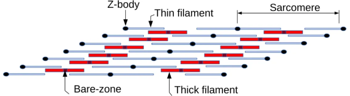
For cross-striated muscle it is possible to explain the shape of the length-tension curve based on the dimensions of the thick filaments, thin filaments, bare zones and the Z disk thickness (Gordon et al., 1966). When the sarcomere is most extended, there is no overlap between the myofilaments and thus the myosin heads cannot interact with the thin filaments; the muscle cannot produce force. At shorter lengths, the thin filaments overlap with the myosin heads on the thick filaments to produce force. The amount of force the muscle produces
In addition to the effects of this change in overlap on the tension, in obliquely-striated muscle there is an additional effect because the angle of striation and thus amount of stagger between adjacent myofilaments changes (shearing) as the muscle elongates and shortens. Although early studies interpreted shearing in obliquely-striated muscle as a separate and supplemental mechanism responsible for shortening (Rosenbluth, 1967), it was recognized subsequently that changes in the angle of striation are due simply to the geometry of an essentially constant volume fiber (Lanzavecchia, 1977). The effects of this shearing on the length-tension behavior of obliquely-striated muscle, however, remain unclear. The goal of this study was to use mathematical modeling to explore the implications of shearing for the
mechanics of obliquely-striated muscle. In addition, the model provides a means of predicting the effects of changes in myofilament dimensions and organization on the mechanics of this important striation type.
Methods
The model was created in MATLAB r2014b (The Mathworks, Natick, MA) (App. 1). Broadly, the model calculates the amount of force a single sarcomere could produce based on the overlap of the filaments. It starts by calculating this force for a sarcomere that is the length of one thick filament and increases the length by a unit equivalent to the distance between myosin heads until the sarcomere no longer produces force.
filaments is calculated by dividing the height of the sarcomere by the number of filaments. The length of the imaginary line that would be produced if there was a line connecting all of the Z-bodies on one side of the sarcomere was calculated by dividing the height of the sarcomere by the sine of the striation angle. This length was called p. The coordinates of the thin filaments and the myosin heads were calculated as indicated in Figure 5.
Once the coordinates are determined, the right thick filament is compared to the right thin filaments on either side of it. If the right thick filament overlaps with a right thin filament, but not with the corresponding left thin filament, a value of 1 is added to the total force for that length (Fig. 6). This is repeated on the left hand side and for the thin filaments on one n lower than the thick filament. When both right and left thin filaments are present, no force is added to reflect the hypothesis that the thin filaments interfere with each other, preventing cross-bridges from forming (Gordon et al., 1966).
For each new sarcomere length, the height of the sarcomere was recalculated by dividing the area of the sarcomere by the new length. Additionally, the angle of striation was recalculated by taking the inverse sine of the height of the sarcomere divided by p.
Results
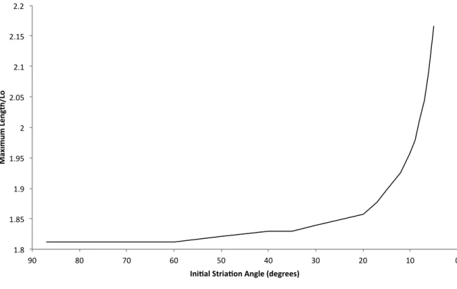
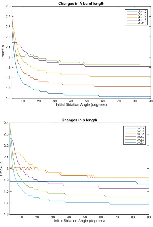
The smaller the initial striation angle (the greater the stagger between thick filaments), the greater the maximum length normalized by L0 at which the muscle is able to produce force (Fig. 6).
Because of the rounding and tangent functions in the model (App. 1), there are jumps in the model that change depending on the values of the parameters (Fig. 7). Additionally, below an initial striation angle of about 3 or 4 degrees, there are spacing issues in the model that cause the muscle to no longer behave in a biologically relevant way (Fig. 8).
Discussion
Though changes in myofilament and bare zone lengths can affect the shape of the length-tension curve, changes in the initial angle of striation increases the maximum length at which the muscle can produce force in the most predictable way. This suggests that obliquely-striated muscle could have evolved in order to increase the operating length of the muscle. This might be especially important to animals without hinged skeletons. If an animal has a hinged skeleton it can use the skeleton as a lever to amplify the force or displacement created by the muscle. Absent such a system, animals must have muscle that is capable of operating over the full length of the animal. Thus in animals with hydrostatic skeletons, it would be advantageous for the longitudinal muscle to be obliquely-striated and capable of long extensions.
The primary limitation to my model is that most of the parameters considered in the model (filament length, filament spacing, bare zone length, the spacing of the myosin heads, and the initial angle of striation) are not known for obliquely-striated muscle from most species, though some of these parameters like filament length and spacing are known for Hirudo
species and then predict the length-tension curve. However, it is possible to determine which variables are likely to have the largest effects on the length-tension curve. Because these
parameters are difficult to collect, as a next step it would be a better to focus not on determining their values for additional species but on collecting additional length-tension curves and
kinematic data in order to gain a broader understanding of obliquely-striated muscle. Additionally my model is two-dimensional and sarcomeres are actually
three-dimensional. I maintain a constant area of the muscle by adjusting the height of the sarcomere as the length of the sarcomere changes. In reality, the depth of the sarcomere could also be
changing. Because at every length I recalculate the angle of striation based on the length and height of the sarcomere, if much of the change in dimension occurs in the depth dimension rather than the height dimension, it is possible that the angle is not changing in a realistic way.
My model is expressed in terms of parameters that scientists can measure experimentally. Some of these parameters could be expressed in terms of each other. For example, all lengths including the length of the sarcomere, thin filaments, and bare-zone, could be expressed in terms of the length of the thick filament. A future version of this model might want to express all lengths as a ratio in order to better explore the effects of varying these lengths.
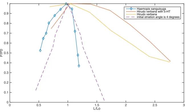
Comparing Literature and Model Results: Leeches
note that many of the parameters considered in my model are known only for Hirudo medicinalis
Figure 5. Obliquely-striated sarcomeres with dimensions considered in the model. L = the length of the sarcomere, b = the length of two thin filaments and one Z body, x = the horizontal distance between two vertically adjacent thin filaments, d = distance between two thin filaments, n = number of filament pairs, BZ = the length of the bare zone, A = the length of a thick filament, theta = angle of striation.
b
A BZ
L
Z body
(n-1)x b/2+(n-1)x L-b/2+nx
L+nx
d
x
θ n = 1
n = 2 n = 3
L-BZ+nx 2
L+BZ+nx 2
L-A +nx
Figure 6. Diagram of one set of thick and thin myofilaments. Dark blue, vertical lines indicate myosin heads that produce force. On the left side of the sarcomere, these are myosin heads that overlap with the left thin filaments and do not overlap with the right thin filaments. On the right side of the sarcomere, these are myosin heads that overlap with the right thin filaments and do not overlap with the left thin filaments. Black, vertical lines indicate myosin heads that do not produce force because they overlap with both left and right thin filaments (B).
Bare-zone
Thin filament
Thick filament
Z-body
Myosin head
producing force
Bare-zone
Thin filament
Thick filament
Z-body
Myosin head
producing force
Myosin head
producing no force
A
Figure 7. Maximum length/ Lo changes with initial striation angle. Small initial angles of striation increase the maximum length at which the muscle can produce force.
1.8$ 1.85$ 1.9$ 1.95$ 2$ 2.05$ 2.1$ 2.15$ 2.2$
0$ 10$
20$ 30$
40$ 50$
60$ 70$
80$ 90$
Ma
xi
m
um
'L
en
gt
h/
Lo
'
Ini1al'Stria1on'Angle'(degrees)'
Figure 8. Effects of varying parameters on inflection points in the model
Initial Striation Angle (degrees)
10 20 30 40 50 60 70 80 90
Lmax/Lo 1.6 1.7 1.8 1.9 2 2.1 2.2 2.3 2.4
2.5 Changes in A band length
A=1.2 A=1.4 A=1.6 A=1.8 A=2.0
Initial Striation Angle (degrees)
10 20 30 40 50 60 70 80 90
Lmax/Lo 1.6 1.7 1.8 1.9 2 2.1 2.2 2.3
2.4 Changes in b length
Initial Striation Angle (degrees)
10 20 30 40 50 60 70 80 90
Lmax/Lo
1.8 1.85 1.9 1.95 2 2.05 2.1 2.15 2.2 2.25
2.3 Changes in BZ length
BZ=.1 BZ=.14 BZ=.18 BZ=.22 BZ=.26 BZ=.3
Initial Striation Angle (degrees)
10 20 30 40 50 60 70 80 90
Lmax/Lo
1.7 1.8 1.9 2 2.1 2.2
2.3 Changes in initial height of the sarcomere
Initial Striation Angle (degrees)
10 20 30 40 50 60 70 80 90
Lmax/Lo 1.8 1.85 1.9 1.95 2 2.05 2.1 2.15 2.2 2.25
2.3 Changes in myosin head spacing
mh=.01 mh=.014 mh=.018 mh=.022 mh=.026 mh=.03
Initial Striation Angle (degrees)
10 20 30 40 50 60 70 80 90
Lmax/Lo 1.8 1.85 1.9 1.95 2 2.05 2.1 2.15 2.2 2.25
2.3 Changes in n
Figure 9. Comparing literature and model results for leeches. All parameters for the dashed purple curve, including the initial striation angle, were assigned experimental values from Lanzavecchia (1985) when possible.
L/Lo
0 0.5 1 1.5 2 2.5 3
P/P0
0 0.1 0.2 0.3 0.4 0.5 0.6 0.7 0.8 0.9 1
Haemopis sanguisuga Hirudo verbana with 5-HT Hirudo verbana
CHAPTER 3: CONCLUSIONS AND FUTURE DIRECTIONS Given the scarcity of evidence for super elongation, it would be interesting for researchers to consider alternative explanations for oblique striation. For instance, obliquely-striated muscle may be an adaptation for hydrostatic skeletons. Because animals with hydrostatic skeletons do not have the option of manipulating the displacement output of the muscle by varying the relative lengths of input and output lever arms, it is possible that oblique striation evolved as a way of accommodating the large dimensional changes common in invertebrates. This explanation is the most compelling in animals with highly organized oblique and cross-striated muscle like cephalopods. However, for other animals, that lack cross-cross-striated muscle or lack highly organized obliquely-striated muscle, it is possible that oblique striation is simply an intermediate form between smooth and cross-striated muscle. The sarcomeres of cross-striated muscle allow the muscle to amplify velocity by organizing the sarcomeres in series. Animals without cross striation, are unable to take advantage of this in a systematic manner, and may have evolved obliquely-striated muscle as a solution.
In order to understand the function and evolution of obliquely-striated muscle it would be helpful to consider the variation within obliquely-striated muscle from various phyla.
significance of the angle of striation, up to this point, scientists have been acting on the
assumption that these two different structures should have the same function because they have the same name. However, it is challenging to classify the types of obliquely-striated muscle with the information available in the literature. On the two-dimensional level, it is hard to determine the angle of striation, the lengths of the filaments, and the exact proteins present in the
sarcomeres. In general, only a few micrographs are published in each study, and even with access to all of the micrographs taken, it is difficult to piece together a 3D structure from 2D images. It would be useful to have complete three-dimensional reconstructions, perhaps through the use of an electron microscope capable of sectioning during the imaging process.
Though obliquely-striated muscle has been documented in over 80 species (Table 1), many of these species are difficult to obtain or manipulate. Some of the species like the
APPENDIX 1: MATLAB PROGRAM FOR PREDICTING THE LENGTH TENSION CURVE OF OBLIQUELY-STRIATED MUSCLE
function[LTcurve] = sarc_sim(angle,angle_changing,animate,moviename)
% assign filament dimensions in microns
A = 1.6; % length of the thick filament
I = 2.05; % length of two thin fiaments and a z-line or body
BZ = 0.20; % length of the bare-zone
mh_spacing = 0.02; % spacing between myosin heads
n = 30; % number of thin filaments
h_initial = 6; % initial height of the sarcomere
area = A*h_initial; % area is the initial length of the sarcomere (A) times
the intial height of the sarcomere (h)
p = h_initial/sin(angle/180*pi); % other side of the parallelogram
% create figure for the animation
if animate
fig = figure;
hold on
h1 = []; % dummy handle for some figure graphic
end
% create movie container if called for
if exist('moviename','var')
mov = VideoWriter([moviename,'.avi']);
open(mov);
end
% find the force at each sarcomere length
LF = zeros(round(2*I/mh_spacing)+1,2); % create an array in which to store
Length and Tension l = 1;
for L = A:mh_spacing:A+4*I
h = area/L; % calculate the height of the sarcomere
d = h/n; % calculate the distance between the filaments based on area and
length.
% if the angle is changing, calculate the angle of striation
if angle_changing
theta = asin(h/p);
else
theta = angle/180*pi;
end
x = d/tan(theta); % calculate the lateral distance between the start of
thin filaments
hold the locations of the left myosin heads % fill in array for myosin heads
for i = 1:n-1
k = 1;
for j = (L-A)/2 + x*i:mh_spacing:(L-BZ)/2 + x*i
TFL(i,k,1) = j;
TFL(i,k,2) = (i-1)*d; k = k+1;
end
end
TFR = zeros(n-1,round((A-BZ)/2/mh_spacing)+1,2); % create an array to
hold the locations of the right myosin heads % fill in array for myosin heads
for i = 1:n-1
k = 1;
for j = (L+BZ)/2 + x*i:mh_spacing:(L+A)/2 + x*i
TFR(i,k,1) = j;
TFR(i,k,2) = (i-1)*d; k = k+1;
end
end
tfl = zeros(3,n); % create an array to hold the locations of the left
thin filaments
% fill in array for the thin filaments
for i = 1:n
tfl(1,i) = (i-1)*x;
tfl(2,i) = I/2 + (i-1)*x; tfl(3,i) = (i-1)*d;
end
tfr = zeros(3,n); % create an array to hold the locations of the right
thin filaments
% fill in array for the thin filaments
for i = 1:n
tfr(1,i) = L-I/2+i*x; tfr(2,i) = L+i*x; tfr(3,i) = (i-1)*d;
end
% compare the arrays to calculate the force
Fsumn = zeros(1,n); % create an empty array to count the force produced
by each filament set
for i = 1:n-1 % for each filament
for k = 1:round((A-BZ)/2/mh_spacing)+1 % along all possible positions
if (tfl(1,i) <= TFL(i,k,1)) && (TFL(i,k,1) <= tfl(2,i)) % if the left myosin head overlaps with the left thin filament
if (tfr(1,i) <= TFL(i,k,1)) && (TFL(i,k,1) <= tfr(2,i)) % if they left myosin head overlaps with the right thin filament
Fsumn(i) = Fsumn(i); % keep the force the same because
the thin filaments are interfering with each other
else
Fsumn(i) = Fsumn(i) + 1; % if the thin filaments do not
end
if (tfl(1,i+1) <= TFL(i,k,1)) && (TFL(i,k,1) <= tfl(2,i+1)) % check the thin filament on the other side of the left myosin head
if (tfr(1,i+1) <= TFL(i,k,1)) && (TFL(i,k,1) <= tfr(2,i+1)) Fsumn(i) = Fsumn(i);
else
Fsumn(i) = Fsumn(i) + 1;
end
end
if (tfr(1,i) <= TFR(i,k,1)) && (TFR(i,k,1) <= tfr(2,i)) %do the same for the right myosin heads
if (tfl(1,i) <= TFR(i,k,1)) && (TFR(i,k,1) <= tfl(2,i)) Fsumn(i) = Fsumn(i);
else
Fsumn(i) = Fsumn(i) + 1;
end
end
if (tfr(1,i+1) <= TFR(i,k,1)) && (TFR(i,k,1) <= tfr(2,i+1)) if (tfl(1,i+1) <= TFR(i,k,1)) && (TFR(i,k,1) <= tfl(2,i+1)) Fsumn(i) = Fsumn(i);
else
Fsumn(i) = Fsumn(i) + 1;
end end end end
% store the length and total force in the LF array LF(l,1) = L;
LF(l,2) = sum(Fsumn);
% create/update the animation if called for
if animate
if isempty(h1) == false % delete graphics from the previous
delete(h1); delete(h2); delete(h3); delete(h4); delete(h5); end
% plot new components in the bottom panel
subplot(2,1,1);
hold on
h1 =
plot(TFL(1:n-1,1:round((A-BZ)/2/mh_spacing)+1,1),TFL(1:n-1,1:round((A-BZ)/2/mh_spacing)+1,2),'g*');
h2 =
q = tfl(1:2,1:n); w = tfl(3,1:n); w(2,:) = tfl(3,1:n); h3 = plot(q,w);
r = tfr(1:2,1:n); s = tfr(3,1:n); s(2,:) = tfr(3,1:n); h4 = plot(r,s);
axis([0 A+2*I 0 h_initial]);
% Adjust the length tension curve for P0 and L0
[m,q] = size(LF);
maxForce = max(LF(1:m,2));
LTcurve(1:m,2) = LF(1:m,2)/maxForce;
[r,c] = find(LTcurve == 1); [x,y] = size(r);
L0 = sum(r(1:x))/x;
LTcurve(1:m,1) = LF(1:m,1)/LF(round(L0),1);
% plot new components in bottom panel
subplot(2,1,2);
hold on
h5 = plot(LTcurve(1:m,1),LTcurve(1:m,2),'g*');
axis([0 4 0 1])
% add frame to the movie if we're creating one
if exist('moviename','var')
% Write each frame to the file
currFrame = getframe(gcf); writeVideo(mov,currFrame);
end
else
% Adjust the length tension curve for P0 and L0
[m,q] = size(LF);
maxForce = max(LF(1:m,2));
LTcurve(1:m,2) = LF(1:m,2)/maxForce;
[r,c] = find(LTcurve == 1); [x,y] = size(r);
L0 = sum(r(1:x))/x;
LTcurve(1:m,1) = LF(1:m,1)/LF(round(L0),1);
end
if LF(l,2) == 0 % if the force is 0, stop the program
break
end
l = l+1;
end
% close the movie if we created one
if exist('moviename','var') close(mov);
REFERENCES
Amsellem, J. and Nicaise, G. (1980). Ultrastructural study of muscle cells and their connections in the digestive tract of Sepia officinalis. J. Submicr. Cytol. 12, 219–231.
Bone, Q. and Ryan, K. P. (1974). On the structure and innervation of the muscle bands of Doliolum (Tunicata: Cyclomyaria). Proc. R. Soc. Lond. B. 187, 315–27.
Bone, Q., Pulsford, A. and Chubb, A. D. (1981). Squid mantle muscle. J. Mar. Biol. Ass. U.K.
61, 327–342.
Burr, A. H. and Gans, C. (1998). Mechanical significance of obliquely striated architecture in nematode muscle. Biol. Bull. 194, 1–6.
Candia Carnevali, M. D. and Saita, A. (1985a). Muscle system organization in the
echinoderms: II. Microscopic anatomy and functional significance of the muscle‐ligament‐ skeleton system in the arm of the comatulids (Antedon mediterranea). J. Morphol. 185, 59– 74.
Candia Carnevali, M. D. and Saita, A. (1985b). Muscle system organization in the
echinoderms : III . Fine structure of the contractile apparatus of the arm flexor muscles of the comatulids (Antedon mediterranea). J. Morphol. 185, 75–87.
Candia Carnevali, M. D., Saita, A. and Fedrigo, A. (1986). An unusual Z-system in the obliquely striated muscles of crinoids: three-dimensional structure and computer simulations. J. Muscle Res. Cell Motil. 7, 568–578.
Clément, P. and Amsellem, J. (1989). The skeletal muscles of rotifers and their innervation.
Hydrobiologia 186/187, 255–278.
De Eguileor, M. and Valvassori, R. (1977). Studies on the helical and paramyosinic muscles. VII. Fine structure of body wall muscles in Sipunculus nudus. J. Submicr. Cytol. 9, 363– 372.
De Eguileor, M., Cotelli, F., Valvassori, R., Brivio, M. and di Lernia, L. (1988). Functional significance of intermediate filament meshwork in annelid helical muscles. J. Ultrastruct. Mol. Struct. Res. 100, 183–193.
Duvert, M. (1969). Ultrastructure des myofibrilels dans les muscles longitudinaux du tronc de sagitta setosa (chaetognath). C.R. Acad. Sci. Paris 268, 2452–2454.
Duvert, M. and Salat, C. (1980). The primary body-wall musculature in the arrow-worm Sagitta setosa (Chaetognatha): an ultrastructural study. Tissue Cell 12, 723–738.
Epstein, H. F., Miller, D. M., Ortiz, I. and Berliner, G. C. (1985). Myosin and paramyosin are organized about a newly identified core structure. J. Cell Biol. 100, 904–915.
Eshleman, W. P., Wilkens, J. L. and Cavey, M. J. (1982). Electrophoretic and electron microscopic examination of the adductor and diductor muscles of an articulate brachiopod, Terebratalia transversa. Can. J. Zool. 60, 550–559.
Faller, A. (1964). Zur frage der struktur der glatten muskelzellen von Hirudo medicinalis L.
Zeitschrift für Zellforsch. 63, 799–815.
Gerry, S. P. and Ellerby, D. J. (2011). Serotonin modulates muscle function in the medicinal leech Hirudo verbana. Biol. Lett. 7, 885–888.
Gerry, S. P., Daigle, A. J., Feilich, K. L., Liao, J., Oston, A. L. and Ellerby, D. J. (2012). Serotonin as an integrator of leech behavior and muscle mechanical performance. Zool. 115, 255–260.
Gordon, A. M., Huxley, A. F. and Julian, F. J. (1966). The variation in isometric tension with sarcomere length in vertebrate muscle fibres. J. Physiol. 184, 170–192.
Hanson, J. and Lowy, J. (1957). Structure of smooth muscles. Nature 180, 906–909.
Hanson, J. and Lowy, J. (1961). The structure of the muscle fibres in the translucent part of the adductor of the oyster Crassostrea angulata. Proc. R. Soc. Lond. B. 154, 173–196.
Heumann, H. G. and Zebe, E. (1967). Über feinbau und funktionsweise der fasern aus dem hautmuskelschlauch des regenwurms Lumbrics terrestris L. Zeitschrift für Zellforsch. 79, 131–150.
Hope, W. D. (1969). Fine Structure of the Somatic Muscles of the Free-Living Marine
Nematode Deontostoma californicum Steiner and Albin , 1933 ( Leptosomatidae ). Proc. Helm. Soc. Wash. 36, 10–29.
Kawaguti, S. and Ikemoto, N. (1957a). Electron microscopy on the smooth muscle from the body wall of the earthworms, Pheretima communissima and Eisenia foetida. Biol. J. Okayama Univ.
Kawaguti, S. and Ikemoto, N. (1957b). Electron microscopy of the smooth muscle of a cuttlefish, Sepia esculenta. Biol. J. Okayama Univ. 3, 196–208.
Kawaguti, S. and Ikemoto, N. (1958a). Electron microscopy on the smooth muscle of the leech, Hirudo nipponia. Biol. J. Okayama Univ. 4, 79–91.
Kawaguti, S. and Ikemoto, N. (1958b). Electron microscopy on the adductor muscle of the scallop, Pecten albicans. Biol. J. Okayama Univ. 191–205.
Kawaguti, S. and Ikemoto, N. (1959). Electron microscopy of the adductor muscle of the thorn oyster, Spondylus cruentus. Biol. J. Okayama Univ. 5, 73–87.
Kier, W. M. (1985). The Musculature of Squid Arms and Tentacles: Ultrastructural Evidence for Functional Differences. J. Morphol. 185, 223–239.
Kier, W. M. (1989). The fin musculature of cuttlefish and squid (Mollusca, Cephalopoda): morphology and mechanics. J. Zool. Lond. 217, 23–38.
Kier, W. M. (1991). Squid Cross-Striated Musclce: The Evolution of A Specialized Muscle Fiber Type. Bull. Mar. Sci. 49, 389–403.
Kier, W. M. and Curtin, N. A. (2002). Fast muscle in squid (Loligo pealei): contractile properties of a specialized muscle fibre type. J. Exp. Biol. 205, 1907–16.
Kier, W. and Leeuwen, J. (1997). A kinematic analysis of tentacle extension in the squid Loligo pealei. J. Exp. Biol. 200, 41–53.
Kuga, H. and Matsuno, A. (1988). Ultrastructural investigations on the anterior adductor muscle of a brachiopoda, Lingula unguis. Cell Struct. Funct. 13, 271–279.
Lanzavecchia, G. (1977). Morphological modulations in helical muscles (Aschelminthes and Annelida). Int. Rev. Cytol. 51, 133–186.
Lanzavecchia, G. (1981). Morphofunctional and phylogenetic relations in helical muscles. Boll. Zool. 48, 29–40.
Lanzavecchia, G. (1985). Superelongation in helical muscles of leeches. J. Muscle Res. Cell Motil. 6, 569–584.
Lanzavecchia, G. and Arcidiacono, G. (1981). Contraction mechanism of helical muscles: experimental and theoretical analysis. J. Submicr. Cytol. 13, 253–266.
Lanzavecchia, G. and de Eguileor, M. (1987). Analysis and reconstruction of unusual
obliquely striated fibres in Lumbriculids (Annelida, Oligochaeta). J. Muscle Res. Cell Motil.
8, 209–219.
Lanzavecchia, G., Valvassori, R., de Eguileor, M. and Lanzavecchia, P. (1979). Three-dimensional reconstruction of the contractile system of the Nematomorpha muscle fiber. J. Ultrastruct. Res. 66, 201–223.
Lanzavecchia, G., Valvassori, R. and de Eguileor, M. (1994). Body wall muscles in oligochaetes. Hydrobiologia 278, 179–188.
Lartillot, N. and Philippe, H. (2008). Improvement of molecular phylogenetic inference and the phylogeny of Bilateria. Philos. Trans. R. Soc. Lond. B. Biol. Sci. 363, 1463–72.
MacRae, E. K. (1965). The fine structure of muscle in a marine turbellarian. Zeitschrift für Zellforsch. 68, 348–362.
Matsuno, A. (1987). Ultrastructural studies on developing oblique-striated muscle cells in the cuttlefish, Sepiella japonica Sasaki. Zool. Sci. 4, 53–59.
Matsuno, A. (1988). Ultrastructural differences between the anterior and posterior adductors of the strawberry cockle, Fragum unedo. Tissue Cell 20, 283–290.
Matsuno, A. and Kawamura, Y. (1991). Cell types differing in thick myofilament diameter in obliquely striated muscle of a polychaete annelid, Neanthes. Tissue Cell 23, 481–487. Matsuno, A. and Kuga, H. (1989). Ultrastructure of muscle cells in the adductor of the boring
clam Tridacna crocea. J. Morphol. 200, 247–253.
Mattisson, A., Nilsson, S. and Fänge, R. (1974). Light microscopical and ultrastructural organization of muscles of Priapulus caudatus (Priapulida) and their responses to drugs, with phylogenetic remarks. Zool. Scr. 3, 209–218.
Mill, P. J. and Knapp, M. F. (1970). The fine structure of obliquely striated body wall muscles in the earthworm, Lumbricus terrestris Linn. J. Cell Sci. 7, 233–261.
Miller, J. B. (1975). The length-tension relationship of the dorsal longitudinal muscle of a leech.
J. Exp. Biol. 62, 43–53.
Milligan, B., Curtin, N. and Bone, Q. (1997). Contractile properties of obliquely striated muscle from the mantle of squid (Alloteuthis subulata) and cuttlefish (Sepia officinalis). J. Exp. Biol. 200, 2425–2436.
Morrison, C. M. and Odense, P. H. (1974). Ultrastructure of some pelecypod adductor muscles. J. Ultrastruct. Res. 49, 228–251.
Nunzi, M. G. and Franzini-Armstrong, C. (1981). The Structure of Smooth and Striated Portions of the Adductor Muscle of the Valves in a Scallop. J. Ultrastruct. Res. 76, 134– 148.
Paniagua, R., Royuela, M., García-Anchuelo, R. M. and Fraile, B. (1996). Ultrastructure of invertebrate muscle cell types. Histol. and Histopathol. 181–201.
Plenk, H. (1924). Nachweis von Querstreifung in säumtlichen Muskelfasern von Ascaris megalocephala. Anat. Embryol. 358–388.
Pucci, I. and Afzelius, B. A. (1962). An electron microscope study of sarcotubules and related structures in the leech muscle. J. Ultrastruct. Res. 7, 210–224.
Rieger, R. M. and Mainitx, M. (1977). Comparative fine structure study of the body wall in Gnathostomulida and their phylogenetic position between Platyhelminthes and
Aschelminthes. Zeitschrift für Zool. Syst. und Evol. 15, 9–35.
Rieger, R. M., Ruppert, E., Rieger, G. E. and Shoepfer-Sterrer, C. (1974). On the fine structure of gastrotrichs with description of Chordodasys antennatus sp. Zool. Script. 3, 219–237.
Roberts, T. J. and Azizi, E. (2010). The series-elastic shock absorber: tendons attenuate muscle power during eccentric actions. J. Appl. Physiol. 109, 396–404.
Röhlich, P. (1962). The fine structure of the muscle fiber of the leech Hirudo medicinalis. J. Ultrastruct. Res. 7, 399–408.
Rosenbluth, J. (1965a). Ultrastructural organization of obliquely striated muscle fibers in Ascaris lumbricoides. J. Cell Biol. 25, 495–515.
Rosenbluth, J. (1965b). Smooth muscle: An ultrastructural basis for the dynamics of its contraction. Science (80-. ). 148, 1337–1339.
Rosenbluth, J. (1967). Obliquely striated muscle: III. Contraction mechanism of ascaris body muscle. J. Cell Biol. 34, 15–33.
Rosenbluth, J. (1968). Obliquely striated muscle: IV. Sarcoplasmic reticulum, contractile apparatus, and endomysium of the body muscle of a polychaete, Glycera, in relation to its speed. J. Cell Biol. 36, 245–259.
Rosenbluth, J., Szent-Györgyi, A. G. and Thompson, J. T. (2010). The ultrastructure and contractile properties of a fast-acting, obliquely striated, myosin-regulated muscle: the funnel retractor of squids. J. Exp. Biol. 213, 2430–2443.
Royuela, M. A. R., Fraile, B., Garcia-anchuelo, R. and Paniagua, R. (1995). Ultrastructurally different muscle cell types in Eisenia foetida (AnneIida, OIigochaeta). J. Morphol. 224, 87– 96.
Royuela, M., Paniagua, R. and Rivier, F. (1999). Presence of invertebrate dystrophin-like products in obliquely striated muscle of the leech, Pontobdella muricata (Annelida, Hirudinea). Histochem. J. 31, 603–608.
Royuela, M., Hugon, G., Rivier, F., Paniagua, R. and Mornet, D. (2001).
Dystrophin-associated proteins in obliquely striated muscle of the leech Pontobdella muricata (Annelida , Hirudinea). Histochem. J. 33, 135–139.
Saita, A., Carnevali, M. D. C. and Canonaco, M. (1982). Muscle system organization in the echinoderms I. Intervertebral muscles of Ophioderma longicaudum (Ophiuroidea). J. Submicr. Cytol. 14, 291–304.
Tashiro, N. and Yamamoto, T. (1971). The phasic and tonic contraction in the longitudinal muscle of the earthworm. J. Exp. Biol. 55, 111–122.
Teuchert, G. (1974). Aufbau und Feinstruktur der Muskelsysteme von Turbanella cornuta Remane (Gastrotricha, Macrodasyoidea). Mikrofauna d. Meeresb. 39, 1–26.
Thompson, J. T., Szczepanski, J. A. and Brody, J. (2008). Mechanical specialization of the obliquely striated circular mantle muscle fibres of the long-finned squid Doryteuthis pealeii.
J. Exp. Biol. 211, 1463–1474.
Thompson, J. T., Taylor, K. R. and Gentile, C. (2010a). Gradients of strain and strain rate in the hollow muscular organs of soft-bodied animals. Biol. Lett. 6, 482–485.
Thompson, J. T., Bartol, I. K., Baksi, A. E., Li, K. Y. and Krueger, P. S. (2010b). The ontogeny of muscle structure and locomotory function in the long-finned squid Doryteuthis pealeii. J. Exp. Biol. 213, 1079–1091.
Thompson, J. T., Shelton, R. M. and Kier, W. M. (2014). The length-force behavior and operating length range of squid muscle vary as a function of position in the mantle wall. J. Exp. Biol. 217, 2181–2192.
Valvassori, R., de Eguileor, M. and Lanzavecchia, G. (1981). Studies on the helical and paramyosinic muscle VIII. Ultrastructural analysis of body wall muscles from Mermis sp. J. Ultrastruct. Res. 76, 82–88.
Ward, S. M., McKeer, G. and Allen, J. M. (1986). Structure and ultrastructure of muscle systems within Grillotia erinaceus metacestodes (Cestoda: Trypanorhyncha). Parasitology
93, 587–597.
Wissocq, J. C. (1967). Etude ultrastructurale de l’évolution des muscles longitudinaux lors de la stolonisation expérimentale de Syllis amica (Quatrefages) (Annélide Polychète). Zeitschrift für Zellforsch. 83, 449–467.
Wissocq, J. C. and Boilly, B. (1977). Muscles à simple striation oblique et à striation