International Journal of Pharmaceutical Research&Allied Sciences, 2017, 6(2):194-203
Research Article
CODEN(USA) : IJPRPM
ISSN : 2277-3657
194
Subconjunctival Bevacizumab versus Mitomycin c adjunctive to trabeculectomy
in primary open angle glaucoma
Eslam Mohamed, Msc, Prof. Dr. Hassan LotfyFahmy, Prof. Dr. Mohamed Sayed,
AbdelsalamAbdallah, MD, Mahmoud Abd El-Radi,
Department of Ophthalmology, Faculty of Medicine, Assiut University, Egypt
*
Corresponding authors
Eslam Mohamed
Department of Ophthalmology, Faculty of Medicine, Assiut University, Egypt
Email: [email protected]
ABSTRACT
Purpose: to compare efficacy and safety of mitomycin C versus bevacizumab as adjunctive to trabeculectomy in primary open angle glaucoma Setting: A prospective, comparative, open-label study was conducted at Assiut university hospitals, Assiut, Egypt, from January 2016 to January 2017.
Methods: Thirty eyes from 30 patients with uncontrolled primary open angle glaucoma were enrolled. 15 eyes underwent trabeculectomy with subconjunctival bevacizumab injection (1.25 mg/0.1 mL), and 15 eyes underwent trabeculectomy with MMC (0.02% for 3 minutes). The primary outcome measure wasthe intraocular pressure (IOP) while the secondary outcome measures were number of IOP-lowering medications, and bleb morphologic features (based on the Indiana Bleb Appearance Grading Scale).
Results: follow-up times for the MMC and bevacizumab groups were 6thmonthsmonths. The mean preoperative IOP in the bevacizumab group improved from 29.80 ± 2.83 mm Hg with 2.67 ± 0.98antiglaucoma medications to 14.13 ± 5.58mm Hg with 1.00 ± 1.13 antiglaucoma medications at the last visit (P < .001 and P < .001, respectively). The mean preoperative IOP in the MMC group improved from 28.40 ± 2.06mm Hg with 2.40 ± 0.99 antiglaucoma medications to 12.27 ± 4.85 mm Hg with 0.87 ± 1.06antiglaucoma medications at the final visit (P <.001 and P < .001, respectively). There was no statistically significant difference in the IOP between the 2 groups at the last visit (P < 0.250). The cumulative probabilities of total success at the last follow-up according to Kaplan- Meier analysis were 86.7% and 79.9% in MMC and bevacizumab groups, respectively.
Conclusions: Subconjunctival Bevacizumab adjunctive to trabeculectomy effectively control IOP and its effect was comparable to Mitomycin C
Key words:MitomycinC, Bevacizumab, trabeculectomy and primary open angle glaucoma
INTRODUCTION
Filtration surgery represents a crucial and efficient surgical procedure to lower the intraocular pressure (IOP) in glaucoma patients especially those who don’t respond or resist medical treatment with different anti-glaucoma drugs and /or argon laser trabeculoplasty (ALT) (1).
195
related to the severity of conjunctival vascularization, the tortuosity of vessels, and the migration and proliferation of fibroblast(2).
Scarring and adhesions may occur at different levels but mostly between conjunctiva and episcleral tissue as well as between the scleral flap and underlying sclera with deposition of dense collagen fibers and fibroblast formation & contraction leading to obstruction of aqueous outflow at the filtering site (3).
Many antifibrotic agents have been used in adjunct to filteration glaucoma surgeries to decrease fibrosis, scarring thus increasing the surgical success rate of such procedures. 5-Flourouracile (5FU) and the more powerful Mitomycin-C were the most common antimitotic agentsused to modulate wound healing after filtration surgery. However their use was associated with number of undesirable serious side effects such as increased risk of endophthalmitis, corneal toxicity, scleral melting and chronic hypotonywith leakage&overfiltration(4).
Many therapeutic approaches are currently under experimental & clinical research for the purpose of wound healing modulation with special attention to lower the risk of the previously mentioned devastating undesirable side effects.
Vascular endothelial growth factor (VEGF), which is a potent angiogenicinducer, plays an important role in wound healing process with increasing evidence of promoting collagen deposition and fibroblast formation (5-7).
Many authors reported that VEGF levels was higher in the aqueous humor of glaucoma patients than normal individuals with strong evidence that the use of anti-VEGF drugs as an adjunctive to trabeculectomy could decrease fibroblast proliferation and scar formation (8).
Bevacizumab (Avastin), a humanized monoclonal antibody, is a common worldwide used anti-VEGF agent in ocular diseases with vascular pathologies. The potent antiangiogenic effects of bevacizumabencourageophthalmic surgeons to use it as a wound healing modulator in glaucoma surgeries (9,10).
In this study, we aimed to compare the outcome of trabeculectomy with adjunctive bevacizumab with that of trabeculectomy with adjunctive MMC.
PATIENT AND METHOD
This prospective interventional study was conducted at Assiutuniversity hospital from October 2015 to January 2017.
Thirty eyes of 30 patients underwent trabeculectomy modulated with MMC or bevacizumab in patients with inadequately controlled POAG.
Written informed consent was obtained from all subjects and institute ethics committee clearance was obtained for the study.
The inclusion criteria were: patients with POAG in the age group above 40 years who had inadequately controlled IOP [IOP > 21 mmHg with maximal glaucoma medications (three or more drugs)] or intolerance to glaucoma medications.
The exclusion criteria were:Patients with pseudoexfoliation, uveitis, any kind of trauma, angle recession, angle closure glaucoma, any kind of refractive surgery, drug allergy to MMC or bevacizumab (systemic or topical).
The patients were categorized according to the adjunctive substance used into group I (Trabeculectomy with adjunctive subconjuntivalMitomycin C MMC 0.02% (0.2 mg/mL) and group II (Trabeculectomy with adjunctive subconjunctival bevacizumab 1.25 mg/ 0.05 ml).
196
Interactive Threshold Algorithm) strategy and retinal nerve fiber layer analysis (RNFL) with optical coherence tomography (OCT).
All surgeries were carried out under either general or retrobulbar anesthesia by the same surgeon. Each patient underwent a fornix-based trabeculectomy involving the use of a half thickness trapezoidal scleral flap (3 ×2 mm) in the supranasal quadrant. For all eyes in the MMC group, after creating the scleral flap, MMC 0.02% (0.2 mg/mL) was applied using multiple thin sponges under the scleral flap and between the sclera and Tenon capsule for 3 minutes. The sponges then were removed and the surgical field was irrigated with copious amounts of balanced salt solution. Sclerectomy was performed with a Kelly-Descemet punch, and peripheral iridectomy was performed with a Vannas scissors (Katena Products, Inc. Denville, New Jersey,USA). The scleral flap was closed with two (10-0 nylon sutures). Trabeculectomy in Group II eyes differed from Group I in the use of subconjunctival bevacizumab (1.25 mg/ 0.05 ml) with 30 G needle at the end of surgery without using MMC. The site of needle entrance was at least 8 mm away from the site of injection to prevent any leakage. Post-operative regimen included preservative free topical dexamethasone (0.1%) and (0.3%) four times per day for 4 weeks. Cycloplegic drug (cyclopentolate) was given twice per day for one week and once per day for another week after the surgery.
The postoperative assessment included BCVA, IOP, slit lamp biomicroscopy, bleb evaluation and grading based on Indiana Bleb Grading System (MBGS) and fundus examination by 90D on each visit (one day, one week, four weeks, eight weeks, three months and six months). Visual field and OCT for RNFL was also done at the end of 6th month follow-up.
The primary outcome measure was IOP while the secondary outcome measures were number of IOP-lowering medications, and bleb morphologic features (based on the Indiana Bleb Appearance Grading Scale).
Bleb photographs were obtained at the 6th months follow up visit and evaluated according to Indiana bleb grading scale in terms of extension, height, vascularization and leakage.
Complete success was defined as an IOP of 21 mmHg (from 5mmHg to 21 mmHg) or less and at least 20% reduction in preoperative pressure, without any anti-glaucoma medications.
Qualified success was defined as following IOP of 21 mmHg or less and at least 20% reduction in preoperative pressure, with reduction of at least 2 medications.
Failure of treatment was defined as IOP of more than 21 mmHg on 2 consecutive visits after 3 months or needing further glaucoma surgery tocontrol the IOP. Needling without the injections of antifibrotic agents and anterior chamber formation were not considered to be failure of surgery.
Statistical Analysis:
The statistical analysis was done by SPSS software version 20.0 (SPSS, Inc, USA). Mann-Whitney test was used to compare means among groups.
Results
Patient baseline characteristics:
The study included thirty eyes of 30 patients (13 males & 17 females) who were categorized into two groups: Group I (n= 15) included trabeculectomy modulated with MMC c and Group II (n = 15) had trabeculectomy with bevacizumab.
197
Age (years) Group I(n= 15) Group II(n= 15) P-value
Mean ± SD 55.80 ± 8.39 56.00 ± 8.12
0.739
Range 43.0 - 65.0 45.0 - 67.0
Preoperative and postoperative IOP:
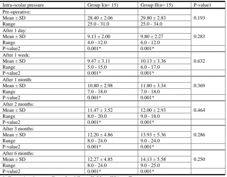
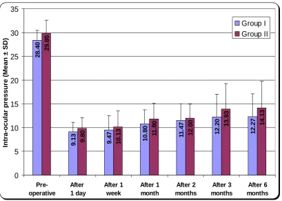
The mean preoperative IOP in the mitomycinc group improved from 28.40 ± 2.06mm Hg with2.40 ± 0.99 glaucoma medication to 12.27 ± 4.85mmgh with 0.87 ± 1.06 glaucoma medications at 6th months followup (p˂0.01).
The mean preoperative IOP in the bevacizumab group improved from 29.80 ± 2.83mm Hg with 2.67 ± 0.98 medication to 14.13 ± 5.58 mm Hg with 1.00 ± 1.13 glaucoma medications at 6th months follow up (p˂0.01) (Table 2).
Table (2): Intra-ocular pressure
Intra-ocular pressure Group I(n= 15) Group II(n= 15) P-value1 Pre-operative:
0.193
Mean ± SD 28.40 ± 2.06 29.80 ± 2.83
Range 25.0 - 31.0 25.0 - 34.0
After 1 day:
0.283
Mean ± SD 9.13 ± 2.00 9.80 ± 2.27
Range 4.0 - 12.0 6.0 - 12.0
P-value2 0.001* 0.001*
After 1 week:
0.632
Mean ± SD 9.47 ± 3.11 10.13 ± 3.36
Range 5.0 - 15.0 6.0 - 17.0
P-value2 0.001* 0.001*
After 1 month:
0.369
Mean ± SD 10.80 ± 2.98 11.80 ± 3.34
Range 7.0 - 18.0 7.0 - 18.0
P-value2 0.001* 0.001*
After 2 months:
0.464
Mean ± SD 11.47 ± 3.52 12.00 ± 2.93
Range 8.0 - 20.0 9.0 - 18.0
P-value2 0.001* 0.001*
After 3 months:
0.286
Mean ± SD 12.20 ± 4.86 13.93 ± 5.36
Range 8.0 - 24.0 9.0 - 24.0
P-value2 0.001* 0.001*
After 6 months:
0.250
Mean ± SD 12.27 ± 4.85 14.13 ± 5.58
Range 8.0 - 24.0 9.0 - 25.0
P-value2 0.001* 0.001*
1: Comparison between Group I and Group II (Mann-Whitney Test
198
Bleb morphology:
There were no significant differences found in bleb morphological features according to Indiana bleb grading scale in regard to height, extension, vascularity and leakage from the bleb (Tables 3,4 &5).
Table (3): Height
Height
Group I (n= 15)
Group II
(n= 15) P-value
No. % No. %
Flat 1 6.7 1 6.7 1.000
Low 5 33.3 8 53.3 0.269
Medium 6 40.0 3 20.0 0.427
High 3 20.0 3 20.0 1.000
Table (3) shows height of the bleb of studied groups. it shows that most cases are low to moderate height in group 1(low height 33.3%, moderate height 40% and group 11(low height 53.3%, moderate height 20%) with no statistical significant difference between studied groups.
Table (4): Extent
Extent
Group I (n= 15)
Group II
(n= 15) P-value
No. % No. %
0 – 1 1 6.7 2 13.3 0.543
1 – 2 3 20.0 2 13.3 0.624
2 – 4 8 53.3 9 60.0 0.713
> 4 3 20.0 2 13.3 0.624
Table (4) shows extent of the bleb according to clock hours. It shows no statistical significant difference between group 1 and group 11 according to the extent.
28.
40
9.
13 9.47 10.
80 1 1. 47 12. 20 12. 27 29. 80 9. 80 10.
13 11.
80 12. 00 13. 93 14. 13 0 5 10 15 20 25 30 35 Pre-operative After 1 day After 1 week After 1 month After 2 months After 3 months After 6 months Intra-ocula r pre ss
ure (Mean ±
SD)
199 Table (5) Vascularity
Vascularity
Group I (n= 15)
Group II
(n= 15) P-value
No. % No. %
Mild 10 66.7 10 66.7 1.000
Moderate 1 6.7 2 13.3 0.543
Avascular white 2 13.3 0 0.0 0.483
Avascular cystic 2 13.3 3 20.0 0.624
Table (5) shows that according to vascularity of the bleb most of cases are of mild vascularity in group 1 and group 11(66.7%) with no statistically significant difference between studied groups according to vascularity.
Two cases in group 1 develop avascular cystic bleb while 3 cases in group 11 develop avascular cystic bleb with IOP more than 21 mmgh after 4 weeks from surgery needling was done after 3 months one in each group failed to improve high IOP while other cases had IOP less than 21mmgh with 1-2 medications.
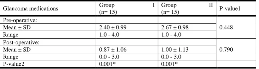
Anti-glaucoma medications:
Table (6): Glaucoma medications
Glaucoma medications Group I
(n= 15)
Group II
(n= 15) P-value1
Pre-operative:
0.448
Mean ± SD 2.40 ± 0.99 2.67 ± 0.98
Range 1.0 - 4.0 1.0 - 4.0
Post-operative:
0.790
Mean ± SD 0.87 ± 1.06 1.00 ± 1.13
Range 0.0 - 3.0 0.0 - 3.0
P-value2 0.001* 0.001*
Table (6):1- Comparison between Group I and Group II (Mann-Whitney Test)
2: Comparison between Pre-operative and Post-operative (Wilcoxon Signed Ranks Test) * Statistical significant difference (P < 0.05)
The number of postoperative antiglaucoma used in group 1 was lower than group 2 but with no statistically significant difference (p>0.05) between studied groups as regards pre-operative glaucoma, also in post-operative 6th months but in comparison between pre-operative and post-operative there is statistically significant difference (p<0.05) in group I, and group 11
Table (7): Success rate
Success rate
Group I (n= 15)
Group II
(n= 15) P-value
No. % No. %
Failure 2 13.3 3 20.0 0.624
Qualified 6 40.0 5 33.3 0.705
Complete 7 46.7 7 46.7 1.000
200
Figure(1) moderatevascularity, moderateblebheightwith extension from 2-4 clockhours in bivacizumab group
201
Figure(3)Avascular white, flat bleb in mitomycin group
Figure(4)lowblebheight, mildvascularitywith extension from 1-2 clockhours in bevacizumab group
DISCUSSION
The study compared the mean changes in IOP, bleb morphology, success and failure rate pre and postoperatively between MMC and Bevacizumab(avastin) groups.
There was no significant difference in postoperative IOP control between group I and group II despite that mean postoperative IOP was lower in group I than group II at each visit and at last follow up.
Bevacizumab group (group II) require more post-operativeantiglaucoma medications to control IOP than group I with no significant difference between the two groups.
Jaya Kaushika et al. reported comparable results to our study regarding IOP control between mitomycin C and bevacizumab groups at one year follow up as Themeanpreoperative IOP in the MMC group improved from 22.64 ± 0.90 mm Hg with 3.1 ± 0.8 glaucoma medications to 13.84 ± 0.73 mm Hg with 0.17 ± 0.38 glaucoma medications at one year (P < 0.001). The mean preoperative IOP in the bevacizumab group improved from 22.41 ± 1.08 mm Hg with 3.2 ± 0.9 glaucoma medications to 13.68 ± 0.79 mm Hg with 0.11 ± 0.32 glaucoma medications at one year (P < 0.001) with significant difference in peripheral bleb vascularity and peripheral non bleb vascularity in bevacizumab group but he uses Moorefield bleb grading scale for bleb morphological assessment instead of Indiana bleb grading scale in our study (9).
202
glaucoma for six months follow up and they found that there was a significant difference between the two groups regarding IOP control, there study uses bevacizumab concentration 2.5% instead of 1.25% in our study also cases with pseudo exfoliation (2ry open angle glaucoma) were in included in there study and the age above 18 years old while in our study only cases with primary open angle glaucoma were included and the age above 40 years old (10).
1-Year Comparative Follow-up Study done byJulide, Forty-two patients with primary open angle glaucoma underwent trabeculectomy, 21 with subconjunctival bevacizumab (2.5 mg/0.1 mL), and 21 with topical MMC (0.2 mg/mL for 3 min). He reported that the mean preoperative IOP in the bevacizumab group improved from 23.9±2.7mm Hg to 13.9±2.8mm Hg 12 months (P<0.001 and P<0.001, respectively) while the mean preoperative IOP in MMC group improved from 22.9±2.6mm Hg to 12.2±3.2mmHg at 12 months (P<0.001 and P<0.001, respectively) with no significant difference between the two groups, Encapsulated bleb was seen in 2 (10%) patients in MMC group and in 6 (29%) patients in bevacizumab group (P=0.23).
These results are in agreement with our study regarding IOP and bleb morphology.
The difference between this study and our study is that bevacizumab concentration is 2.5% in this study while bevacizumab concentration in our study is 1.25%; also bleb morphology grading was done using Moorefield bleb grading scale (11).
Twelve individuals (7 males; 5 females) with a diagnosis of POAG or CACG underwent trabeculectomy with subconjunctival bevacizumab with a recorded intraocular pressure (IOP) of more than 21 mmHg (between 10 AM and 12 PM),in Nonrandomized, open-label, prospective, interventional case series study done by Dilrajet al., with a mean follow-up of 182 days found that a successful trabeculectomy with respect to IOP control was observed in 11 eyes (92%), with an average IOP reduction of 52% with significant reduction of IOP postoperatively which is in agreementwith our results(12).
In summary, the present study found that bevacizumab was safe and effective as adjunctive to trabeculectomy in primary open angle glaucoma for 6th months follow up time and its effect was comparable to MitomycinC, the main limitations to this study were the small sample size and short follow up time so further studies on large number of populations for longer periods are needed.
References:
1. Schwartz, K., Budenz, D., Current management of glaucoma. Curr. Opin. Ophthalmol2004.15, 119–126.
2. 2-Hitchings RA, Grierson I. Clinicopathological correlation in eyes with failed fistulizing surgery. Trans
OphthalmolSoc U K 1983;103(Pt 1):84–88.
3. 3-Addicks EM, Quigley HA, Green WR, Robin AL. Histologic characteristics of filtering blebs in
glaucomatous eyes. Arch Ophthalmol 1983;101(5):795–798.)
4. 4-Justyna Jurkowska-Dudzińska, EwaKosior-Jarecka,and TomaszŻarnowski.Comparison of the use of 5
-FU and bevacizumab in primarytrabeculectomy: results at one year,* Clinical and Experimental
Ophthalmology, 8 May 2011
5. 5-CAT-152 0102 Trabeculectomy Study Group. A phase III study of subconjunctival human
anti-transforming growth factor beta (2) monoclonal antibody (CAT-152) to prevent scarring after first-time
trabeculectomy. Ophthalmology 2007;114: 1822–1830.)
6. 6-Lama PJ, Fechtner RD. Antifibrotics and wound healing inglaucoma surgery. SurvOphthalmol
2003;48(3):314 –346.
7. 7-Li J, Zhang YP, KirsnerRS.Angiogenesis in wound repair:angiogenic growth factors and the extracellular
203
8. 8-Li Z, Van Bergen T, Van de Veire S, etalInhibition of vascular endothelial growth factor reduces scar
formation after glaucoma filtration surgery. Invest Ophthalmol Vis Sci2009;50(11):5217–5225.)
9. 9-Jaya Kaushik, Jitendra Kumar Singh Parihar, Vaibhav Kumar Jain, SandeepGupta, PramodNath,
PrashantDurgapal&Jagat Ram,Efficacy of Bevacizumab Compared to
MitomycinCModulatedTrabeculectomy in Primary Open AngleGlaucoma: A One-Year Prospective
RandomizedControlledStudy,Current Eye Research.2016
10. 10-Naveed Nilforushan, Maryam Yadgari, ShahinKhosh Kish, And NarimanNassiriSubconjunctival
Bevacizumab Versus Mitomycin C Adjunctive to Trabeculectomy Am J Ophthalmol 2012;153:352–357
11. 11-Julide U. Akkan, D* and SemihCilsim, Role of Subconjunctival Bevacizumab as an Adjuvant to
Primary Trabeculectomy: A Prospective Randomized Comparative 1-Year Follow-up Study, J Glaucoma
2015;24:1–8).
12. 12-Dilraj S. Grewal, 1,2 Rajeev Jain, MD,1 Harsh Kumar, 1 Satinder Pal Singh Grewal, Evaluation of
Subconjunctival Bevacizumab as an Adjunct to Trabeculectomy, Ophthalmology 2008;115:2141–2145 ©