R E V I E W
Open Access
Dynamic foot function as a risk factor for lower
limb overuse injury: a systematic review
Geoffrey J Dowling
1, George S Murley
1,2*, Shannon E Munteanu
1,2, Melinda M Franettovich Smith
3, Bradley S Neal
4,5,
Ian B Griffiths
4, Christian J Barton
2,4,5,6and Natalie J Collins
7Please see related article: http://www.jfootankleres.com/content/7/1/55
Abstract
Background:Dynamic foot function is considered a risk factor for lower limb overuse injuries including Achilles tendinopathy, shin pain, patellofemoral pain and stress fractures. However, no single source has systematically appraised and summarised the literature to evaluate this proposed relationship. The aim of this systematic review was to investigate dynamic foot function as a risk factor for lower limb overuse injury.
Methods:A systematic search was performed using Medline, CINAHL, Embase and SportDiscus in April 2014 to identify prospective cohort studies that utilised dynamic methods of foot assessment. Included studies underwent methodological quality appraisal by two independent reviewers using an adapted version of the Epidemiological Appraisal Instrument (EAI). Effects were expressed as standardised mean differences (SMD) for continuous scaled data, and risk ratios (RR) for nominal scaled data.
Results:Twelve studies were included (total n = 3,773; EAI 0.44 to 1.20 out of 2.00, representing low to moderate quality). There was limited to very limited evidence for forefoot, midfoot and rearfoot plantar loading variables (SMD 0.47 to 0.85) and rearfoot kinematic variables (RR 2.67 to 3.43) as risk factors for patellofemoral pain; and plantar loading variables (forefoot, midfoot, rearfoot) as risk factors for Achilles tendinopathy (SMD 0.81 to 1.08). While there were significant findings from individual studies for plantar loading variables (SMD 0.3 to 0.84) and rearfoot kinematic variables (SMD 0.29 to 0.62) as risk factors for‘non-specific lower limb overuse injuries’, these were often conflicting regarding different anatomical regions of the foot. Findings from three studies indicated no evidence that dynamic foot function is a risk factor for iliotibial band syndrome or lower limb stress fractures. Conclusion:This systematic review identified very limited evidence that dynamic foot function during walking and running is a risk factor for patellofemoral pain, Achilles tendinopathy, and non-specific lower limb overuse injuries. It is unclear whether these risk factors can be identified clinically (without sophisticated equipment), or modified to prevent or manage these injuries. Future prospective cohort studies should address methodological limitations, avoid grouping different lower limb overuse injuries, and explore clinically meaningful representations of dynamic foot function.
Keywords:Biomechanics, Plantar pressures, Kinematics, Prospective studies, Musculoskeletal diseases, Review
* Correspondence:[email protected] 1
Department of Podiatry, Faculty of Health Sciences, La Trobe University, Melbourne, Australia
2
Lower Extremity and Gait studies program, Faculty of Health Sciences, La Trobe University, Melbourne, Australia
Full list of author information is available at the end of the article
JOURNAL OF FOOT AND ANKLE RESEARCH
© 2014 Dowling et al.; licensee BioMed Central. This is an Open Access article distributed under the terms of the Creative Commons Attribution License (http://creativecommons.org/licenses/by/4.0), which permits unrestricted use, distribution, and reproduction in any medium, provided the original work is properly credited. The Creative Commons Public Domain Dedication waiver (http://creativecommons.org/publicdomain/zero/1.0/) applies to the data made available in this article, unless otherwise stated.
Dowlinget al. Journal of Foot and Ankle Research (2014) 7:53
Introduction
Overuse injuries of the lower limb associated with inten-sive weight bearing exercise are a significant problem for athletes and military recruits, with estimated incidence of running-related injuries reported to range from 20% to 79% [1]. Lower limb overuse injuries are generally recognised as having multifactorial aetiologies [2]. Some of the most common injuries, such as Achilles tendino-pathy, medial tibial stress syndrome, patellofemoral pain and lower limb stress fractures, are reported to be more prevalent in those with altered foot function [3,4].
The potential mechanisms linking variations in dynamic foot function with lower limb overuse injury may be related to altered lower limb biomechanics and subse-quent changes in tissue stress [5]. This is supported by laboratory-based research using uninjured participants, which suggests that variations in foot posture (flat- and normal-arched feet) are associated with systematic dif-ferences in lower limb kinematics [6-8], kinetics [4,9,10], muscle function [11-16] and tendon morphometry [17].
While laboratory-based research is important for un-derstanding potential mechanisms linking foot function and lower limb overuse injury, field-based prospective studies are required to determine whether foot function is a risk factor for lower limb overuse injury. Our accompanying systematic review [18] found that static measures indicating greater foot pronation were associ-ated with an increased risk of patellofemoral pain and medial tibial stress syndrome. However, the small effects suggest that static measures may not adequately represent dynamic foot function. A substantial number of prospective studies have utilised a variety of meas-urement techniques in order to quantify dynamic foot function and its relationship with lower limb overuse injury [19-46]. However, it is unclear if there are con-sistent findings across different measures, or whether particular foot function characteristics are risk factors for specific overuse injuries. Enhanced knowledge re-garding this may lead to the development of targeted preventative strategies.
Therefore, the aim of this systematic review was to: (i) identify and appraise the current evidence for the prospective link between dynamic foot posture and lower limb overuse injury; and (ii) provide guidance for future research in this area. This review represents the second component of a two-part systematic review on foot posture-related risk factors for lower limb overuse injury.
Methods
The systematic review protocol was developed in consultation with guidelines provided by the Preferred Reporting of Systematic Reviews and Meta-Analysis (PRISMA) Statement [47].
Search strategy
MEDLINE, CINAHL, Embase and SPORTDiscus were searched from inception until April 2014. Medical Subject Headings (MeSH) were exploded to include relevant subheadings, in addition to keywords specific to the research question (Additional file 1). The search was limited to adult human participants and English language publications. To ensure identification of all relevant studies, reference lists of appropriate narrative and systematic reviews were hand searched, and discus-sion with field experts (e.g. physiotherapists, podiatrists) was conducted regarding known important publications. A cited reference search for each included paper was also completed in Google Scholar.
Eligibility criteria
All studies identified by the search strategy were exported to Endnote version X5 (Thomson Reuters, Philadelphia), by a single investigator (GJD). Abstracts and then full text versions were reviewed by two authors (GJD, MMFS) to determine eligibility. Discrepancies were resolved in consultation with a third reviewer (GSM). Initial eligi-bility criteria were: (i) prospective cohort study design; (ii) quantitative measurement of foot posture or func-tion at baseline (static or dynamic); and (iii) prospect-ive collection of specific or non-specific lower limb overuse injury surveillance data over a specified time period. Specific lower limb overuse injuries were defined as injuries with a single diagnosis, while non-specific lower limb overuse injuries included injuries without a specific diagnosis or where multiple overuse types of injuries were pooled by the study reviewed. After retrieval of studies that fulfilled the initial eligi-bility criteria, suitable studies were separated into those that investigated dynamic measures of foot func-tion (i.e. measured during walking or running), and those that investigated static measures of foot posture. This review focused on dynamic measures as risk factors, while static measures are addressed in the accompanying review [18].
Quality assessment
to excellent intra-rater (Kappa coefficient range 52 to 60), and inter-rater reliability (Kappa coefficient = 90% [95% CI; 87 to 92%]) [48]. For the purpose of this re-view, the wording of all 43 items was modified slightly to improve clarity and rater interpretation. No items were removed or modified, in order to maintain validity (Additional file 2).
Two raters (GJD, NJC) independently evaluated each study while blind to author and publication details. For any discrepancies in assessment of items between the two raters, a meeting occurred and consensus was achieved. To evaluate the overall quality of the studies, average scores across the 43 items were calculated, with a max-imum possible score of two (i.e. as individual items are scored‘0’,‘1’or‘2’, the maximum‘average’score across 43 items is two). A ranking system was used to evaluate the quality of evidence, whereby studies were classified as being high (EAI≥1.4), moderate (EAI 1.1 to <1.4), or low quality (EAI < 1.1) [47].
Data management
Two investigators (GJD, GSM) extracted data regarding study characteristics, including publication details (year, author, country), participant characteristics (number of injured and uninjured, age, sex, inclusion and exclusion criteria, population [i.e. military]) and study methods (dynamic foot function measurement, examiner details, injury outcome, duration of study and covariates inves-tigated). To facilitate calculation of effects, means and standard deviations (SD) were extracted for injured and uninjured participants for continuous foot function variables, while raw counts were extracted for nominal variables.
Where appropriate data was not provided in the pub-lication, authors were contacted with a request to pro-vide additional data. Where studies described specific variables but did not publish data, it was recorded as
‘not reported’(NR) and, for the purpose of the analysis, assumed that the variable investigated was not signifi-cantly different between the injured and the uninjured population.
Statistical methods
Inter-rater reliability of the raters’EAI scores was evalu-ated using a descriptive analysis. Differences between rater scores for “Yes”, “Partial”, “No”, and “Unable to determine” were calculated, with a difference of zero indicating perfect agreement and a difference of 1 indi-cating near perfect. The rating “not applicable” was excluded from analysis because no interpretation was required for this rating.
For continuous foot function variables, standardised mean differences (SMD) were calculated as the dif-ference between injured and uninjured group means,
divided by the pooled standard deviation [49]. SMDs and 95% confidence intervals (CI) were calculated using the‘Effect Size Calculator’from the Centre for Evaluation and Monitoring [50]. Interpretation of the SMD was based on previous recommendations, where > 1.2 was considered large, 0.6 to 1.2 moderate, and < 0.6 small [51]. For nominal scaled foot function variables, risk ratios (RR) and 95% CI were calculated using the
‘Confidence Interval Calculator’ from the Physiother-apy Evidence Database (PEDro) [52]. This was repre-sented as the number of participants with lower limb overuse injury in the group with the associated factor (e.g. delayed time to peak force), divided by participants with lower limb overuse injury in the group without the associated factor. A RR > 1.0 indicated that the lower limb overuse injury was more likely to be found in participants with the risk factor present. A small effect was indicated by a RR≥2.0, and a large effect≥4.0 [53]. Effects were considered statistically significant if the associated 95% CI did not contain zero for the SMD, or one for RR.
Evidence-based recommendations
In order to provide recommendations based on statis-tical findings, while incorporating the methodological quality of included papers, a scale regarding levels of evidence was utilised, based on previous work by van Tulder et al. [54].
Strong evidence: pooled results derived from three or more studies, including a minimum of two high quality studies that are statistically homogenous; may be asso-ciated with a statistically significant or non-significant pooled result.
Moderate evidence: statistically significant pooled results derived from multiple studies that are statisti-cally heterogeneous, including at least one high quality study; or from multiple moderate quality or low quality studies which are statistically homogenous.
Limited evidence:results from one high quality study or multiple moderate or low quality studies that are sta-tistically heterogeneous.
Very limited evidence: results from one moderate quality study or one low quality study.
No evidence: pooled results insignificant and derived from multiple studies regardless of quality that are sta-tistically heterogeneous.
Results Search results
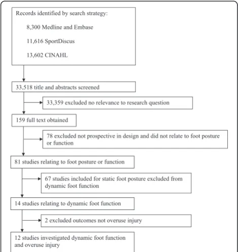
Across the two parts of this systematic review (static foot posture and dynamic foot function), a total of 33,518 citations were retrieved from the electronic database search. Following the sequential review of titles, abstracts and full texts, as well as removing studies
that were not prospective cohort studies, 80 studies were eligible (Figure 1). Of these, 12 studies investigated dynamic foot function variables, and were included in this part of the review [27,29,35,38-46]. Due to incon-sistencies in outcomes measured, pooling of data was not possible.
Quality assessment
Quality scores ranged from 0.44 to 1.20 (out of a possible total score of 2.00) (Additional file 3). With the exception of one moderate quality study [43], all studies were rated as low quality [27,29,35,37-42,44-46]. In terms of inter-rater reliability across 35 items included in the quality assessment, 24 items had perfect or near perfect agreement between raters. That is, these items were awarded the same score or there was a maximum of one point difference in scoring. For a further 10 items, the raters had near perfect agreement for 80% of the articles reviewed. Item 10 (‘reporting of adverse effects’) displayed the lowest agreement, with perfect or near perfect agreement for only 5/12 studies. Per-centage agreement across the 35 items ranged from 17 to 100%.
All studies clearly reported the aim and objective (item 1) and that foot posture was measured prospect-ively before longer-term follow up of injury (item 28) [27,29,35,38-46]. Eleven studies clearly defined the assessment of foot function (item 2) [27,29,35,38-45] and eight studies clearly defined the lower limb overuse
injury of interest (item 3) [29,35,39,41-45]. None of the included studies provided an adequate description of all intrinsic or extrinsic covariates or how these were adjusted for in the analysis (items 11, 12, 13, 36 and 37) (e.g. footwear worn, skill level or playing surface). Fur-thermore, no study provided an adequate report of the reliability and validity of foot function or injury out-come measurement of interest (items 25, 26, 31 and 32). Three studies provided an adequate standardisa-tion procedure for assessing foot funcstandardisa-tion (item 27) [39,42,45] and five studies reported standardisation of injury outcome (item 33).
Clear reporting of all data was present in four studies (items 14 and 15) [29,39,40,46]. However, the remaining seven studies primarily reported data only for significant relationships [27,35,38,42-45], while one study did not report any data [41]. Only one study reported effects for all results (odds or risk ratios) (item 16) [29]. With re-spect to generalisability of results, nine studies received a score of“Partial”(item 43) as results were deemed to be applicable to similar population groups to those investigated [29,35,38-45].
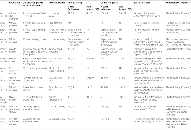
Study characteristics
The 12 included studies incorporated a total of 3,773 participants. Table 1 presents a summary of study char-acteristics. The participant population varied, with five studies investigating military personal [27,29,39,41,43], five studies investigating runners [38,40,42,44,46], and two studies investigating cohorts of physical therapy students [35,45]. The types and incidence of lower limb overuse injuries reported were: tibial and femoral stress fractures, 8.7 to 10.0% [29,39]; iliotibial band syndrome, 9.4% [29,40]; patellofemoral pain, 4.0 to 17.0% [27,29,42-44]; medial tibial stress syndrome, 7.9% [41]; Achilles tendinopathy, 5.1 to 15.8% [29,44]; and non-specific lower limb overuse injuries, 14.0 to 20.6% [35,38,45].
Prior to prospective investigation, eight of the 12 studies investigated dynamic plantar loading (i.e. plantar pressure) [29,35,38,41-45], six investigated kinematic variables [27,35,39,40,45,46] and one investigated rearfoot joint moments [45] (Additional files 4 and 5). A large number of plantar pressure variables were evaluated. Baseline mea-sures of foot function were commonly performed during unshod gait [27,29,35,38,39,41-44], although four stud-ies obtained measures during shod gait [29,40,45,46]. Gait was assessed during treadmill walking at 5 kilome-ters per hour [27,39], or during overground walking or running at a self-selected speed [29,35,38,40-46]. Only four studies that investigated overground running re-ported mean values of the speed at which participants were observed, ranging between 3.3 to 3.7 metres per second [35,40,45,46].
8
1
Table 1 Summary of study characteristics
Population Observation period (activity, duration)
Injury outcome Injured group Uninjured group Gait assessment Foot function measure
N total (n females)
Age (mean ± SD)
N total (n females)
Age (mean ± SD)
Hesar et al., [38]
Athletics club members
3 running sessions/week; 10 weeks
LL overuse injury
27 (22) 41 ± 8 104 (89) 39 ± 11 Barefoot; 15 m runway;
self-selected running speed
Plantar loading (Footscan)
Hetsroni et al., [27]
Military personal
4 month basic training course
Patellofemoral pain
NR NR NR NR Barefoot; treadmill running
at 5 km/hr
Rearfoot kinematics (Ariel Dynamics Inc.)
Hetsroni et al., [39]
Military personal
4 month basic training course
Tibial and femoral stress fractures
Dependent on outcome variable investigated
NR Dependent on
outcome variable investigated
NR Barefoot; treadmill running
at 5 km/hr
Rearfoot kinematics (Ariel Dynamics Inc.)
Kaufman et al., [29]
Military personal
25 week training course LL overuse injury Dependent on outcome variable investigated
NR Dependent on
outcome variable investigated
NR Boots and barefoot;
self-selected walking speed (no mean or range presented)
Plantar pressure ratios– dynamic arch index (<4.14 cavus, >8.10 planus)
Noehren et al., [40]
Female runners
Individual non-specified running programs over a 2 year period
Iliotibial band syndrome
18 (18) 26 Dependent on
outcome variable investigated
28 ‘Standard running shoe’; running along a 25 runway at a speed of 3.7 m/s
Rearfoot kinematics (Vicon)
Noehren et al., [46]
Female runners
Individual non-specified running programs over a 2 year period
Patellofemoral pain
15 (15) 27 ± 10 15 (15) 27 ± 10 ‘Standard running shoe’(Nike,
Pegasus); running along a 25 run way at a speed of 3.7 m/s
Rearfoot kinematics (Vicon)
Sharma et al., [41]
Male infantry recruits
26 week military training Medial tibial stress syndrome
37 (0) NR 239 (0) NR Barefoot; self selected walking
speed (no mean or range presented)
Plantar loading (Footscan)
Thijs et al., [42]
Novice recreational runners
10 week start to run programme
Patellofemoral pain
17 (16) 39 ± 10 85 (NR) 37 ± 9 Barefoot; walking at a self-chosen,
moderate velocity (no mean or range presented)
Plantar loading (Footscan)
Thijs et al., [43]
Military personal
6 week basic military training
Patellofemoral pain
36 (19) 19 ± 2 48 (NR) 19 ± 1 Barefoot; walking at a self-chosen,
moderate velocity (no mean or range presented)
Plantar loading (Footscan)
Van Ginckel et al., [44]
Novice runners
10 week start to run programme
Achilles tendinopathy
10 (2) 38 ± 11 53 (45) 40 ± 9 Barefoot; self-selected jogging
pace (no mean or range presented)
Plantar loading (Footscan)
Willems et al., [35]
Physical education students
University physical education course
LL overuse injury 46 (29) NR 167 (NR) NR Barefoot; 3.3 m/s within a
boundary of 0.17 m/s
Plantar loading (Footscan)/ ankle, knee and hip kinematics and kinetics (Proreflex)
Willems et al., [45]
Physical education students
University physical education course
LL overuse injury 46 (29) NR 167 (NR) NR ‘Neutral running shoe’; 3.3 m/s
within a boundary of 0.17 m/s
Plantar loading (Footscan)/ ankle, knee and hip kinematics and kinetics (Proreflex)
LL = lower limb; NR = not reported.
Dynamic foot function variables as risk factors for lower limb overuse injuries
We found evidence supporting foot function as a risk factor for lower limb overuse injuries. There waslimited to very limited evidencesupporting (i) plantar loading and kinematic variables as risk factors for patellofemoral pain;
(ii) plantar loading variables for Achilles tendinopathy; and (iii) plantar loading and kinematic variables for vari-ous non-specific lower limb overuse injuries. This is illus-trated in Figure 2. For a complete reference of significant and non-significant findings for all injuries investigated, refer to Additional files 4 and 5.
A)
B)
C)
D)
Patellofemoral pain
Plantar loading variables
There was limited evidence for plantar loading vari-ables as a risk factor for patellofemoral pain, see Figures 2A and B. Participants who developed patello-femoral pain had earlier relative time to peak force in the lateral heel (SMD −0.56, 95% CI −1.09 to −0.37) and greater peak force in the second (0.65, 0.12 to 1.17) and third (0.60, 0.07 to 1.12) metatarsal regions during running [42]. Those who developed patellofe-moral pain also demonstrated greater lateral centre of pressure (COP) displacement (−0.47, −0.90 to −0.03) and lower maximal displacement velocity of the med-iolateral COP (−0.85,−1.29 to −0.39) during the ‘ fore-foot contact phase’of walking [43].
Kinematic variables
There was very limited evidence for kinematic vari-ables as a risk factor for patellofemoral pain, see Figure 3A. A single study [27] investigated rearfoot kinematics, reporting opposite findings for the left and right sides. Greater pronation velocity on the left was a significant risk factor for patellofemoral pain development (quartile 4 versus quartile 3: RR 3.43 95% CI 1.32 to 8.96). Conversely, reduced pronation velocity of the right foot was a significant predictor of patellofemoral pain development (quartile 4 versus quartile 3: 0.38, 0.15 to 0.92). The authors did not specify whether the outcome (i.e. greater or reduced pronation velocity) was related to the side affected by patellofemoral pain.
Achilles tendinopathy
Plantar loading variables
There was very limited evidence for plantar loading variables as a risk factor for mid-portion Achilles ten-dinopathy, evaluated in one study [44], see Figure 2C. Participants who developed Achilles tendinopathy ex-hibited significantly earlier time to peak force in the medial heel (SMD −0.716, 95% CI −1.39 to −0.02) and lateral heel (−1.08, −1.77 to −0.37), and delayed time to initial contact in the second metatarsal region (−1.00, −1.69 to −0.29). They also demonstrated greater peak force (0.84, 0.14 to 1.52) and a higher absolute force time integral (0.81, 0.11 to 1.49) in the fifth metatarsal region. In addition, those that developed Achilles tendi-nopathy displayed less anterior-posterior center of force (COF) displacement for the whole foot (−0.95,−1.64 to −0.25), greater laterally directed force in the forefoot at
‘forefoot flat’(−0.88,−1.57 to−0.18) and a more poster-ior COF position at ‘last foot contact’ (−0.95, −1.63 to −0.24). During forefoot push-off, those that devel-oped Achilles tendinopathy displayed more posterior COF displacement (−0.75,−1.43 to−0.05).
Non-specific lower limb overuse injuries
There waslimited evidence for plantar loading variables as a risk factor for non-specific lower limb overuse injur-ies, see Figure 2D.
Plantar loading variables - discrete plantar regions
Participants who developed a non-specific lower limb overuse injury exhibited delayed initial lateral heel contact
Figure 3Kinematic risk factors for: (A) patellofemoral pain; and (B) non-specific injuries.
(SMD 0.60, 95% CI 0.35 to 0.86) and terminal heel contact in the second and third metatarsal region (0.43, 0.18 to 0.68; 0.37, 0.12 to 0.62, respectively) [35]. In the fifth metatarsal region, an increase in peak force (0.52, 0.09 to 0.95 [38]) and absolute force-time integral (0.57, 0.14 to 1.00 [38]), as well as delayed time until initial contact (0.32, 0.07 to 0.57 [35]) were risk factors for non-specific overuse injury. However, contrary to these find-ings, Willems and colleagues reported lower fifth meta-tarsal region peak pressure (−0.44,−0.70 to−0.19) [35] and absolute impulse (−0.31,−0.56 to−0.05 [45]; −0.42, −0.67 to −0.17 [35]) in those who developed non-specific lower limb overuse injuries.
Plantar loading variables - time-specific gait events
At first foot contact, participants who developed a non-specific lower limb overuse injury had a more laterally directed COP (SMD−0.47, 95% CI−0.73 to −0.22) [45] and a more anterior COP position (0.31, 0.06 to 0.56) [35]. At first metatarsal contact, participants who devel-oped a non-specific lower limb overuse injury had greater lateral force as indicated by three mediolateral regional force ratios (−0.55, −0.97 to −0.12;−0.57,−0.99 to−0.13;−0.59,−1.02 to−0.16) [38]. At forefoot flat, there was a lower velocity of the medio-lateral (−0.64, −1.07 to −0.21) and anterior-posterior displacement of the COF (−0.46, −0.88 to−0.03); and a more anterior COF position (0.61, 0.18 to 1.04) in those that developed non-specific lower limb overuse injuries [38]. Willems et al. [35,45] reported greater medial pressure as indi-cated by two pressure ratios (0.47, 0.22 to 0.72 [35]; 0.40, 0.09 to 0.59 [45]) and a more medially directed COP (0.38, 0.13 to 0.63) [35]. At heel-off, participants who developed a non-specific lower limb overuse injury had a more laterally directed COF (−0.70, −1.13 to −0.27) [38]. Contrary to this finding, Willems et al. [35,45] reported greater medial pressure, as indicated by two pressure ratios (0.33, 0.07 to 0.58 [35]; 0.33, 0.08 to 0.58 [45]) in those who developed overuse injuries. At last foot contact, participants who developed a non-specific overuse injury had a more laterally directed COP (−0.81, −1.07 to −0.55) [35], and more posterior COP position (−0.53,−0.79 to−0.28) [35].
Plantar loading variables (phase-specific gait events)
During the initial contact phase, participants who devel-oped a non-specific lower limb overuse injury had a more laterally directed plantar force (SMD−0.43, 95% CI−0.85 to −0.001) [38]. Contrary to this finding, Willems et al. [45] reported a more medially directed pressure, as indi-cated by one pressure ratio (0.57, 0.31 to 0.82) and a more medially directed COP displacement (0.61, 0.36 to 0.86).
Hesar et al. [38] found that, during the forefoot con-tact phase, participants who developed a non-specific
lower limb overuse injury had greater lateral COF dis-placement (−0.84,−1.27 to−0.40). Contrary to this find-ing, Willems et al. [35,45] reported a greater medial pressure (0.54, 0.29 to 0.79) [35] and a more medially directed COP displacement (0.58, 0.33 to 0.83 [35]; 0.31, 0.05 to 0.56 [45]).
Participants who developed a non-specific lower limb overuse injury had a more laterally directed COF dis-placement (−0.61, −1.03 to −0.17 [38]) during the foot flat phase, and a more medially directed COF during the forefoot push off phase (0.52, 0.09 to 0.94 [38]). Contrary to this latter finding, Willems et al. [35,45] reported a more laterally directed pressure during forefoot push off, as indicated by one pressure ratio (−0.35,−0.60 to−0.09) [45], and a more laterally directed COP displacement (−0.84,−1.09 to−0.58 [35];−0.37,−0.62 to−0.12 [45]).
Kinematic variables
There was limited evidence for kinematic variables as a risk factor for non-specific lower limb overuse injuries, see Figure 3B. For the rearfoot segment, participants who developed a non-specific lower limb overuse injury exhibited a greater maximal eversion position (SMD 0.37, 95% CI 0.12 to 0.62) [35], eversion excursion (0.36, 0.10 to 0.61 [35]; 0.31, 0.06 to 0.56 [45]), mean eversion velocity (0.37, 0.12 to 0.62) [35], time to maximal ever-sion (0.39, 0.14 to 0.64) [45], maximal everever-sion velocity (0.39, 0.14 to 0.64 [35]; 0.29, 0.03 to 0.54 [45]), mean inversion velocity (0.44, 0.18 to 0.69) [35], maximal re-inversion velocity (0.41, 0.16 to 0.66) [45], and mean re-inversion velocity (0.31, 0.06 to 0.56) [45].
In the forefoot segment, participants who developed a non-specific lower limb overuse injury exhibited greater maximal abduction velocity (0.62, 0.37 to 0.88) [35] and abduction excursion (0.36, 0.10 to 0.61 [35]; 0.31, 0.06 to 0.56 [45]). One study derived a three-dimensional pronation angle from eversion, abduction and dorsi-flexion excursions, and reported that participants who developed a non-specific lower limb overuse injury ex-hibited greater three-dimensional pronation excursion (0.49, 0.23 to 0.74) [45].
Other lower limb overuse injuries
There was no evidence supporting dynamic foot func-tion as a risk factor for any other lower limb overuse injury. Non-significant effects were found for iliotibial band syndrome [29,40] and stress fractures [29].
Discussion
dynamic foot function are associated with an increased risk of patellofemoral pain, Achilles tendinopathy and non-specific lower limb overuse injury [35,38,42-45]. Notably, significant findings reported across the studies had small to moderate effect sizes, and many 95% confi-dence intervals included zero, indicating non-significant findings.
Plantar pressure patterns associated with patellofe-moral pain differed for walking and running gait. Risk factors in walking gait included greater lateral COP dis-placement and lower maximal disdis-placement of the medio-lateral COP [42], whereas for running gait risk factors included earlier time to peak force in the lateral heel and greater peak force in the second and third metatarsal region [43]. While it is difficult to suggest a mechanism linking these plantar pressure differences with the development of patellofemoral pain, Thijs et al. [42,43] speculated that these findings may indicate a resultant reduction in foot pronation during the loading phase of gait, and subsequent reduction in shock attenu-ation at the foot. This could increase transfer of ground reaction forces to more proximal structures, such as the patellofemoral joint.
Plantar pressure patterns associated with Achilles tendinopathy were evident from one study investigating jogging gait, and included earlier time to peak force in the lateral heel, less posterior COF displacement/more posterior COF position, greater laterally directed force and delayed time to initial contact in the second meta-tarsal region [44]. Van Ginkel and colleagues [44] spec-ulated that these findings may indicate a more lateral foot roll-over following heel strike and diminished forward force transfer from the rearfoot to the forefoot. It is plausible that differences in force transfer across the foot may lead to altered loading of the Achilles tendon and contribute to injury, but this requires fur-ther evaluation.
Another consideration is that increased lateral loading at the foot is an adaptive response to proximal mechan-ics that increase medial lower limb loading. Prospective studies have shown that increased hip adduction during overground running [46] and increased hip internal rota-tion when landing from a drop jump [22] are risk factors for the development of patellofemoral pain. Further-more, cross-sectional studies have reported deficits in neuromuscular control of the hip in those with patello-femoral pain [55-61] and Achilles tendinopathy [62,63]. Further research is required to better understand the relationship between proximal and distal mechanics during gait, and risk of overuse injury development.
In contrast to evidence we found regarding plantar pressure, we found very few kinematic risk factors for lower limb overuse injuries. Our search strategy iden-tified only one study that investigated kinematic risk
factors for patellofemoral pain, which presented contra-dictory findings, no prospective studies that investigated kinematic risk factors for Achilles tendinopathy and two studies that reported differences in rearfoot eversion and forefoot abduction as risk factors for non-specific injuries [35,45]. Whilst cross-sectional findings indicate differences in foot kinematics in people with patellofemoral pain [64] and Achilles tendinopathy [49], we found a lack of prospective kinematic data to indicate the temporal relationship between foot kinematics and overuse in-jury. Thus, at this time it is difficult to draw conclu-sions as to whether altered foot kinematics is a clear risk factor for lower limb overuse injuries.
In addition to necessitating more kinematic studies, consideration needs to be given to the method of meas-uring foot kinematics. Considering that overuse injuries generally involve cumulative exposure to load, it is plausible that those who develop overuse injuries dem-onstrate subtle kinematic differences that are not detect-able by current kinematic measures. This is supported by previous findings regarding a lack of biomechanical coupling of plantar pressure indices and angular move-ments recorded between the calcaneus and the tibia [65]. Further studies are required to increase under-standing of this relationship, which could be achieved using more sophisticated three-dimensional and multi-segment foot modeling techniques, and more clinically applicable measures of foot function.
Not surprisingly, it was difficult to identify a system-atic pattern of plantar loading and kinemsystem-atic risk factors for the category of ‘non-specific injuries’. For example, significant risk factors were evident for greater lateral and medial directed COP, as well as increases and decreases in pressure-related outcomes in the fifth metatarsal region. While these findings indeed add evi-dence of a relationship between dynamic foot function and lower limb injury, the nature of the relationship is unpredictable, and likely relates to the variability of injuries evaluated under the term ‘non-specific injur-ies’. Therefore, with the advancement and availability of diagnostic algorithms and imaging for lower limb injury, future research should avoid pooling all injur-ies, and instead focus efforts on exploring conditions that are discrete and well-defined. This is likely to enhance identification of injury-specific risk factors.
Interestingly, we found no evidence that dynamic foot function is a risk factor for iliotibial band syndrome or lower limb stress fractures including the foot. Findings from Noehren et al. [46] indicated that aberrant hip me-chanics may be a stronger risk factor for iliotibial band syndrome than dynamic foot function. They reported that increased hip adduction during running, but not rearfoot eversion, was a predictor of patellofemoral pain development in a cohort of 400 female runners [46].
This is logical given the proposed mechanism of iliotibial band syndrome, where increased tension on the iliotibial band compresses the lateral femoral epicondyle [46].
The lack of foot specific injuries (e.g. plantar fasciitis, metatarsal stress fracture) associated with dynamic foot function is another unexpected finding. Although Kaufman et al. [29] reported that dynamic pes planus in shoes, measured as the ratio of midfoot contact area to total contact area, was a significant predictor of lower limb stress fracture (one third of which involved the foot), our effect size calculations were not significant. This is because the authors set significance at 0.10, whereas we used the more conventional alpha of 0.05. Because of the large number of variables evaluated, this is the more conservative approach to reduce the risk of type II error. An earlier study also reported that pronated foot type (i.e. static foot posture) was a significant risk factor for metatarsal stress fractures, while a supinated foot type was a risk factor for tibial and femoral stress frac-tures [66]. However, the static x-ray measure of foot type used in this study may not correlate with dynamic foot function. It is plausible that lower limb stress frac-tures are more a function of bony overload due to the application of external loads, rather than the biomech-anical characteristics of the foot. This is in part sup-ported by the use of military cohorts in both studies [29,66]. The influence of dynamic foot function on the development of lower limb stress fractures should be investigated in civilian populations to ascertain this.
Plantar loading variables were the most abundant risk factor identified for lower limb injury, albeit a relatively low risk with small to moderate effect sizes. In terms of the clinical application of these findings, it is difficult to map the plantar pressure risk factors to specific static foot types. De Cock et al. [67] reported that participants with low arched feet had a more laterally directed COP across the gait cycle. This is consistent with our plantar pressure findings relating to patellofemoral pain and Achilles tendinopathy. Conversely, Wong et al. [68] investigated the effect of foot morphology on center-of-pressure excursion during barefoot walking. Their find-ings indicated that more supinated foot types displayed a larger area of lateral COP excursion, and, conversely, more pronated foot types displayed a smaller area of lateral COP excursion. However, these findings were taken over the entire gait cycle, rather than the discrete phases evaluated in the prospective studies included in this review. In light of the volume of studies that use plantar pressure measures to evaluate dynamic foot function, there is a clear need for further studies to investigate methods of transferring plantar pressure information to clinically relevant measures.
Nevertheless, having some limited knowledge of the pattern of plantar loading risk factors may serve to
inform the design of new and existing interventions that may redistribute or counter-balance plantar loading pat-terns observed in people at risk of injury. For example, arch-contoured foot orthoses alter plantar pressure sys-tematically by reducing pressure in the forefoot and heel regions, and redistributing pressure to the midfoot [69]. With this in mind, there is evidence from pooled data from randomised clinical trials (RCTs) that foot orthoses are effective in preventing lower limb overuse injuries [70], as well as evidence from high-quality RCTs that foot orthoses reduce symptoms associated with patello-femoral pain [71]. In the absence of evidence regarding kinematic effects, our findings suggest that foot orthoses may exert their clinical effects by redistributing plantar pressure (i.e. alter the magnitude, location and temporal patterns of reaction forces at the foot-orthosis interface). However, this requires further investigation.
Whilst this review has highlighted specific measures of dynamic foot function that are risk factors for lower limb overuse injuries, there are several limitations to the identification of these risk factors in a clinical practice setting. Firstly, while findings indicate the direction of altered plantar loading that may increase the risk of development of Achilles tendinopathy or patellofemoral pain, there are no reported thresholds of when an individual is deemed at risk (e.g. peak force in forefoot region exceeding 150 N). Future investigations are re-quired to establish clinical guidelines and screening criteria for these risk factors. Secondly, the assessment of plantar pressures and three-dimensional kinematics requires expensive and sophisticated equipment that is not readily available in clinical practice settings, as well as specialised training in performing and processing these measurements. Future studies should investigate the translation of these laboratory-based measures to clinically applicable measures.
of included studies was generally poor. This was largely related to inadequate reporting of foot function measures, covariates, and non-significant results. Thus, the findings should be considered with this in mind. In order to enhance the overall quality of research in this field, future prospective studies should comply with published guidelines for minimum standards of reporting [74].
Conclusion
This systematic review identified very limited evidence, with small to moderate effect sizes, that dynamic foot function during walking and running is a risk factor for patellofemoral pain, Achilles tendinopathy, and non-specific lower limb overuse injuries. More lateral plantar loading patterns were found to be risk factors for patel-lofemoral pain and Achilles tendinopathy. Findings from three studies indicate that there is no evidence that dynamic foot function is a risk factor for iliotibial band syndrome or lower limb stress fractures. At present, it is unclear whether these risk factors can be identified clinically (without sophisticated equipment), or modified to prevent or manage overuse injuries. Future prospect-ive studies should address methodological limitations, avoid grouping different lower limb injuries in analyses, and explore clinically meaningful representations of dynamic foot function.
Additional files
Additional file 1:Search strategy.
Additional file 2:Epidemiological Appraisal Instrument used to rate the quality of the 12 included studies.
Additional file 3:Results from quality assessment using the Epidemiological Appraisal.Instrument (12 included studies). Additional file 4:Presentation of plantar loading variables across the 12 studies.
Additional file 5:Presentation of kinematic and kinetic variables across the 12 studies.
Competing interests
The authors declare that they have no competing interests.
Authors’contributions
GSM, MMFS, BSN, IBG, CB, SEM and NJC conceived the idea for this review. GSM, MMFS, BSN, IBG designed and piloted the search strategy. GJD undertook the search. Title and abstracts were reviewed by GJD and GSM. Quality appraisal was undertaken by GJD and NJC. Study information and data was extracted by GJD and GSM. The manuscript was drafted by GJD, GSM, MMFS, BSN, IBG, CB, SEM, and NJC. All authors have read and approved the final manuscript.
Author details 1
Department of Podiatry, Faculty of Health Sciences, La Trobe University, Melbourne, Australia.2Lower Extremity and Gait studies program, Faculty of
Health Sciences, La Trobe University, Melbourne, Australia.3School of Physiotherapy, Australian Catholic University, Brisbane, Australia.4Pure Sports
Medicine, London, UK.5Centre for Sports and Exercise Medicine, Queen Mary University of London, London, UK.6Complete Sports Care, Melbourne,
Australia.7Department of Mechanical Engineering, Melbourne School of Engineering, The University of Melbourne, Melbourne, Australia.
Received: 27 August 2014 Accepted: 18 November 2014
References
1. Gent RN, Siem D, Middelkoop M, Os AG, Bierma-Zeinstra SM, Koes BW: Incidence and determinants of lower extremity running injuries in long distance runners: a systematic review.Br J Sports Med2007,41:469–480. 2. Murphy DF, Connolly DA, Beynnon BD:Risk factors for lower extremity
injury: a review of the literature.Br J Sports Med2003,37:13–29. 3. Neely FG:Biomechanical risk factors for exercise-related lower limb
injuries.Sports Med1998,26:395–413.
4. Teyhen DS, Stoltenberg BE, Collinsworth KM, Giesel CL, Williams DG, Kardouni CH, Molloy JM, Goffar SL, Christie DS, McPoil T:Dynamic plantar pressure parameters associated with static arch height index during gait.
Clin Biomech (Bristol, Avon)2009,24:391–396.
5. McPoil TG, Hunt GC:Evaluation and management of foot and ankle disorders: present problems and future directions.J Orthop Sports Phys Ther1995,21:381–388.
6. Buldt AK, Murley GS, Butterworth P, Levinger P, Menz HB, Landorf KB:The relationship between foot posture and lower limb kinematics during walking: A systematic review.Gait Posture2013,38:363–372. 7. Levinger P, Murley GS, Barton CJ, Cotchett MP, McSweeney SR, Menz HB:
A comparison of foot kinematics in people with normal- and flat-arched feet using the Oxford Foot Model.Gait Posture2010,32:519–523. 8. Cobb SC, Tis LL, Johnson JT, Wang YT, Geil MD, McCarty FA:The effect of
low-mobile foot posture on multi-segment medial foot model gait kinematics.Gait Posture2009,30:334–339.
9. Cavanagh PR, Morag E, Boulton AJ, Young MJ, Deffner KT, Pammer SE:The relationship of static foot structure to dynamic foot function.J Biomech
1997,30:243–250.
10. Burns J, Crosbie J, Hunt A, Ouvrier R:The effect of pes cavus on foot pain and plantar pressure.Clin Biomech (Bristol, Avon)2005,20:877–882. 11. Cornwall MW, McPoil TG:The influence of tibialis anterior muscle activity
on rearfoot motion during walking.Foot Ankle Int1994,15:75–79. 12. Gray EG, Basmajian JV:Electromyography and cinematography of leg and
foot (“normal”and flat) during walking.Anat Rec1968,161:1–15. 13. Hunt AE, Smith RM:Mechanics and control of the flat versus normal foot
during the stance phase of walking.Clin Biomech (Bristol, Avon)2004, 19:391–397.
14. Keenan MA, Peabody TD, Gronley JK, Perry J:Valgus deformities of the feet and characteristics of gait in patients who have rheumatoid arthritis.J Bone Joint Surg Am1991,73:237–247.
15. Murley GS, Landorf KB, Menz HB, Bird AR:Effect of foot posture, foot orthoses and footwear on lower limb muscle activity during walking and running: A systematic review.Gait Posture2009,29:172–187.
16. Murley GS, Menz HB, Landorf KB:Foot posture influences the electromyographic activity of selected lower limb muscles during gait.
J Foot Ankle Res2009,2:1–9.
17. Murley GS, Tan JM, Edwards RM, De Luca J, Munteanu SE, Cook JL:Foot posture is associated with morphometry of the peroneus longus muscle, tibialis anterior tendon, and Achilles tendon.Scand J Med Sci Sports2014, 24:535–41.
18. Neal BS, Griffiths IB, Dowling GJ, Murley GS, Munteanu SE, Franettovich Smith MM, Collins NJ, Barton CJ:Foot posture as a risk factor for lower limb overuse injury: a systematic review and meta-analysis.J Foot Ankle Res.2014,7:55.
19. Bennett JE, Reinking MF, Pluemer B, Pentel A, Seaton M, Killian C:Factors contributing to the development of medial tibial stress syndrome in high school runners.J Orthop Sports Phys Ther2001,31:504–510. 20. Bennett JE, Reinking MF, Rauh MJ:The relationship between isotonic
plantar flexor endurance, navicular drop, and exercise-related leg pain in a cohort of collegiate cross-country runners.Int J Sports Phys Ther2012, 7:267–278.
21. Beynnon BD, Renstrom PA, Alosa DM, Baumhauer JF, Vacek PM:Ankle ligament injury risk factors: a prospective study of college athletes.
J Orthop Res2001,19:213–220.
22. Boling MC, Padua DA, Marshall SW, Guskiewicz K, Pyne S, Beutler A: A prospective investigation of biomechanical risk factors for patellofemoral pain syndrome: the joint undertaking to monitor and prevent ACL injury (JUMP-ACL) cohort.Am J Sports Med2009, 37:2108–2116.
23. Buist I, Bredeweg SW, Lemmink KA, van Mechelen W, Diercks RL:Predictors of running-related injuries in novice runners enrolled in a systematic training program: a prospective cohort study.Am J Sports Med2010, 38:273–280.
24. Burne SG, Khan KM, Boudville PB, Mallet RJ, Newman PM, Steinman LJ, Thornton E:Risk factors associated with exertional medial tibial pain: a 12 month prospective clinical study.Br J Sports Med2004,38:441–445. 25. Burns J, Keenan AM, Redmond A:Foot type and overuse injury in triathletes.
J Am Podiatr Med Assoc2005,95:235–241.
26. Cain LE, Nicholson LL, Adams RD, Burns J:Foot morphology and foot/ankle injury in indoor football.J Sci Med Sport2007,10:311–319.
27. Hetsroni I, Finestone A, Milgrom C, Sira DB, Nyska M, Radeva-Petrova D, Ayalon M:A prospective biomechanical study of the association between foot pronation and the incidence of anterior knee pain among military recruits.J Bone Joint Surg Br2006,88:905–908.
28. Hubbard TJ, Carpenter EM, Cordova ML:Contributing factors to medial tibial stress syndrome: a prospective investigation.Med Sci Sports Exerc
2009,41:490–496.
29. Kaufman KR, Brodine SK, Shaffer RA, Johnson CW, Cullison TR:The effect of foot structure and range of motion on musculoskeletal overuse injuries.
Am J Sports Med1999,27:585–593.
30. Moen MH, Bongers T, Bakker EW, Zimmermann WO, Weir A, Tol JL, Backx FJ: Risk factors and prognostic indicators for medial tibial stress syndrome.
Scand J Med Sci Sports2012,22:34–39.
31. Plisky MS, Rauh MJ, Heiderscheit B, Underwood FB, Tank RT:Medial tibial stress syndrome in high school cross-country runners: incidence and risk factors.J Orthop Sports Phys Ther2007,37:40–47.
32. Rauh MJ, Macera CA, Trone DW, Reis JP, Shaffer RA:Selected static anatomic measures predict overuse injuries in female recruits.Mil Med
2010,175:329–335.
33. Reinking MF:Exercise-related leg pain in female collegiate athletes: the influence of intrinsic and extrinsic factors.Am J Sports Med2006, 34:1500–1507.
34. Reinking MF, Austin TM, Hayes AM:Exercise-related leg pain in collegiate cross-country athletes: extrinsic and intrinsic risk factors.J Orthop Sports Phys Ther2007,37:670–678.
35. Willems TM, De Clercq D, Delbaere K, Vanderstraeten G, De Cock A, Witvrouw E:A prospective study of gait related risk factors for exercise-related lower leg pain.Gait Posture2006,23:91–98.
36. Winfield AC, Moore J, Bracker M, Johnson CW:Risk factors associated with stress reactions in female Marines.Mil Med1997,162:698–702. 37. Yates B, White S:The incidence and risk factors in the development of
medial tibial stress syndrome among naval recruits.Am J Sports Med
2004,32:772–780.
38. Ghani Zadeh Hesar N, Van Ginckel A, Cools A, Peersman W, Roosen P, De Clercq D, Witvrouw E:A prospective study on gait-related intrinsic risk factors for lower leg overuse injuries.Br J Sports Med2009,43:1057–1061. 39. Hetsroni I, Finestone A, Milgrom C, Ben-Sira D, Nyska M, Mann G, Almosnino
S, Ayalon M:The role of foot pronation in the development of femoral and tibial stress fractures: a prospective biomechanical study.Clin J Sport Med2008,18:18–23.
40. Noehren B, Davis I, Hamill J:ASB Clinical Biomechanics Award Winner 2006: Prospective study of the biomechanical factors associated with iliotibial band syndrome.Clin Biomech (Bristol, Avon)2007,22:951–956. 41. Sharma J, Golby J, Greeves J, Spears IR:Biomechanical and lifestyle risk factors for medial tibia stress syndrome in army recruits: A prospective study.Gait Posture2011,33:361–365.
42. Thijs Y, De Clercq D, Roosen P, Witvrouw E:Gait-related intrinsic risk factors for patellofemoral pain in novice recreational runners.Br J Sports Med2008,42:466–471.
43. Thijs Y, Tiggelen D, Roosen P, Clercq D, Witvrouw E:A prospective study on gait-related intrinsic risk factors for patellofemoral pain.Clin J Sport Med2007,17:437–445.
44. Van Ginckel A, Thijs Y, Hesar NG, Mahieu N, De Clercq D, Roosen P, Witvrouw E:Intrinsic gait-related risk factors for Achilles tendinopathy in novice runners: a prospective study.Gait Posture2009,29:387–391. 45. Willems TM, Vitvrouw E, De Cook A, De Clercq D:Gait-related risk factors
for exercise-related lower-leg pain during shod running.Med Sci Sports Exerc2007,39:330–339.
46. Noehren B, Hamill J, Davis I:Prospective evidence for a hip etiology in patellofemoral pain.Med Sci Sports Exerc2013,45:1120–1124.
47. Liberati A, Altman DG, Tetzlaff J, Mulrow C, Gøtzsche PC, Ioannidis JP, Clarke M, Devereaux PJ, Kleijnen J, Moher D:The PRISMA statement for reporting systematic reviews and meta-analyses of studies that evaluate health care interventions: explanation and elaboration.J Clin Epidemiol
2009,62:e1–e34.
48. Genaidy AM, Lemasters GK, Lockey J, Succop P, Deddens J, Sobeih T, Dunning K:An epidemiological appraisal instrument - a tool for evaluation of epidemiological studies.Ergonomics2007,50:920–960. 49. Munteanu SE, Barton CJ:Lower limb biomechanics during running in
individuals with achilles tendinopathy: a systematic review.J Foot Ankle Res2011,4:15.
50. Effect size calculator.http://www.cem.org/evidence-based-education/ effect-size-calculator.
51. Hume P, Hopkins W, Rome K, Maulder P, Coyle G, Nigg B:Effectiveness of Foot Orthoses for Treatment and Prevention of Lower Limb Injuries: A Review.Sports Med2008,38:759.
52. Confidence interval calculator.http://www.pedro.org.au/english/ downloads/confidence-interval-calculator/.
53. Citrome L:Relative vs. absolute measures of benefit and risk: what's the difference?Acta Psychiatr Scand2010,121:94–102.
54. Van Tulder M, Furlan A, Bombardier C, Bouter L, Editorial Board of the Cochrane Collaboration Back Review G:Updated method guidelines for systematic reviews in the cochrane collaboration back review group.
Spine (Phila Pa 1976)2003,28:1290–1299.
55. Dierks TA, Manal KT, Hamill J, Davis IS:Proximal and distal influences on hip and knee kinematics in runners with patellofemoral pain during a prolonged run.J Orthop Sports Phys Ther2008,38:448–456.
56. McKenzie K, Galea V, Wessel J, Pierrynowski M:Lower extremity kinematics of females with patellofemoral pain syndrome while stair stepping.
J Orthop Sports Phys Ther2010,40:625–632.
57. Souza RB, Powers CM:Differences in hip kinematics, muscle strength, and muscle activation between subjects with and without patellofemoral pain.J Orthop Sports Phys Ther2009,39:12–19.
58. Souza RB, Powers CM:Predictors of hip internal rotation during running: an evaluation of hip strength and femoral structure in women with and without patellofemoral pain.Am J Sports Med2009,37:579–587. 59. Willson JD, Davis IS:Utility of the frontal plane projection angle in females
with patellofemoral pain.J Orthop Sports Phys Ther2008,38:606–615. 60. Willson JD, Davis IS:Lower extremity mechanics of females with and
without patellofemoral pain across activities with progressively greater task demands.Clin Biomech (Bristol, Avon)2008,23:203–211.
61. Willson JD, Kernozek TW, Arndt RL, Reznichek DA, Scott Straker J: Gluteal muscle activation during running in females with and without patellofemoral pain syndrome.Clin Biomech (Bristol, Avon)2011,26:735–740. 62. Azevedo LB, Lambert MI, Vaughan CL, O'Connor CM, Schwellnus MP:
Biomechanical variables associated with Achilles tendinopathy in runners.Br J Sports Med2009,43:288–292.
63. Franettovich Smith MM, Honeywill C, Wyndow N, Crossley KM, Creaby MW: Neuromotor control of gluteal muscles in runners with achilles tendinopathy.Med Sci Sports Exerc2014,46:594–599.
64. Barton CJ, Levinger P, Menz HB, Webster KE:Kinematic gait characteristics associated with patellofemoral pain syndrome: a systematic review.
Gait Posture2009,30:405–416.
65. Cornwall MW, McPoil TG:Reliability and validity of center-of-pressure quantification.J Am Podiatr Med Assoc2003,93:142–149.
66. Simkin A, Leichter I, Giladi M, Stein M, Migrom C:Combined effect of foot arch structure and an orthotic device on stress fractures.Foot Ankle Int
1989,10:25–29.
67. De Cock A, Vanrenterghem J, Willems T, Witvrouw E, De Clercq D:The trajectory of the centre of pressure during barefoot running as a potential measure for foot function.Gait Posture2008,27:669–675. 68. Wong L, Hunt A, Burns J, Crosbie J:Effect of foot morphology on
center-of-pressure excursion during barefoot walking.J Am Podiatr Med Assoc2008,98:112–117.
69. Redmond AC, Landorf KB, Keenan AM:Contoured, prefabricated foot orthoses demonstrate comparable mechanical properties to contoured, customised foot orthoses: a plantar pressure study.J Foot Ankle Res2009, 2:20.
70. Collins N, Bisset L, McPoil T, Vicenzino B:Foot orthoses in lower limb overuse conditions: a systematic review and meta-analysis.Foot Ankle Int
71. Collins N, Crossley K, Beller E, Darnell R, McPoil T, Vicenzino B:Foot orthoses and physiotherapy in the treatment of patellofemoral pain syndrome: randomised clinical trial.BMJ2008,337:a1735.
72. Hong Y, Wang L, Li JX, Zhou JH:Comparison of plantar loads during treadmill and overground running.J Sci Med Sport2012,15:554–560. 73. Kluitenberg B, Bredeweg SW, Zijlstra S, Zijlstra W, Buist I:Comparison of
vertical ground reaction forces during overground and treadmill running. A validation study.BMC Musculoskelet Disord2012,13:235. 74. von Elm E, Altman DG, Egger M, Pocock SJ, Gotzsche PC, Vandenbroucke JP,
Initiatives:The Strengthening the Reporting of Observational Studies in Epidemiology (STROBE) statement: guidelines for reporting observational studies.Lancet2007,370:1453–1457.
Submit your next manuscript to BioMed Central and take full advantage of:
• Convenient online submission
• Thorough peer review
• No space constraints or color figure charges
• Immediate publication on acceptance
• Inclusion in PubMed, CAS, Scopus and Google Scholar
• Research which is freely available for redistribution
Submit your manuscript at www.biomedcentral.com/submit