Hippocampus and basal forebrain volumes modulate effects of
anticholinergic treatment on delayed recall in healthy older adults
Stefan J. Teipel1,2, Davide Bruno3, Michel J. Grothe1, Jay Nierenberg4,5, Nunzio Pomara4,5
1German Center for Neurodegenerative Diseases (DZNE) – Rostock/Greifswald, Rostock,
Germany
2Department of Psychosomatic Medicine, University of Rostock, Rostock, Germany
3Department of Psychology, Liverpool Hope University, Liverpool, UK
4Nathan Kline Institute for Psychiatric Research, Orangeburg, NY, USA
5Department of Psychiatry, School of Medicine, New York University, New York City, NY,
USA
Running Title: Anticholinergic treatment effects on delayed recall
© 2015. This manuscript version is made available under the CC-BY-NC-ND 4.0 license http://creativecommons.org/licenses/by-nc-nd/4.0/
Manuscript requirements:
Title: 128 spaces
Abstract: 150
Text: 1502
References: 20
Figures 2
Corresponding Author: Stefan J. Teipel, M.D.
Department of Psychosomatic Medicine University of Rostock,
and DZNE Rostock, Gehlsheimer Str. 20, 18147 Rostock, Germany Tel.: 01149-381-494-9470 Fax: 01149-381-494-9472
Abstract
INTRODUCTION: Volumes of hippocampus and cholinergic basal forebrain are associated with delayed recall performance and may modulate the effect of a muscarinic receptor antagonist on delayed recall in healthy volunteers.
METHODS: We studied 15 older adults before and after oral administration of a single dose of 1mg or 2mg of the preferential M1 muscarinic receptor antagonist trihexyphenidyl (Artane™) or placebo in a double-blind randomized cross-over design. Hippocampus and basal forebrain volumes were measured using MRI.
RESULTS: We found a significant interaction between treatment and hippocampus volume and a trend level effect between treatment and anterior basal forebrain volume on task performance, with an attenuation of the association between volume size and performance with trihexyphenidyl.
Introduction
Administration of trihexyphenidyl hydrochloride (Artane™) and other muscarinic receptor antagonists has been reported to decrease delayed recall in healthy volunteers [1-3]. These findings are consistent with cholinergic transmission playing a key role in attention and memory tasks that require effort and concentration [4]. The main cholinergic input to the human cerebral cortex arises from the basal forebrain cholinergic nuclei [5]. The
hippocampus is involved in the coherent representation of a memory, a requirement for successful retrieval [6]. The main cholinergic input to the hippocampus arises from the most anterior subnuclei of the cholinergic basal forebrain [7], termed Ch1 and Ch2 according to Mesulam’s nomenclature [8].
In the present study, we examined whether the volumes of the anterior basal
Participants and Methods
The study included 15 healthy elderly individuals (8 women), mean age was 66.9 (SD 3.7) years, ranging between 62 and 74 years, and mean education was 16.7 (SD 2.3) years. Individuals did not take medications known to affect cognitive functioning, such as
neuroleptics or antidepressants, at least 2 weeks before beginning the study and had a negative urine toxicology screen. Further details of recruitment have been described before [9]. All subjects were only examined if they gave their written informed consent. The study has been carried out in accordance with The Code of Ethics of the World Medical
Association (Declaration of Helsinki).
Neuropsychological testing and Study Design
Participants were recruited as part of a study on the effects of APOE variants on the response to trihexyphenidyl [9]. Delayed recall (after 15 minutes) was tested using the Buschke Selective Reminding Test [10], administered before as well as 1, 2.5 and 5 hours after drug/placebo administration. Each subject participated in the 3-week, double-blind, randomized, placebo-controlled study, with sessions taking place one week apart: treatment conditions were placebo, 1.0mg and 2.0mg of trihexyphenidyl. To exclude training effects within and across treatment sessions, twelve parallel word lists were used, in a randomized rotational basis across sessions. The same list was used across subjects in each of the 12 assessment periods (treatment (3) X test time (4)).
MRI data acquisition and analysis
The acquisition was performed on a 1.5 T Siemens Vision system (Erlangen,
MRI data processing followed procedures described previously for hippocampus [11] and basal forebrain [12] volumetry, implemented in SPM8 and the VBM8-toolbox in Matlab. Basal forebrain subregions [8] were determined according to a map from an in cranio post mortem MRI scan and histology of a single individual’s brain, as previously described [12]. The total intracranial volume (TIV) was used in the statistical model to account for differences in head size, and was calculated as the sum of the total segmented gray matter, white matter and cerebrospinal fluid volumes in native space. We selected volumes of left and right hippocampus as well as of most anterior basal forebrain nuclei, Ch1 and Ch2 according to Mesulam’s nomenclature [8], that provide the main cholinergic innervation of the hippocampus [7].
Statistical analysis
We determined the effect of treatment on delayed recall across subjects using a mixed effects model with subject-related random effects, controlling for age and sex. For time after drug intake (0 to 5 hours), we compared a linear with a second order polynomial term. The model fit was compared between the two nested models (first vs. second order
polynomial term for time) using Akaike’s information criterion (AIC) [13].
To test for an interaction between volumes of basal forebrain and hippocampus, respectively, and the drug effect, we selected performance at the time of peak drug effect (1 to 2.5 hours post ingestion [14]); we also tested for main effects of volume and drug, and controlled for total intracranial volume, sex and age in all analyses. Significance of
parameters was determined using t-statistics with degrees of freedom determined according to the Satterthwaite approximation.
Results
In the basic model across all time points, we found a significant linear effect of time on delayed recall performance (t=-4.6, df=162, p<0.001). There was no significant main effect of treatment (t=-1.68, df=162, p=0.09). The second order polynomial model improved the fit over the linear model (AIC=837.7 for the linear model, and AIC=813.1 for the
polynomial model).
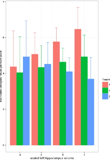
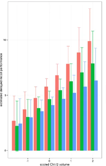
For hippocampus, considering the peak drug effect time points only, we detected a significant volume by treatment interaction (left and right hippocampus: t=-2.1, df=72, p=0.04; Figure 1) as well as a significant main effect of treatment (t=-3.2, df=72, p=0.002). With anterior basal forebrain volume, we observed a trend level significant interaction effect (t=-1.8, df=72, p=0.074; Figure 2), and a significant main effect of treatment (t=-3.2, df=72, p=0.002). AIC of nested models indicated moderately improved fit for the complex models compared with the basic model: basic model AIC = 423.4; full model with Ch1/2, AIC = 421.3; full model with left hippocampus, AIC = 422.3; full model with right hippocampus, AIC = 422.2.
Discussion
We found a decline in delayed recall performance in response to the muscarinic antagonist trihexyphenidyl, consistent with previous studies [1-3] and predictions. We used a mixed effects model to take blocking of observations within subjects and inter-individual variation in performance levels into account [15]. The time activity curve of the drug was best modeled by a second order polynomial consistent with the previously reported central
pharmacodynamics of trihexyphenidyl [14]. Under placebo, hippocampal and anterior basal forebrain volumes were positively associated with delayed recall performance. The
treatment-induced decline of performance was associated a reduced or even revers association between regional volume and task performance with a higher dose of the muscarinic receptor antagonist (Figures 1 and 2). The treatment-induced reductions of performance, compared with placebo, were more pronounced at higher volumes. This finding suggests the presence of a possible floor effect for performance at smaller volumes whereby performance levels only decrease slightly as a consequence of medication since cholinergic blocking is less effective when the system and its input areas in the hippocampus are already impaired.
The numerically similar effects for the hippocampus and the cholinergic system would indicate that cholinergic input towards the hippocampus from the anterior basal forebrain (the main source of cholinergic projections to the hippocampus [7]) only partially regulates
hippocampus-related determinants of delayed recall performance [16]. Due to the limited number of participants in our sample, however, we did not formally test for such a potential mediation effect.
mainly represented a trait marker of reserve capacity, and if the level of anticholinergic effect were just above the threshold necessary to elicit functional effects. Here, however, we examined a sample of older adults where a low dose of the drug already induced significant decline in performance. This suggests that variation in cholinergic system integrity serves at least partially as a state marker for functional performance, and at smaller volumes the blockade of muscarinic receptors occurs in an already impaired cholinergic system. To better resolve this question, one would need to study the interaction between anticholinergic
treatment, and hippocampus and basal forebrain volumes in healthy young adults where variation in volume is expected not to be rate limiting for task performance.
Increased atrophy of the cholinergic basal forebrain and decline of cholinergic functioning over and above the effects of normal aging have been associated with cognitive decline and the development of dementia in neurodegenerative conditions such as
Alzheimer’s disease and Lewy body disorders[17]. Controlled anticholinergic stress tests have been proposed as prognostic markers for the development of dementia in the elderly [18, 19]. MRI-based measurements of the structural integrity of the cholinergic basal forebrain and its functionally relevant target areas may provide important neurobiological additional information to such pharmacological stress tests.
Acknowledgements
References:
[1] Guthrie SK, Manzey L, Scott D, Giordani B, Tandon R. Comparison of central and
peripheral pharmacologic effects of biperiden and trihexyphenidyl in human volunteers. J Clin Psychopharmacol. 2000;20:77-83.
[2] Nakra BR, Margolis RB, Gfeller JD, Grossberg GT, Sata LS. The effect of a single low dose of trihexyphenidyl on memory functioning in the healthy elderly. Int Psychogeriatr. 1992;4:207-14.
[3] Pomara N, Yi L, Belzer K, Facelle TM, Willoughby LM, Sidtis JJ. Retrograde facilitation of verbal memory by trihexyphenidyl in healthy elderly with and without the APOE epsilon4 allele. Eur Neuropsychopharmacol. 2010;20:467-72.
[4] Dumas JA, Newhouse PA. The cholinergic hypothesis of cognitive aging revisited again: cholinergic functional compensation. Pharmacol Biochem Behav. 2011;99:254-61.
[5] Mesulam MM. The cholinergic innervation of the human cerebral cortex. Prog Brain Res. 2004;145:67-78.
[6] Opitz B. Memory function and the hippocampus. Front Neurol Neurosci. 2014;34:51-9. [7] Mesulam MM, Geula C. Nucleus basalis (Ch4) and cortical cholinergic innervation in the human brain: observations based on the distribution of acetylcholinesterase and choline acetyltransferase. J Comp Neurol. 1988;275:216-40.
[8] Mesulam MM, Volicer L, Marquis JK, Mufson EJ, Green RC. Systematic regional differences in the cholinergic innervation of the primate cerebral cortex: distribution of enzyme activities and some behavioral implications. Annals of neurology. 1986;19:144-51. [9] Pomara N, Willoughby LM, Wesnes K, Sidtis JJ. Increased anticholinergic challenge-induced memory impairment associated with the APOE-epsilon4 allele in the elderly: a controlled pilot study. Neuropsychopharmacology. 2004;29:403-9.
[11] Grothe MJ, Schuster C, Bauer F, Heinsen H, Prudlo J, Teipel SJ. Atrophy of the
cholinergic basal forebrain in dementia with Lewy bodies and Alzheimer's disease dementia. J Neurol. 2014.
[12] Kilimann I, Grothe M, Heinsen H, Alho EJ, Grinberg L, Amaro E, Jr., et al. Subregional basal forebrain atrophy in Alzheimer's disease: a multicenter study. J Alzheimers Dis. 2014;40:687-700.
[13] Sakamoto Y, Ishiguro M, Kitagawa G. Akaike Information Criterion Statistics. Boston: D. Reidel Publishing Company; 1986.
[14] Burke RE, Fahn S. Pharmacokinetics of trihexyphenidyl after short-term and long-term administration to dystonic patients. Annals of neurology. 1985;18:35-40.
[15] Kenward MG, Jones B. Design and Analysis of Cross-Over Trials. In: Rao CR, Miller JP, Rao DC, editors. Epidemiology and Medical Statistics. New York: Elsevier; 2008. p. 464-90. [16] Muir JL. Acetylcholine, aging, and Alzheimer's disease. Pharmacol Biochem Behav. 1997;56:687-96.
[17] Grothe MJ, Schuster C, Bauer F, Heinsen H, Prudlo J, Teipel SJ. Atrophy of the
cholinergic basal forebrain in dementia with Lewy bodies and Alzheimer's disease dementia. J Neurol. 2014;261:1939-48.
[18] Pomara N, Nolan K, Halpern G. Scopolamine-induced impairment as a potential predictor of Alzheimer's disease in individuals with Apolipoprotein E type 4 alleles. Neurochem Res. 1995;20:1519-20.
[19] Snyder PJ, Lim YY, Schindler R, Ott BR, Salloway S, Daiello L, et al. Microdosing of scopolamine as a "cognitive stress test": rationale and test of a very low dose in an at-risk cohort of older adults. Alzheimers Dement. 2014;10:262-7.
Figure legends:
Figure 1: Association of delayed recall performance with left hippocampus volume at
different levels of treatment
Using the “effects” library in R, we computed the fitted values and standard errors for delayed recall under the model for the interaction term of treatment by (mean centered) volume with the values of the other predictors being fixed at typical values, i.e. for an interval scaled covariate at its mean, and for a factor at its proportional distribution in the data, as described in [20].
Treatment 1mg = 1 mg trihexyphenidyl Treatment 2mg = 2mg trihexyphenidyl
Figure 2: Association of delayed recall performance with anterior basal forebrain
(Ch1/2) volume at different levels of treatment