250
Comparative Analysis Of Brain Tumor Detection
Using Deep Learning Methods
K. Rajesh Babu, U. Sai Deepthi, A. Sudha Madhuri, P. Sai Prasad, Syed shameem
Abstract: Brain tumor means growth of abnormal cells in brain. It occurs mainly due to the damaged DNA. Human brain has more than 10 billion working cells. The damaged brain cells are diagnosed themselves by splitting to make more cells. In advanced stages brain tumor is most dangerous disease which cannot be cured. Hence, it should be detected in the early stages with the help of MRI (Magnetic Resonance Image).So; there will be more changes to the patient to survive. For detecting tumor we have used MRI grey scale images. The MRI grey scale image based brain tumor detection is much complicated task due to difficulty and inconsistency of brain tumors. We proposed a model to detect tumor with the help of machine learning techniques. We used CNN and ANN to detect the brain tumor .And in CNN we are using two types of dataset un-augmented and augmented. The main goal of the paper is to compare two models accuracy, precision, sensitivity, specificity and based on these parameters the best model will be predicted.
Index Terms: MRI; Brain Tumor; Deep Learning Techniques; ANN; CNN; Un-augmented; Augmented. —————————— ——————————
1.
INTRODUCTION
BRAIN tumors are group, or bulk of unusual cells in the human brain. It is very crucial to detect the brain tumor as early as possible [3]. Brain tumors are classified as malignant (cancerous) tumors and benign (non-cancerous) tumors. Malignant tumors are categorized as primary and secondary tumors, primary tumors are which have the starting point in the brain and secondary tumor which extent from one location to another location. And benign (non-cancerous) tumors are of two types Adenomas and Fibromas. Benign (non-cancerous) tumors are also called as low grade and malignant (cancerous) tumors is known as high- grade. The brain tumor detection is done with the help of MRI’s, CAT, Ultrasound, X-rays and many more. In this paper we are using brain MRI images for detecting brain tumor. There are 4 grades in brain tumor
Grade I - In this grade the brain tumors are slowly growing and are more likely to spread. And they can be cured by surgery. Grade II - In this grade tumors are less likely to increase and spread. And they often grow after the treatment.
Grade III – In this grade the tumors are more likely to develop quickly.
Grade IV – In this grade the tumors are actively dividing in to more number of cells. And will be growing as fast as it can, it will be the last grade of tumor and for the effected person too. Why brain tumor problem occurs? It occurs due to inaccurate location area of tumor[1].And there is no any specific reason for the abnormal growth of tumor and it mainly caused by damaged DNA .
The tumor imaging can be done by CT scanning i.e. computer tomography, Ultra sound, X-Rays, MRI scanning. MRI is used to detect the brain cancer by evaluating, For infinite tumor cases it takes more time.[3] Brain tumor can be treated by using surgery apart from this radiation and also chemotherapy can be used to diminish the progress of tumors, but cannot be detached materially. MRI is one of the most common tests used to diagnoses the brain tumors which provide detailed images of brain. The MRI works asFirstly, the patient is put in a magnetic field and then a radio transmitter sends a radio wave through patient body where the scan is required and then the radio wave shakes the protons that are present in patient body and when the radio wave ends the proton continues to shake for some time and after then creates a new radio wave which is captured by an antenna (coil) at finally a computer algorithm turns it into a MRI images.
2
RELATED
WORK
Natarajan et al. [1] projected a model that states that the MRI brain images are initial done with pre-processing by using the median filter and after that, the segmentation part of a given image is done with the support of segmentation using threshold and also by morphological methods are applied on the model and then the region where tumor is present using subtraction of image. And this image subtraction approach gives the correct tumor shape as an image. Komal Sharma et al. [2] researched a model where the accuracy was 90% but the drawback in this is the dataset considered less number of samples and the training data taken is in less number. Aaswas Sawant et al. [3] said that the accuracy was a bit more which are compared to the above models and he also used tensor flow but the main drawback is the model is created for CPU based tensor flow. Mahmoud Al-Ayyoub et al. [4] studied the comparison of naive Bayes, J48 decision tree and neural network are done, the drawback in this is this approach is a traditional one. N. Subash et al. [5] proposed brain tumor classification using machine learning. In this, they have made a comparison for K-nn, SVM the accuracy of the model is 0.95.The accuracy is more in this model due to the automated intelligent system results. And also the error rate is minimized due to this model. Automation of MRI greyscale image classification based on SVM will be the most prominent one which aids the physician to make the final decision. And this is only meant for single layer it cannot be extended. Ben Athiwaratkun et al. [6] proposed Feature Representation in Convolutional Neural networks states that Random Forest and SVM can be used with features from CNN to yield better a ____________________
K. Rajesh Babu is currently pursuing Ph.D. program in electronics and communication engineering in KLEF deemed to be University, Country, PH-09885995375. E-mail:
U. Sai Deepthi is currently pursuing bachelor’s degree program in electronics and communication engineering in KLEF deemed to be University, India, PH-06305405615. E-mail:
A. Sudha Madhuri is currently pursuing bachelor’s degree program in electronics and communication engineering in KLEF deemed to be University, India, PH-08074234102. E-mail: [email protected]
P. Sai Prasad is currently pursuing bachelor’s degree program in electronics and communication engineering in KLEF deemed to be University, India, PH-07842310968. E-mail:
prediction accuracy compared to the original CNN. Training Random Forest takes a significant amount of memory with only 25K training samples on 400 trees. In addition, for dataset with large number of classes such as ImageNet (1000 classes). H. E. M. Abdalla et al. [7] in the study used to design an algorithm that discovers the brain tumor automatically with the help of ANN. The algorithm has designed an algorithm and also implemented and tested with the MRI dataset taken from the internet source. In this they proposed image segmentation techniques like feature extraction which gives more accuracy when compared to others. The accuracy of the proposed task is 0.99.The drawback for this model is Boundary insufficiencies and also the computational time is more. Kalpana u. Rathod et al. [8] proposed MRI brain tumor detection using ANN. This paper describes detection and Classification of Brain tumor Using ANN approach namely, Backpropagation network (BPNs). The proposed technique is developed for the diagnosing of tumor from MRI grey scale images of the brain. The further scope of the system is to improved ANN architecture by using other approach. Sangeetha.C et al.[9] proposed a model used modified K-means clustering which is used to segment an image. Clustering is a unsupervised learning model. The Modified K-means is used to increase the effectiveness and efficiency of brain tumor segmentation. Mohammad Havaei et al.[10] states that considering brats 2013 data this model managed to improve the accuracy and speed but the drawback for this is the dataset is only considering positive tumor samples. Sérgio Pereira et al. [11] in a model said that MRI images are used for brain tumor segmentation using CNN. They started with the processing method which contains bias field correction. Sajid Iqba et al. [12] research paper discusses about the architecture for segmentation of tumor with the support of multi modal images. Different algorithms are modeled for improvisation in performance metrics. Pablo Ribalta Lorenz et al. [13] researched about that the FCNN is trained on different size of patches, and also provided an algorithm for segmentation. And keeping all this models as a reference we have made a comparative analysis of brain tumor detection using machine learning. In this we try to compare the accuracy of CNN (convolution neural network) and ANN (artificial neural network). And the brief description will be given in further pages.
2.1 DATASET
One of the major challenges of this project is to find the suitable dataset and that to fully label. Bio medical images are hard to find due to privacy issues. But the dataset we have used for this model was taken from BRATS (Brain Tumor Segmentation) dataset from 2015. BRATS are a challenge that has been stared from 2012 and has been done every year. For our model we have taken 550 image samples which consist of tumor and no tumor. The images are MRI scans. This has to classes:
NO- no tumor ,encoded as 0
YES- tumor, encoded as 1.
The whole dataset for brain tumor detection are divided into two parts training dataset and testing dataset. And each set consist of both tumor and no tumor images. The whole 100% data is divided as 70% of the total dataset is given for training of the model the model, and the continuing 30% is given for testing the model. We are modelling the model with the help of dataset by using python tensor flow. This gives more accuracy and also less computational time.
3
METHODOLOGY
In this paper we performed comparative analysis of machine learning models for brain tumor detection. We are considering two machine learning architectures –ANN - Artificial Neural Networks, CNN – Convolution Neural Networks.
3.1 Artificial Neural Network
Computational algorithm is a type of ANN. Those are not
identical to biological neural networks. The nodes are collection
of connected units. It acts same as natural neural network (human brain).
Fig.1 Biological Neural Network
Fig.2. ANN model.
The Artificial Neural Network acts as given below:
Dendrites - Inputs
Soma - Activation Function
Axons – Outputs
252 activation function. Activation functions are of two types. They
are Linear function, Non - Linear function. ANN is adaptive in nature it learns from the experiences. It moulds according to the environment and learns from the past experiences. The model is trained in ANN phase and then the parameters are fixed to the system which is used to solve the problem at that phase. They are two methods for performing ANN as Forward propagation, Back propagation.
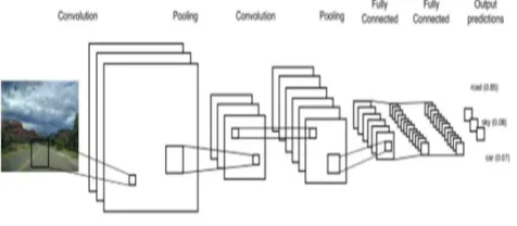
3.2 Convolutional Neural Network
CNN is a type of neural network that are widely used for image recognition and classification purpose. There are 3 classifications in CNN. They are Le Net, Alex Net, Google Net . CNN is mainly used to image classification. We give the image as input but the computer takes it in the form of array of pixels.
Fig 3.Flow chart of CNN
The layers in CNN are:
i. Convolution Layer
The procedure of convolution layer in CNN model is to extract the features of an image which is given as input. A convolution layer has a number of filters that does convolutional operation. A filter is used to slide over input grey scale image to collect the feature map. Convolution of another filter on the same image will give another feature map. With the help of convolution operation local dependencies can be calculated from the given original image. Throughout the training process CNN will get the filter values by its own. The more filters we have, the more image features we get. Network accuracy becomes better at recognizing patterns in undetected images.
ii. ReLU
ReLU is abbreviated as Rectified Linear Unit which is of non-linear process. All the negative pixel values are swapped to zero ReLU is too introduced for non-linearity function in CNN. As we take the physical world data for this model we use ReLU. So we use CNN to learn the data. ReLU can be expressed in the following way:
F(x)=max{0,x}---equation(1)
iii. Pooling
Pooling is a dimensionality reductionality algorithm which decreases the dimension of each feature map. Pooling is of different types. They are Max pooling, Average pooling, Sum pooling. The pooling function replaces the net value by the nearby outputs. In max Pooling, largest element from the window is taken and modifies all the near elements in that particular window and replaced with the largest element. Average or sum
of all the elements in the particular window can also be considered. In this model we are using max pooling since it shows better work.
iv. Fully connected Layer
It acts as a MLP which can be used as activation function to find the output of the given layer. The activation function of a node defines the output of the given input node. The most shared activation function used is softmax function. The output of the both convolution and also including the pooling layers represents good number of the features of the input image. For our method we trained our model with the help of Convolution Neural Network using to different datasets.
They are:
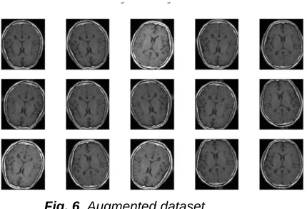
Un-Augmented dataset.
Augmented dataset.
Fig 4.Un-augmented dataset
Fig. 6 Augmented dataset
The main differences between the two datasets are in un-augmented dataset the image samples which we given will be same. For training the model we need more data so to acquire that data we take the help of augmented dataset. In this the image is given to the model in different directions. We do augmentation to increase the size of dataset. This helps in the increase of accuracy.
The formulas for calculating the performance metrics are T1= True Positive
F1= False Negative T2= True Negative F2= False Positive
Accuracy is the ability to determine the correctness or closeness of tumor. It is calculated using following formula
Accuracy = (T2+ T1)/ (T2+ T1+ F1+ F2)
Precision refers to the closeness/ correctness of two or more than two values. The formula for precision can be given by
Precision = T1/ (T1 + F2)
Sensitivity means ability to determine the tumor or any affected
are correctly. The value can be obtained using following equation
Sensitivity = T1/ (T1 +F1)
Specificity refers to the ability for the correctness or correct determination of tumor. The value can be obtained using following equation
Specificity = T2/ (T2 + F2)
The comparative analysis of these four terms is given below.
4
RESULTS
AND
DISCUSSIONS
The input we have given for the model is the grey scale images obtained from the dataset related to brain tumor obtained from the internet. The images from the dataset are split into two sets training set and testing set. Out of 550 images 450 images are used for model training and the remaining images are used for testing the model. In the given images have both tumor and no tumor images. In this paper we compare the accuracy of brain tumor detection model by using ANN and CNN.
(a) (b) (c) (d)
Fig.7 Output images of CNN and ANN (a) original image
(b) ANN (c) CNN (Un-augmented) (d) CNN (Augmented)
The accuracy we got by using basic ANN is 89.6%,precision is 90.4% , sensitivity is 88.5%, specificity is 90.8% in ANN we only use one layer we can’t apply ANN for more than one layers which results in less accuracy ,precision, sensitivity , specificity values. So for that we modelled the model by using CNN .In CNN too we have modelled the dataset by using un-augmented and augmented data for the better results. We used CNN for 3 layers and CNN with un-augmented dataset got the accuracy as 92.3%, precision is 95.2%, sensitivity is 93.4%, specificity is 91.8% and CNN with augmented dataset the accuracy is 94.1%, precision is 96%, sensitivity is 95.5% and specificity is 93.2%. And the epochs (trainings) we have given are 150. When considering the four parameters accuracy, precision, sensitivity and specificity the model using CNN with augmented dataset gives best results.
Tabel 1. Accuracy,Precision,Sensitivity,Specificity values
5 CONCLUSION
The current study developed a comparative analysis of brain tumor detection using machine learning methods. The greyscale image dataset is given to the model in both augmented and un-augmented forms to compare the better result. We also included ANN in this to know the accuracy of ANN model which is only used for one layer which is the major drawback in ANN .As ANN has only one layer the accuracy, precision, sensitivity, specificity are less for ANN model. For more than one layer we have used CNN even in CNN we used both augmented and un-augmented dataset. In those two models Accuracy, precision, sensitivity, specificity of augmented dataset using CNN has more values and the layers of CNN we used are also three (3). By comparing all this models we conclude that model using CNN with augmented dataset gives more accurate results. In future more work could be extended by including optimization for the given models which helps the model to give more accurate results with less computational time.
6
ACKNOWLEDGEMENT
254
RFERENCES
[1] Natarajan P, Krishnan.N, Natasha Sandeep Kenkre, Shraiya Nancy, Bhuvanesh Pratap Singh, "Tumor Detection using threshold operation in MRI Brain Images" , IEEE International Conference on Computational Intelligence and Computing Research, 2014.
[2] Brain cancer detection from MRI: machine learning Approach (tensor flow).
[3] Aaswad Sawant#1, Mayur Bhandari #2, Ravikumar Yadav#3, Rohan Yele#4, Mrs. Sneha Bendale#5 2018. [4] Al-Ayyoub, Mahmoud & Husari, Ghaith & Darwish, Omar
& Alabed, Ahmad. (2014). Machine learning approach for brain tumor detection.
[5] N. Subash and J. Rajeesh, "Brain Tumor Classification Using Machine Learning" In International Science Press, I J C T A, 8(5), 2015, pp. 2335-2341
[6] Athiwaratkun, Ben & Kang, Keegan. (2015). Feature Representation in Convolutional Neural Networks. (2015).
[7] H. E. M. Abdalla and M. Y. Esmail, "Brain Tumor Detection by using Artificial Neural Network," 2018 International Conference on Computer, Control, Electrical, and Electronics Engineering (ICCCEEE), Khartoum, 2018, pp. 1-6.
[8] Kalpana U. Rathod and Prof. Y. D. KapseResearch, 8, (06), 33558-33564.research article MRI brain tumor detection using artificial neural network.
[9] Ms. Sangeetha C., * Ms. Shahin A.,brain tumor segmentation using artificial neural network.
[10]Brain Tumor Segmentation Using Convolutional Neural Networks in MRI Images Sérgio Pereira*, Adriano Pinto, Victor Alves, and Carlos A. Silva*.
[11]Iqbal S, Ghani MU, Saba T, Rehman A. Brain tumor segmentation in multi-spectral MRI using convolutional neural networks (CNN). Microsc Res Tech. 2018; 00:1 – 9.