Reverse Transcriptase PCR Assay
Todd N. Wylie,a,bKristine M. Wylie,a,bRichard S. Buller,aMaria Cannella,aGregory A. Storcha
Department of Pediatrics, Washington University School of Medicine, St. Louis, Missouri, USAa; McDonnell Genome Institute, Washington University School of Medicine,
St. Louis, Missouri, USAb
We have developed and evaluated a real-time reverse transcriptase PCR (RT-PCR) assay for the detection of human enterovirus D68
(EV-D68) in clinical specimens. This assay was developed in response to the unprecedented 2014 nationwide EV-D68 outbreak in the
United States associated with severe respiratory illness. As part of our evaluation of the outbreak, we sequenced and published the
ge-nome sequence of the D68 virus circulating in St. Louis, MO. This sequence, along with other GenBank sequences from past
EV-D68 occurrences, was used to computationally select a region of EV-EV-D68 appropriate for targeting in a strain-specific RT-PCR assay.
The RT-PCR assay amplifies a segment of the VP1 gene, with an analytic limit of detection of 4 copies per reaction, and it was more
sensitive than commercially available assays that detect enteroviruses and rhinoviruses without distinguishing between the two,
in-cluding three multiplex respiratory panels approved for clinical use by the FDA. The assay did not detect any other enteroviruses or
rhinoviruses tested and did detect divergent strains of EV-D68, including the first EV-D68 strain (Fermon) identified in California in
1962. This assay should be useful for identifying and studying current and future outbreaks of EV-D68 viruses.
H
uman enterovirus D68 (EV-D68) was first isolated from
sam-ples obtained in California in 1962 from four children with
pneumonia and bronchiolitis (
1
). The type strain isolated from
one of these children was designated the Fermon strain.
Subse-quently, only small numbers of EV-D68 cases were reported until
the early 2000s (
2
). However, from 2008 to 2012, outbreaks in
Japan, the Philippines, the Netherlands, and the United States
(Georgia, Pennsylvania, and Arizona) have revealed EV-D68 as an
emerging pathogen capable of causing severe respiratory illness
(
2–7
). During the 2014 enterovirus/rhinovirus season in the
United States, EV-D68 circulated at an unprecedented level (
5
).
From August 2014 to January 2015, Centers for Disease Control
and Prevention (CDC) and state public health laboratories
con-firmed a total of 1,153 cases of respiratory illness caused by
EV-D68, with
ⱖ
14 deaths. The spectrum of disease was diverse. Cases
of flaccid paralysis have been reported in association with EV-D68
infection, but as of the time of this report, a causal relationship has
not been proven (
8
). The infected individuals were primarily
chil-dren and resided in 49 states and the District of Columbia (
5
). The
CDC also reported that there were likely millions of EV-D68
in-fections in which the etiology was not determined (
5
).
In mid-August of 2014, hospitals in Missouri and Illinois
no-ticed an increased number of patients with severe respiratory
ill-ness and reported the presence of EV-D68 (
6
). We also observed
this pattern at St. Louis Children’s Hospital in St. Louis, MO.
Because efforts to define the outbreak were hampered by the lack
of a test for EV-D68 that did not require nucleotide sequencing,
we undertook the development of a rapid and specific reverse
transcriptase PCR (RT-PCR) assay. We began by sequencing the
genome of a representative EV-D68 isolate from St. Louis to
ob-tain the sequence information required to define an assay with
optimal sensitivity and specificity (
9
). EV-D68 causes respiratory
illness, and the virus can be found in respiratory secretions, such
as saliva, nasal mucus, or sputum (
7
), of an infected person.
Therefore, an appropriate assay would primarily focus on
evalu-ating respiratory disease due to EV-D68 by targeting
nasopharyn-geal and other respiratory specimens.
The development goals for our EV-D68 RT-PCR assay
in-cluded (i) avoiding false-positive detection of closely related
en-teroviruses and rhinoviruses, (ii) increasing clinical and analytical
sensitivity compared to those of other available assays, and (iii)
retaining capability for sensitive detection of all known EV-D68
variants.
MATERIALS AND METHODS
Local specimens.After the EV-D68 outbreak was identified in August
2014 (6), clinical specimens testing positive for enterovirus/rhinovirus
with the BioFire FilmArray respiratory virus panel (BioFire Diagnostics, Inc., Salt Lake City, UT) were provided for further testing by the Diagnos-tic Virology Laboratory at St. Louis Children’s Hospital, consistent with a protocol for testing of deidentified residual clinical specimen material approved by the Washington University Human Research Protection Of-fice. Fourteen enterovirus/rhinovirus-positive specimens from the 2014
season were identified as containing EV-D68 by sequencing of the 5=
-nontranslated region of each virus (10). Extracts of total nucleic acid were
prepared from 100-l aliquots of original specimen using a bioMérieux
NucliSENS easyMAG automated extractor (bioMérieux, Durham, NC).
Received6 April 2015Returned for modification6 May 2015 Accepted2 June 2015
Accepted manuscript posted online10 June 2015
CitationWylie TN, Wylie KM, Buller RS, Cannella M, Storch GA. 2015. Development and evaluation of an enterovirus D68 real-time reverse transcriptase PCR assay. J Clin Microbiol 53:2641–2647.doi:10.1128/JCM.00923-15.
Editor:M. J. Loeffelholz
Address correspondence to Gregory A. Storch, [email protected].
Supplemental material for this article may be found athttp://dx.doi.org/10.1128 /JCM.00923-15.
Copyright © 2015, Wylie et al. This is an open-access article distributed under the terms of theCreative Commons Attribution-Noncommercial-ShareAlike 3.0 Unported license, which permits unrestricted noncommercial use, distribution, and reproduction in any medium, provided the original author and source are credited.
doi:10.1128/JCM.00923-15
on May 16, 2020 by guest
http://jcm.asm.org/
Challenge panel from New York State Department of Health.We received a challenge panel from the New York State Department of Health (courtesy of Kirsten St. George and Daryl Lamson). The included viruses are shown in Table S1 in the supplemental material. This panel included nucleic acid extracts prepared using the NucliSENS easyMAG automated extractor from clinical specimens containing the following viruses, iden-tified at the Wadsworth Laboratory by VP1 sequencing: coxsackievirus
A16 (n⫽2) and 21 (n⫽2), echovirus 18 (n⫽2) and 30, and enterovirus
71 (n⫽2). The panel also included a collection of 20 EV-D68 viruses
selected to represent a range of sequence variants. A review of the VP1 sequences from this panel showed 93.8% to 99.4% sequence identity com-pared to the St. Louis 2014 strain. In comparison, the 1962 Fermon strain (see below) had 84.4% identity to the St. Louis 2014 strain in the se-quenced VP1 region.
Challenge set from Children’s Hospital Colorado.We also received a challenge set from Children’s Hospital Colorado (courtesy of Christine Robinson) consisting of frozen aliquots of cultures positive for the follow-ing viruses: coxsackievirus A7 and 9; coxsackievirus B1 to 5; echoviruses 1, 3, 4, 5, 6, 11, 19, and 30; and enteroviruses 68 (Fermon), 70, and 71. Most of these viruses were obtained originally from the American Type Culture Collection (ATCC). Others were derived from clinical specimens that had been typed by the Centers for Disease Control (Christine Robinson, per-sonal communication). All viruses received are shown in Table S1 in the supplemental material. Total nucleic acid extracts were prepared at Wash-ington University.
Washington University samples.Our Special Projects Laboratory at Washington University provided an additional panel of challenge viruses. These viruses had been detected in patient specimens from research
proj-ects carried out in the 5 years prior (11). Viruses in this panel had been
typed based on sequencing a region of the 5=-nontranslated region (10).
Total nucleic acid extracts were prepared using either the NucliSENS easy-MAG automated extractor or Roche MagNA Pure compact system (Roche Diagnostics GmbH, Germany). Viruses included echovirus 14, coxsackievirus A16, and 59 rhinoviruses from species A to C. The rhino-virus types and extraction methods are shown in Table S1 in the supple-mental material. We verified that our assay could amplify EV-D68 from total nucleic acid prepared on both extraction platforms.
EV-D68 St. Louis 2014 genome sequence.As previously described
(9), we used high-throughput sequencing on the Illumina HiSeq 2500 to
obtain one complete and eight partial sequences (GenBank accession no.
KM881710.2, BioProject no. PRJNA263037) from specimens obtained during the 2014 outbreak in St. Louis. This genome sequence, along with other concurrently sequenced/published 2014 EV-D68 genomes, was used as a baseline for circulating EV-D68 sequence specificity.
PCR amplicon sequence selection.To create an assay with specificity
for EV-D68, we performed comprehensivein silicoanalysis of all viruses in
the NIH GenBank genetic sequence database using ak-mer approach
described below to identify unique and contiguous sequences for
candi-date RT-PCR primers and probes. Thek-mer frequency-based methods
were originally used in whole-genome shotgun assembly algorithms to
remove reads containing frequently occurring subsequences of lengthk
during genome assembly (12,13). We started by creating a consolidated
viral sequence database by collecting all Fasta nucleotide sequences from viruses that infect vertebrate or invertebrate hosts, as found in the follow-ing areas of GenBank: RefSeq, Genome Neighbors, and Influenza Virus Resource. The database contained sequences from 34 viral families, which consisted of 190 annotated viral genera and 337 species. By design, this database contained only a single complete EV-D68 reference genome (St.
Louis [STL] 2014 strain, GenBank accession no.KM881710.2).
Compre-hensivek-mer analysis was performed on the database by indexing and
reporting all 20-mer subsequences using the Tallymer software (14). We
eliminated 20-mers that were not unique in thek-mer pool, thus leaving
20-mers that were unique to EV-D68 and those unique to other viral
species. EV-D68-unique 20-mers were collected using BLAST (15) to
align all unique 20-mers to the EV-D68 reference genome, requiring
100% identity. The EV-D68-specific 20-mers were consolidated into con-tiguous sequences by merging overlapping sequences with the BEDTools
suite of utilities (16). Contiguous sequences ofⱖ60 bp were identified as
promising regions for RT-PCR primer and probe design. Of these, a 141-bp region was selected based on its uniqueness, length, and relative conservation among available EV-D68 nucleotide sequences. Notably, this region was within the VP1 gene that is considered the gold standard
for enterovirus typing (17,18).
Design of oligonucleotide primers and probes.In addition to the VP1 gene sequence represented by our candidate 141-bp region from the St. Louis 2014 strain of D68, we also collected 396 other unique EV-D68 VP1 sequences from GenBank. These nucleotide sequences were
mapped and visualized online using MUSCLE (19) at the National
Insti-tute of Allergy and Infectious Diseases (NIAID) Virus Pathogen Database
and Analysis Resource (ViPR) (http://www.viprbrc.org) website to
pro-duce a multiple-sequence alignment (MSA). Focusing on the candidate 141-bp region within the MSA, we evaluated single nucleotide polymor-phism (SNP) frequencies and identified conserved segments appropriate for primer and probe placement. The GenScript real-time PCR primer design application was used to evaluate primer/probe options. The
crite-ria for ideal amplicon selection included primer sequences ofⱖ20 bp,
PCR amplicons of⬍100 bp in length, and melting temperature (Tm)
within a range of 55 to 70°C.
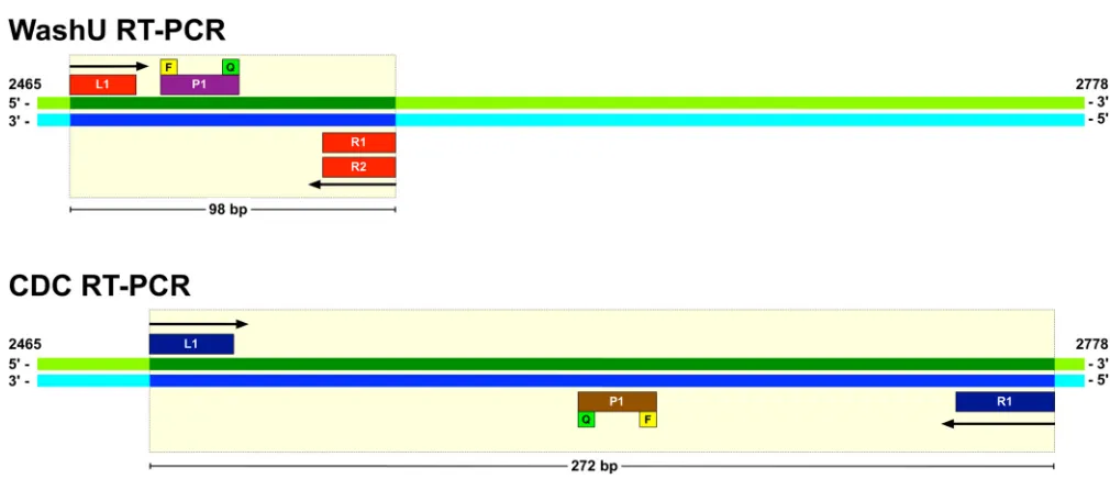
Based on this procedure, we selected an RT-PCR set consisting of two primers and a single probe with complete sequence identity to the 2014 outbreak virus (WashU design 1). To broaden the detection of EV-D68 viruses, we made modifications based on SNP frequencies that included the addition of degenerate bases and a second reverse primer (WashU
design 2). Both designs are shown inTable 1andFig. 1.
Additional specificity analysis.The selected RT-PCR primer and probe sequences were aligned to the GenBank nt database while excluding EV-D68 taxon (taxid 42789) sequences to evaluate possible homology to
non-EV-D68 sequences. Using the NCBI online BLAST interface (20,21)
for highly similar sequence alignment (MegaBlast),⬍20 alignments (90
to 100% identity) were produced, with all having identity to EV-D68 partial coding sequences that had been submitted to the database without full EV-D68 taxon designation (taxid 1193974). Using discontiguous MegaBlast, the top alignments that were not related to EV-D68 had be-tween 70 and 83% sequence identity to EV-D70.
Washington University EV-D68 RT-PCR procedure.Primers and probes for the WashU assays were ordered from Applied Biosystems at Life Technologies (Grand Island, NY). Other reagents included low-EDTA Tris-low-EDTA (TE) buffer, AgPath-ID one-step RT-PCR kit (Life
Technologies), and H2O for negative controls. Mastermixes consisting of
10⫻primer/probe (4M primers/2M probe) were produced for each
assay, and 20l of mastermix was added to each well of a 96-well PCR
plate. For the clinical specimens and controls, 5l of each sample was
added to the reaction mixture. ROX passive reference dye was included in the RT-PCR buffer to normalize well-to-well differences. The reactions were run on the Applied Biosystems 7500 real-time PCR system and
an-alyzed using accompanying threshold cycle (CT) analysis software. The
thermal cycling conditions were 45°C for 10 min, followed by 95°C for 10 min, and then 45 cycles of 95°C for 15 s and 60°C for 45 s.
Modification of the CDC-published EV-D68 assay.In mid-October 2014, the CDC Picornavirus Laboratory made a new EV-D68-specific RT-PCR assay available (Steve Oberste, Centers for Disease Control and Prevention, Atlanta, GA, personal communication). We tested the CDC EV-D68-specific RT-PCR according to the procedure available at that time on the CDC website. In addition, we tested the same assay with Cy5 replacing 6-carboxyfluorescein (FAM) as the probe reporter dye (modi-fied CDC assay) because of concerns for quenching of FAM by the
gua-nine base located at the 5=end of the probe (22) (Rangaraj Selvarangan,
Children’s Mercy Hospital, Kansas City, MO, personal communication). The primers and probes for the CDC assay were ordered from Integrated DNA Technologies, Inc. (Coralville, IA).
on May 16, 2020 by guest
http://jcm.asm.org/
Commercial and laboratory-developed assay testing.Commercial multiplex panels that detect enteroviruses/rhinoviruses were tested ac-cording to the manufacturers’ instructions. These assays included Lu-minex xTAG respiratory viral panel (CE cleared-EU/ROW, analyzed with the IS software version 2.3, which includes the FDA-approved Luminex targets plus additional targets for coronaviruses and parainfluenzavirus type 4 to be used for research purposes only) (Luminex, Austin, TX), the GenMark Dx eSensor respiratory virus panel (GenMark Diagnostics, Inc., Carlsbad, CA), BioFire FilmArray respiratory panel (RP) (BioFire Diag-nostics, Inc., Salt Lake City, UT), Cepheid GeneXpert EV IVD (Cepheid, Sunnyvale, CA), and Focus enterovirus primer pair analyte-specific re-agent (ASR) (Focus Diagnostics, Inc., Cypress, CA).
We also evaluated two laboratory-developed tests (LDTs), the
panen-terovirus assay described by Nijhuis et al. (23), and an assay described by
Piralla et al. (24) that targets the 5=-nontranslated region of EV-D68. To
determine the relative sensitivities of the different LDTs and commercial molecular assays for detecting EV68, material from the original specimen
that yielded the full-length sequence of the St. Louis EV-D68 strain was used. For the Cepheid GeneXpert and BioFire FilmArray assays, which require raw unextracted specimen, a series of 10-fold dilutions of the original specimen were made using universal transport medium (UTM) (Diagnostic Hybrids, Athens, OH) as diluent. Three hundred microliters
of each dilution was then tested in the BioFire assay and 140l was used in
the GeneXpert assay, according to the manufacturers’ instructions. For the LDTs and the GenMark and Luminex xTAG assays, which require
extracted nucleic acids, total nucleic acids were extracted from 100l of
original specimen using a bioMérieux NucliSENS easyMAG automated extractor (bioMérieux, Durham, NC). A series of 10-fold dilutions of the extract were then made using low-EDTA TE as a diluent, and each
dilu-tion was tested in each assay. For the Focus enterovirus ASR assay, 5l of
reaction mix and 5l of EasyMAG nucleic acid extract were added to the
[image:3.585.39.550.77.194.2]wells of a 3M integrated cycler universal disc, and the amplification assay was run using standard Focus Diagnostics assay parameters and a 3M integrated cycler. For the panenterovirus assay, we used the AgPath-ID
TABLE 1WashU EV-D68-specific RT-PCR assay primers and probes
ID by designationa Sequence (5=–3=) Strand Locationb T
m(°C)c Modificationd
WashU design 1e
L1-1 CACTGAACCAGAAGAAGCCA Forward 2475–2494 59.01 NA
R1-1 CCAAAGCTGCTCTACTGAGAAA Reverse 2551–2572 58.93 NA
P1-1 TCGCACAGTGATAAATCAGCACGG Forward 2502–2525 68.39 5=-FAM and 3=-TAMRA
WashU design 2f
L1-2 CAC(T/C)GAACCAGA(A/G)GAAGCCA Forward 2475–2494 58.38–59.01* NA
R1-2 CCAAAGCTGCTCTACTGAGAAA Reverse 2551–2572 58.10–59.75* NA
R2-2 CTAAAGCTGCCCTACTAAG(G/A)AA Reverse 2551–2572 58.10–59.75* NA
P1-2 TCGCACAGTGATAAATCAGCA(T/C)GG Forward 2502–2525 68.39–69.21* 5=-FAM and 3=-TAMRA
a
ID, identification.
bEV-D68 St. Louis (STL) 2014 (GenBank accession no.KM881710.2) subregion positions, 5=-3=orientation. c
Tmranges span all combinations of degenerate bases and mixed primers.
dNA, not applicable; FAM, 6-carboxyfluorescein; TAMRA, 6-carboxytetramethylrhodamine.
e
Distinct single-paired primer design. Amplicon size is 98 bp.
fDegenerate bases and mixed primers included in the design. Amplicon size is 98 bp.
FIG 1WashU and CDC RT-PCR design comparison. WashU and CDC RT-PCR primers and probe locations are illustrated within the VP1 gene of the 2014
outbreak EV-D68 St. Louis (GenBank accession no.KM881710.2) reference genome. The dark green and dark blue areas, as well as the light yellow bounding box,
indicate the regions of the genome targeted by the respective assays and their associated PCR product lengths. Arrows indicate direction of priming for the left (L) and right (R) primers. The yellow squares labeled F (fluorescent reporter) and the green squares labeled Q (quencher) show the relative orientation of the fluorophores on the probes.
on May 16, 2020 by guest
http://jcm.asm.org/
[image:3.585.38.543.458.676.2]one-step RT-PCR kit and recommended cycling conditions, using an Ap-plied Biosystems 7500 real-time PCR system. For the assay targeting the
5=-nontranslated region of EV-D68, we followed the authors’
recom-mended procedures and cycling conditions, using an Applied Biosystems 7300 real-time PCR system.
Analytic limit of detection.A 791-bp region of VP1 containing the amplicon of the WashU assays was reverse transcribed, amplified, and cloned from a clinical sample from the 2014 season from St. Louis using the primers VP1-2325-fwn (GGRTTCATAGCAGCAAAAGATGA) and EV68-VP1-3121-rvni (TAGGYTTCATGTAAACCCTRACRGT), which were
pre-viously described (25). The product was cloned using a TOPO TA cloning kit
(Life Technologies, Grand Island, NY). Sequence was verified by dideoxy sequencing of the plasmid insert. The plasmid was linearized with SpeI prior to its use as a template in the real-time RT-PCR assay. The analytic limit of detection (LOD) was determined by testing up to 10 replicates of dilutions of the linearized cloned VP1-containing plasmid on two separate days. Probit analysis was carried out using the IBM SPSS Statistics Desktop (version 22) software.
RESULTS
Comparison of WashU and CDC assays.
We tested our two
as-says and the two versions of the CDC assay on a set of clinical
samples from the 2014 outbreak (
Table 2
). We also included the
Fermon strain of EV-D68 obtained from the Children’s Hospital
Colorado. The two WashU assays performed similarly on the
sam-ples, with
⬍1 cycle difference between the two assays for 12 of the
14 samples. The published CDC assay (FAM reporter) performed
less well, failing to detect 6 of the 14 samples. However, the
mod-ified CDC assay (i.e., with the substitution of FAM with Cy5)
enabled the detection of all 14 samples. However, the
C
Tvalues
were higher for the modified CDC assay than those for the WashU
assays. The WashU assays but not the CDC assays detected the
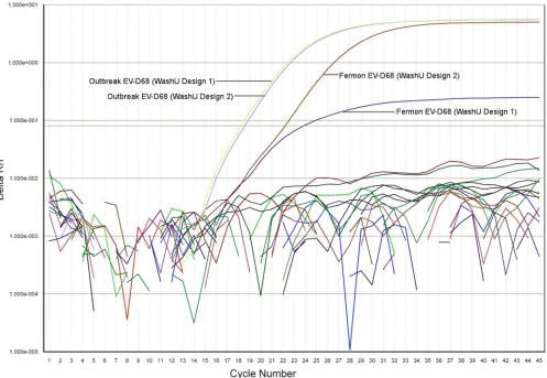
Fermon strain. Strikingly, the WashU design 2 assay detected
Fer-mon 6.7 RT-PCR cycles earlier than WashU design 1 assay, and
the amplification curve indicated improved amplitude and
ampli-fication efficiency (
Fig. 2
).
To follow up on this observation, additional clinical samples
from the 2014 season that had been tested with WashU design 1
were identified for comparison with the modified CDC assay (
Ta-ble 3
). Only the modified assay was used because of its greater
sensitivity. The samples were selected to include 10 from each of 4
categories based on the
C
Tof the WashU assay:⬍22, 22 to 27,
⬎27
to 32, and
⬎
32. Twenty samples negative for EV-D68 were also
tested. In this test, the modified CDC assay detected all of the
samples with
C
Tvalues of
ⱕ
32 but failed to detect those with
C
Tvalues of
⬎32.
Other EV-D68 viruses.
The WashU assays were used to test an
additional 20 specimens positive for EV-D68 from the New York
State Department of Health. Both WashU assays detected EV-D68
in each sample.
Analysis of specificity.
The specificity of the WashU assays was
evaluated using test panels provided by the New York State
De-partment of Health, the Children’s Hospital Colorado, and our
own Special Projects Laboratory. These panels included 4
differ-ent coxsackievirus A viruses, 5 differdiffer-ent coxsackievirus B viruses, 9
different echoviruses, 3 enteroviruses, including EV-D70, which is
the enterovirus that is most closely related to EV-D68, and 59
rhinoviruses representing species A to C. All viruses tested are
shown in Table S1 in the supplemental material. The presence of
viral RNA was confirmed for each of these samples by
amplifica-tion of the nucleic acid extract with an alternative panenterovirus/
rhinovirus real-time RT-PCR assay. The WashU assays did not
amplify any of the test panel viruses.
[image:4.585.39.549.78.310.2]Comparison with laboratory-developed and commercial
as-says.
We compared the sensitivity of the WashU EV-D68 assays
with that of 5 commercial enterovirus assays and 2 LDTs that
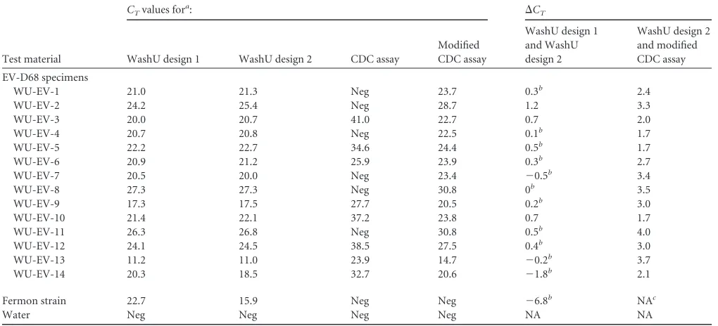
TABLE 2Comparison of WashU and CDC assays using 2014 EV-D68 outbreak specimens and the Fermon strain
Test material
CTvalues fora: ⌬CT
WashU design 1 WashU design 2 CDC assay
Modified CDC assay
WashU design 1 and WashU design 2
WashU design 2 and modified CDC assay EV-D68 specimens
WU-EV-1 21.0 21.3 Neg 23.7 0.3b 2.4
WU-EV-2 24.2 25.4 Neg 28.7 1.2 3.3
WU-EV-3 20.0 20.7 41.0 22.7 0.7 2.0
WU-EV-4 20.7 20.8 Neg 22.5 0.1b 1.7
WU-EV-5 22.2 22.7 34.6 24.4 0.5b 1.7
WU-EV-6 20.9 21.2 25.9 23.9 0.3b 2.7
WU-EV-7 20.5 20.0 Neg 23.4 ⫺0.5b 3.4
WU-EV-8 27.3 27.3 Neg 30.8 0b 3.5
WU-EV-9 17.3 17.5 27.7 20.5 0.2b 3.0
WU-EV-10 21.4 22.1 37.2 23.8 0.7 1.7
WU-EV-11 26.3 26.8 Neg 30.8 0.5b 4.0
WU-EV-12 24.1 24.5 38.5 27.5 0.4b 3.0
WU-EV-13 11.2 11.0 23.9 14.7 ⫺0.2b 3.7
WU-EV-14 20.3 18.5 32.7 20.6 ⫺1.8b 2.1
Fermon strain 22.7 15.9 Neg Neg ⫺6.8b NAc
Water Neg Neg Neg Neg NA NA
a
CT, threshold cycle; Neg, negative. WashU design 1 was a distinct single paired-primer design, and WashU design 2 had degenerate bases and mixed primers included in the design. The CDC assay had a published design with FAM. The modified CDC assay was modified by replacement of FAM with Cy5.
b⌬ CTⱕ0.5. cNA, not applicable.
on May 16, 2020 by guest
http://jcm.asm.org/
detect enteroviruses and/or rhinoviruses but do not specifically
distinguish subtypes (
Table 4
). We prepared 10-fold serial
dilu-tions of a clinical sample from the 2014 St. Louis outbreak and
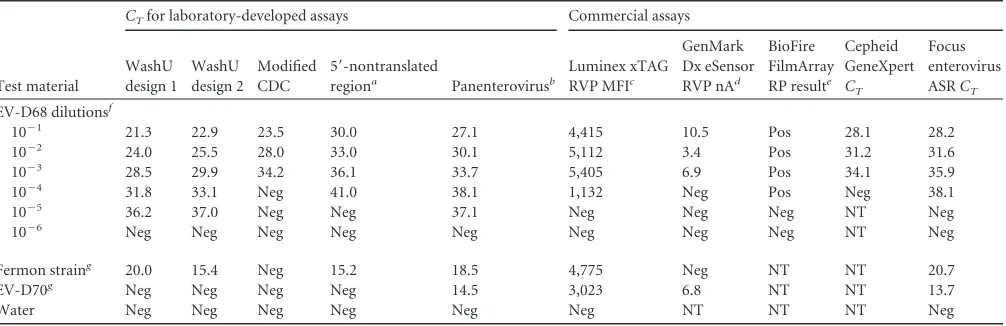
tested each of the assays in parallel. We found that the WashU
assays were able to detect EV-D68 at a dilution of 10
⫺5, which
was 10- to 100-fold more sensitive than the commercial
Lu-minex xTag, GenMark Dx eSensor, Biofire FilmArray, Cepheid
GeneXpert, and Focus enterovirus assays. The LDT targeting
the 5=-nontranslated region of EV-D68 showed equivalent
sen-sitivity for detecting Fermon to that of the WashU design 2
assay; however, it had higher
C
Tvalues overall than those of the
WashU assays for detecting the 2014 outbreak strain and was
10-fold less sensitive in serial dilution testing. Only the
panen-terovirus LDT had comparable sensitivity to the WashU assays.
Analytic sensitivity.
In order to determine the limit of detection
(LOD) of the WashU EV-D68 assay, the cloned 791-bp fragment of
VP1 was serially diluted in a range of 0.14
⫻
10
0to 1
⫻
10
2copies per
reaction and tested with the WashU design 1 assay. Up to 10 replicates
were carried out at each dilution on two separate days. The resulting
95% LOD determined by probit regression analysis was 4 copies per
reaction, with a 95% confidence interval of 3.1 to 6.6 copies.
DISCUSSION
During the summer and fall of 2014, enterovirus D68 circulated at
an unprecedented level in the United States (
4–6
). Because no
molecular test was available for EV-D68-specific identification,
laboratories were forced to rely on amplification and partial
se-quencing of the structural protein genes, VP4 to VP2 or VP1 (
17
,
FIG 2Amplification plot showing WashU RT-PCR assay EV-D68 sensitivity. The PCR amplification cycle number is displayed on thexaxis while log(⌬Rn) is
shown on theyaxis. Rn is the fluorescence of the reporter dye divided by the fluorescence of a passive reference dye.⌬Rn is Rn minus the baseline and is plotted
against PCR cycle number. The light green and light purple lines show detection of the 2014 EV-D68 outbreak strain using the WashU design 1 and design 2 assays, respectively. The brown and dark purple lines show detection of the more distant 1962 Fermon EV-D68 type strain using the WashU design 2 and design 1 assays, respectively. The incorporation of degenerate bases and mixed primers in WashU design 2 shows a significant increase in sensitivity (6.7 cycles earlier
[image:5.585.46.543.67.410.2]detection) for the Fermon type strain (brown line), with minimal decrease in sensitivity to the 2014 outbreak strain (light purple) (⬍0.5 cycles difference).
TABLE 3Comparison of sensitivities of WashU design 1 and modified CDC assays using 2014 EV-D68 outbreak specimens
CTvalue range
(WashU design 1 defined)
No. of samples tested
No. of positive test results for:
WashU design 1
Modified CDC assay
⬍22 10 10 10
22–27 10 10 10
⬎27–32 10 10 10
⬎32 10 10 0
Nega 20 20 20
aNeg, negative.
on May 16, 2020 by guest
http://jcm.asm.org/
[image:5.585.40.287.613.715.2]18
), a much more cumbersome procedure than a specific
real-time RT-PCR assay. The lack of a rapid molecular assay resulted in
vast underrecognition and underreporting of cases of EV-D68
in-fection, because the majority of clinical laboratories did not have
the ability to test specifically for EV-D68. Specific identification of
EV-D68 was primarily from the CDC and state labs. Several
FDA-approved multiplex assays for the detection of respiratory viruses
detect enteroviruses, but these systems are broadly reactive and do
not distinguish between enteroviruses and rhinoviruses; the
re-sults are typically reported as human rhinovirus/enterovirus.
In response to the 2014 nationwide enterovirus D68 outbreak
and the associated increase in severe respiratory illness
presenta-tions, we developed and evaluated a real-time reverse
transcrip-tase PCR assay for the detection of EV-D68 in clinical specimens.
The development of this assay was informed by sequencing the
complete genome of the EV-D68 virus circulating in St. Louis,
MO, during the outbreak. Our RT-PCR primer and probe
se-quences were derived computationally by
k
-mer-mediated
filter-ing of potentially cross-reactive non-EV-D68 viral sequences.
Broad detection of EV-D68 was achieved through
multiple-se-quence alignment review using all published EV-D68 VP1 regions
available through GenBank. Reduced sensitivity for the older and
more distant Fermon EV-D68 type strain, which has only 87.9%
identity to the genome sequence of the St. Louis virus, led us to
modify the assay, which then proved capable of efficiently
ampli-fying more divergent EV-D68 viruses as well.
The CDC released the design and protocol for an
EV-D68-specific RT-PCR on their website as a diagnostic resource for
cli-nicians and health care professionals in mid-October 2014. As
noted within the CDC protocol, the amplicon size of 272 bp is
larger than ideal for a real-time RT-PCR assay. Furthermore, their
selected TaqMan probe had a guanine (G) at the 5=
end linked to
the fluorophore FAM, potentially incurring unwanted
fluores-cence quenching. The replacement of FAM with Cy5 significantly
improved the ability of the CDC assay to detect EV-D68 (
Table 2
).
We evaluated the CDC assay alongside our own, testing against
EV-D68-positive clinical samples (
n
⫽
35). Based on serial
dilu-tion testing of the 2014 outbreak virus, the WashU RT-PCR assays
were 100-fold more sensitive than the published CDC assay, and
the CDC assay failed to detect the Fermon strain. In addition, the
WashU assays were
ⱖ
10-fold more sensitive for detecting EV-D68
than the FDA-approved commercial assays (i.e., Luminex xTAG
RVP, GenMark Dx eSensor RVP, Biofire FilmArray RP, and
Cepheid GeneXpert) for enterovirus/rhinovirus detection (
Table
4
), with the further advantage of specific identification of EV-D68.
The WashU assays showed no evidence of amplification of other
enteroviruses, including the relatively closely related EV-D70
vi-rus, or rhinoviruses.
There are two limitations of this study. First, we were not able
to test the specificity of this assay against every known enterovirus
or rhinovirus subtype. It is possible that the assay cross-reacts with
another subtype, although that is not likely based on
in silico
anal-ysis of the genome sequences. It is also possible that the assay
cross-reacts with a subtype that has yet to be discovered. Second,
although we have tried to show that our assay evaluated a broad
range of EV-D68 strains, EV-D68 strains may exist or emerge with
mutations in the PCR target region that cause the assay to miss
that strain of the virus.
[image:6.585.43.546.77.240.2]The development of another EV-D68-specific RT-PCR by
Pi-ralla et al. (
24
) was communicated in March 2015. This
under-scores the international interest in EV-D68 detection stimulated
by the global reemergence of the virus in 2014. The assay targets a
60-bp region of the 5=-nontranslated region of EV-D68. A
com-parison of the assay to the CDC RT-PCR and commercially
avail-able enterovirus/rhinovirus clinical assays was not reported in
their paper. In our dilution tests, the assay was 10-fold less
sensi-tive in detecting the 2014 outbreak strain of EV-D68 than the
WashU assays. Furthermore, the WashU assays detected the
un-diluted outbreak specimen 7 cycles before the 5=-nontranslated
region-targeting assay reached detection. Because these assays
de-tect completely different segments of the viral genome, they may
have complementary value in future applications.
TABLE 4Comparison of detection of EV-D68 using laboratory-developed and commercial assays
Test material
CTfor laboratory-developed assays Commercial assays
WashU design 1
WashU design 2
Modified CDC
5=-nontranslated
regiona Panenterovirusb
Luminex xTAG
RVP MFIc
GenMark Dx eSensor
RVP nAd
BioFire FilmArray
RP resulte
Cepheid GeneXpert
CT
Focus enterovirus
ASRCT
EV-D68 dilutionsf
10⫺1 21.3 22.9 23.5 30.0 27.1 4,415 10.5 Pos 28.1 28.2
10⫺2 24.0 25.5 28.0 33.0 30.1 5,112 3.4 Pos 31.2 31.6
10⫺3 28.5 29.9 34.2 36.1 33.7 5,405 6.9 Pos 34.1 35.9
10⫺4 31.8 33.1 Neg 41.0 38.1 1,132 Neg Pos Neg 38.1
10⫺5 36.2 37.0 Neg Neg 37.1 Neg Neg Neg NT Neg
10⫺6 Neg Neg Neg Neg Neg Neg Neg Neg NT Neg
Fermon straing 20.0 15.4 Neg 15.2 18.5 4,775 Neg NT NT 20.7
EV-D70g Neg Neg Neg Neg 14.5 3,023 6.8 NT NT 13.7
Water Neg Neg Neg Neg Neg Neg NT NT NT Neg
a
Protocol was as described by Piralla et al (24).
bProtocol as described by Nijhuis et al. (23). The modifications are described in Materials and Methods.
c
Luminex mean fluorescence index (MFI) values: negative,⬍150; equivocal, 150 to 300; positive,⬎300. dGenMark nanoampere (nA) values: positive,⬎3, with⬎100 being strong positive.
e
Pos, positive; Neg, negative; NT, not tested.
fNucleic acid extracted from nasopharyngeal swab from EV-D68-positive patient. See Materials and Methods for details.
g
ATCC strains; total nucleic acid extracted from infected cell culture.
on May 16, 2020 by guest
http://jcm.asm.org/
While there are no specific treatments for EV-D68 and
cur-rently no antiviral targets available, rapid and accurate diagnosis
of current and future EV-D68 infections is of great concern to
clinicians and public health authorities. The EV-D68-specific
RT-PCR assay we have developed can be used for epidemiological
studies of the EV-D68 outbreak and for virus monitoring in
sub-sequent seasons. It is unclear at this time whether typing EV-D68
will be useful for patient management. However, some
FDA-ap-proved multiplex respiratory panels may not detect EV-D68
op-timally or at all. In laboratories using those assays, an additional
assay that detects EV-D68 will be useful for laboratory
documen-tation of EV-D68 infection, which may help with prognosis,
anti-biotic use, and appropriate isolation. The ongoing importance of
improved diagnostic capability for EV-D68 is underscored by the
recent decision by the Department of Health and Human Services
to encourage the development of EV-D68 testing capability by
authorizing the emergency use of new
in vitro
diagnostics for
EV-D68 detection (
http://www.gpo.gov/fdsys/pkg/FR-2015-02-2
7/html/2015-04121.htm
).
ACKNOWLEDGMENTS
We thank Daryl Lamson and Kirsten St. George from the New York State Department of Health and Christine Robinson from Children’s Hospital Colorado for providing enterovirus/rhinovirus samples used in our test-ing; Rangaraj Selvarangan from Children’s Mercy Hospital, Kansas City, MO, Anthony Orvedahl from the Department of Pediatrics at Washing-ton University School of Medicine, and Stephanie Bledsoe from the Clin-ical Virology Laboratory at St. Louis Children’s Hospital for providing EV-D68 residual specimen material; Brandi Herter from the Microbial Genomics Laboratory in the Department of Pediatrics at Washington University for assistance with cloning the fragment of the EV-D68 VP1 gene; and Elena Deych from the division of General Medical Sciences in the Department of Medicine at Washington University for calculating the assay limit of detection.
This study was supported in part by grant R01AI097213 from the National Institute of Allergy and Infectious Diseases awarded to G.A.S. and by institutional funds. Specimen collection was supported in part by grant U01AI077810 from the National Institute of Allergy and Infectious Diseases awarded to Stuart C. Sweet, Washington University School of Medicine, in support of the Clinical Trials in Organ Transplantation in Children (CTOT-C) research program. The funders had no role in study design, data collection and analysis, decision to publish, or preparation of the manuscript.
REFERENCES
1.Schieble JH, Fox VL, Lennette EH.1967. A probable new human
picor-navirus associated with respiratory diseases. Am J Epidemiol85:297–310.
2.Imamura T, Oshitani H.2015. Global reemergence of enterovirus D68 as an important pathogen for acute respiratory infections. Rev Med Virol
25:102–114.http://dx.doi.org/10.1002/rmv.1820.
3.Tokarz R, Firth C, Madhi SA, Howie SRC, Wu W, Sall AA, Haq S, Briese T, Lipkin WI.2012. Worldwide emergence of multiple clades of
enterovirus 68. J Gen Virol. 93(Pt 9):1952–1958.http://dx.doi.org/10
.1099/vir.0.043935-0.
4.Centers for Disease Control and Prevention.2008. Clusters of acute respi-ratory illness associated with human enterovirus 68 —Asia, Europe, and
United States, 2008 –2010. MMWR Morb Mortal Wkly Rep60:1301–1304).
5.Centers for Disease Control and Prevention.2014. Enterovirus D68.
Centers for Disease Control and Prevention, Atlanta, GA.http://www.cdc
.gov/non-polio-enterovirus/outbreaks/EV-D68-outbreaks.html. 6.Centers for Disease Control and Prevention.2014. Severe respiratory illness
associated with enterovirus D68 —Missouri and Illinois, 2014. MMWR
Morb Mortal Wkly Rep 63:798–799.http://www.cdc.gov/MMWR/preview
/mmwrhtml/mm6336a4.htm?s_cid⫽mm6336a4_w.
7.Oberste MS, Maher K, Schnurr D, Flemister MR, Lovchik JC, Peters H,
Sessions W, Kirk C, Chatterjee N, Fuller S, Hanauer JM, Pallansch MA.
2004. Enterovirus 68 is associated with respiratory illness and shares bio-logical features with both the enteroviruses and the rhinoviruses. J Gen
Virol85:2577–2584.http://dx.doi.org/10.1099/vir.0.79925-0.
8.Greninger AL, Naccache SN, Messacar K, Clayton A, Yu G, Somasekar S, Federman S, Stryke D, Anderson C, Yagi S, Messenger S, Wadford D, Xia D, Watt JP, Van Haren K, Dominguez SR, Glaser C, Aldrovandi G, Chiu CY.2015. A novel outbreak enterovirus D68 strain associated with acute flaccid myelitis cases in the USA (2012–14): a retrospective cohort
study. Lancet Infect Dis 15:671– 682. http://dx.doi.org/10.1016/S1473
-3099(15)70093-9.
9.Wylie KM, Wylie TN, Orvedahl A, Buller RS, Herter BN, Magrini V, Wilson RK, Storch GA.2015. Genome sequence of enterovirus D68 from
St. Louis, Missouri, USA. Emerging Infect Dis21:184 –186.
10. Lee WM, Kiesner C, Pappas T, Lee I, Grindle K, Jartti T, Jakiela B, Lemanske RF, Jr, Shult PA, Gern JE.2007. A diverse group of previously unrecognized human rhinoviruses are common causes of respiratory illnesses in infants. PLoS
One2:e966.http://dx.doi.org/10.1371/journal.pone.0000966.
11. Colvin JM, Muenzer JT, Jaffe DM, Smason A, Deych E, Shannon WD, Arens MQ, Buller RS, Lee WM, Weinstock EJ, Weinstock GM, Storch
GA.2012. Detection of viruses in young children with fever without an
apparent source. Pediatrics130:e1455– e1462.http://dx.doi.org/10.1542
/peds.2012-1391.
12. Sutton G, White O, Adams M, Kerlavage A.1995. TIGR Assembler: a new tool for assembling large shotgun sequencing projects. Genome Sci
Technol1:9 –19.http://dx.doi.org/10.1089/gst.1995.1.9.
13. Huson DH, Reinert K, Kravitz SA, Remington KA, Delcher AL, Dew IM, Flanigan M, Halpern AL, Lai Z, Mobarry CM, Sutton GG, Myers
EW.2001. Design of a compartmentalized shotgun assembler for the
human genome. Bioinformatics17(Suppl 1):S132–S139.http://dx.doi.org
/10.1093/bioinformatics/17.suppl_1.S132.
14. Kurtz S, Narechania A, Stein JC, Ware D.2008. A new method to compute K-mer frequencies and its application to annotate large repetitive plant
ge-nomes. BMC Genomics9:517.http://dx.doi.org/10.1186/1471-2164-9-517.
15. Altschul SF, Gish W, Miller W, Myers EW, Lipman DJ.1990. Basic local
alignment search tool. J Mol Biol215:403– 410.http://dx.doi.org/10.1016
/S0022-2836(05)80360-2.
16. Quinlan AR, Hall IM.2010. BEDTools: a flexible suite of utilities for
com-paring genomic features. Bioinformatics26:841– 842.http://dx.doi.org/10
.1093/bioinformatics/btq033.
17. Oberste MS, Maher K, Kilpatrick DR, Flemister MR, Brown BA, Pal-lansch MA.1999. Typing of human enteroviruses by partial sequencing of
VP1 1. J Clin Microbiol37:1288 –1293.
18. Nix WA, Oberste MS, Pallansch MA.2006. Sensitive, seminested PCR amplification of VP1 sequences for direct identification of all enterovirus
serotypes from original clinical specimens. J Clin Microbiol44:2698 –
2704.http://dx.doi.org/10.1128/JCM.00542-06.
19. Edgar RC.2004. MUSCLE: multiple sequence alignment with high
accu-racy and high throughput. Nucleic Acids Res32:1792–1797.http://dx.doi
.org/10.1093/nar/gkh340.
20. Johnson M, Zaretskaya I, Raytselis Y, Merezhuk Y, McGinnis S, Mad-den TL.2008. NCBI BLAST: a better Web interface. Nucleic Acids Res
36:W5–W9.http://dx.doi.org/10.1093/nar/gkn201.
21. Boratyn GM, Camacho C, Cooper PS, Coulouris G, Fong A, Ma N, Madden TL, Matten WT, McGinnis SD, Merezhuk Y, Raytselis Y, Sayers EW, Tao T, Ye J, Zaretskaya I.2013. BLAST: a more efficient
report with usability improvements. Nucleic Acids Res41:W29 –W33.
http://dx.doi.org/10.1093/nar/gkt282.
22. Xiao M, Kwok P-Y.2003. DNA analysis by fluorescence quenching
de-tection. Genome Res13:932–939.http://dx.doi.org/10.1101/gr.987803.
23. Nijhuis M, van Maarseveen N, Schuurman R, Verkuijlen S, de Vos M, Hendriksen K, van Loon AM.2002. Rapid and sensitive routine detection of all members of the genus enterovirus in different clinical specimens by
real-time PCR. J Clin Microbiol40:3666 –3670.http://dx.doi.org/10.1128
/JCM.40.10.3666-3670.2002.
24. Piralla A, Girello A, Premoli M, Baldanti F. 2015. A new real-time RT-PCR assay for detection of human enterovirus 68 (EV-D68) in
respi-ratory samples. J Clin Microbiol53:1725–1726.http://dx.doi.org/10.1128
/JCM.03691-14.
25. Rahamat-Langendoen J, Riezebos-Brilman A, Borger R, van der Heide R, Brandenburg A, Schölvinck E, Niesters HGM.2011. Upsurge of human enterovirus 68 infections in patients with severe respiratory tract infections. J
Clin Virol52:103–106.http://dx.doi.org/10.1016/j.jcv.2011.06.019.