Implementation of Breast Tumor Demarcation in Digital
Mammograms using Local Thresholding and
Mathematical Morphology
Sonali Patil
PhD scholar, Shivaji University faculty, University of Mumbai
k.j.somaiya college of engineering
V.R.Udupi,
PhD. Visvesvaraya TechnologicalUniversity
Gogte Institute of Technology
Poonam M Bhogle
University of Mumbai k.j.somaiya college ofengineering
ABSTRACT
Breast cancer is a deadly disease and should be treated at right time to save life from death. Tumor demarcation from digital mammograms is important for classification of benign and malignant masses. Computer aided systems can provide accurate results for breast tumors which can help radiologists in distinguishing malignant masses from benign. Segmentation is based on the principle of grouping similar regions together based on some pre-defined criteria which helps to separate tumor from breast for further processing. Histograms show almost a perfect distribution of intensity levels of pixels in mammograms. This can be specifically used in thresholding process to enhance the tumor region which further simplifies the segmentation methodology. Detection of breast tumors from digital mammograms can be done in many ways. In this paper, Local Thresholding and Mathematical Morphology methodologies are implemented to segment the breast tumor from digital mammograms. Various morphological operators can be used to extract the tumor from breast. In local thresholding image is divided into different parts and thresholding is applied rather than selecting one threshold for the entire image. In this paper both algorithms are implemented and their respective results are presented and compared.
General Terms
Benign, Malignant and Segmentation.
Keywords
Mathematical Morphology, Local Thresholding, Digital Mammograms.
1.
INTRODUCTION
Human body is made up of many cells which have a certain growth to them as it ages. Cancer is caused when these cells grow in an uncontrolled way which further affects the immune system and damages the other organs resulting to death [1][2]. Breast cancer is the cancer that takes place in the breast most common between females but the study has shown that men also suffer from breast cancer [3]. Most common root cause of breast cancer is milk ducts which carry milk. Tumor starts to grow in the milk ducts further tearing them and spreads in the other parts of the breast. Breast cancer can be most effectively treated when detected at its early stage. In case of breast cancer, mammography is one of the
tumor area [4][5]. However mammography is demanding job for radiologists, and cannot provide consistent results every time. Experience of a radiologist is the most important factor for detection of breast tumor. Mammography can also produce false results, so biopsy is the final procedure for right decision making if one is unsure of the result. Segmentation is the process of grouping areas in an image having similar features. Segmentation methodology can be different for different types of images, there is no general segmentation procedure defined for all images. Segmentation will help in the computer-aided evaluation of the tumors and the distinction of benign and malignant breast tumors.
2.
RELATED WORK
Segementation of breast tumor in digital mammograms can be done in many ways. For further treatment of the breast cancer tumor should be properly segmented from breast for later identification as cancerous or non cancerous. Local thresholding is another technique that can be used for breast tumor segmentation from digital mammograms; here basic principle of segmentation depends on applying different threshold values to different parts of image, rather than using a global threshold value [6][7][8]. Morphological operators like dilation, erosion, opening, closing etc. can be used in a well defined way to demarcate the tumor [9].
2.1
Local Thresholding [6][7][8]
2.1.1 Local Thresholding Algorithm
Step 1: Read the input image.Step 2: Convert the image to grayscale.
Step 3: Divide the image into sub-regions to which local thresholding is to be applied.
Step 4: Draw histogram for each sub-region.
Step 5: Select threshold values for all sub-regions.
Step 6: Pixels having intensity values below threshold should be assigned value as 0 else assign value 255.
Step 7: Perform post processing on the image to get the final segmented image.
2.1.2 The Local Thresholding Algorithm Analysis
Local thresholding selects different threshold for every region. Efficiency of the algorithm depends on the selection of proper threshold value.
2.2
Mathematical Morphology
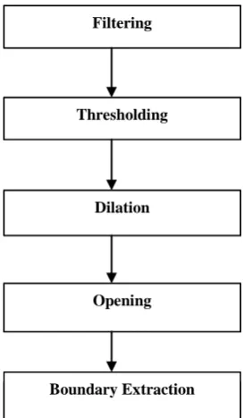
The field of mathematical morphology contributes a wide range of operators to image processing, all based around a few simple mathematical concepts from set theory [9]. The operators are particularly useful for the analysis of binary images and common usages include edge detection, noise removal, image enhancement and image segmentation. Morphological techniques typically probe an image with a small shape or template known as a structuring element. The structuring element is positioned at all possible locations in the image and it is compared with the corresponding neighborhood of pixels. Morphological operations differ in how they carry out this comparison. Morphological operators are used to carry out segmentation in order to extract tumor from breast. Figure 1 shows the flow diagram of mathematical morphology.
[image:2.595.90.227.446.680.2]
Figure 1: Flow diagram for Mathematical Morphology
Filtering
Different types of filters like Gaussian filter, Median filter, Low pass filter and High pass filter can be used here to remove noise and improve the appearance of the image.
Thresholding
Thresholding is the simplest method of image segmentation. A process of creating a black-and-white image out of a grayscale image consisting of setting exactly those pixels to white whose value is above a given threshold, setting the other pixels to black.
Dilation
Dilation is one of the basic operations in mathematical morphology. The value of the output pixel is the maximum value of all the pixels in the input pixel's neighborhood. In a binary image, if any of the pixels is set to the value 1, the output pixel is set to 1.
Opening
Opening is defined as erosion followed by dilation. Morphological Opening generally smoothes the contour of an object and eliminate thin protrusions.
Boundary Extraction
Here all connected pixels whose value is 1 which indicates white is extracted while other pixels are set to zero.
2.2.1 Mathematical Morphology Algorithm
Step 1: Apply the filter on input image depending on the type of noise.Step 2: Histogram is to be plotted for selecting proper threshold value using GINPUT function of MATLAB. Select the threshold value.
1. If pixels of the mammogram image have value less than threshold then assign value 0 to those pixels. 2. Else assign value 255.
Step 3: Apply morphological dilation on the threshold image.
Step 4: Apply morphological opening on dilated image.
Step 5: Extract boundary of the tumor from the thresholded image. Using this boundary to demark the tumor in the original image.
2.2.2 The Mathematical Morphology Algorithm
Analysis
The Morphology algorithm can present a properly segmented breast tumor after performing some post processing on the image. Two key steps of post processing are:
1. Once the boundary extraction is done image should be complemented and subtracted from the original digital mammogram to make the boundary visible on the original mammogram.
2. Tumor from the original image can also be cropped and separately stored to be used in classification by subtracting eroded image from the original image.
3.
DISCUSSION
Table1: Comparative Study of Mathematical Morphology and Local Thresholding.
Algorithm Advantages Disadvantages
Local Thresholding
Less complex in implementation as repetitive steps are to be written.
No pre-processing is done on the image which can affect the segmentation
process if
mammogram is
noisy, this affects the efficiency of the algorithm.
Different threshold values for different regions are to be selected; improper threshold selection can lead to faulty results.
Mathematical Morphology
Only one threshold value is to be selected
Segmentation results may be affected if threshold values are not properly selected from histogram. Morphological
operators yield clear tumor image
Mammogram image may be noisy which can be rectified by filtering stage of the algorithm
4. EXPERIMENTAL RESULTS
This section represents the implementation results of Local thresholding and Mathematical Morphology. All experiments are performed on Intel Core i3-380M processor with 4 GB memory using MATLAB 7.0, with consideration of work folder having handful of at least 100 digital mammogram images obtained from internet and mammography centers. Time required for execution of the two algorithms is as given in Table 2:
Table 2:Execution time for the two algorithms
Algorithm Elapsed Time (sec)
Local Thresholding 141.66577 approx
Mathematical Morphology 10.60900 approx
Graphical output
[image:3.595.353.518.266.421.2]Graphical output in Figure 2 shows the elapsed time presented in terms of bar chart for local thresholding and mathematical morphological.
Figure 2: Execution time for segmentation
Implementation results of Local Thresholding are given in Figure 3:
Figure 3(a): Original Image
(b) (c)
(d) (e)
[image:3.595.357.509.460.663.2]
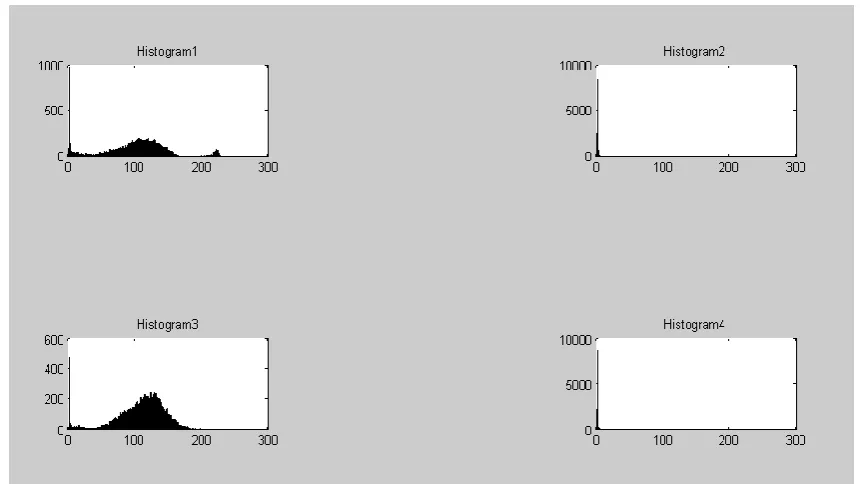
Figure 3(f) Histograms of 4 regions
(g) (h) (i) (j)
Figure 3 (g-j): Image after applying thresholds on 4 regions of image.
(k) (l)
Figure 3(k) Reconstructed Image, (l) Superimposed image
Implementation results of Mathematical Morphology are as given in Figure 4:
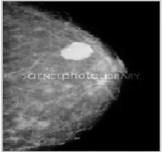
Figure 4(a) Original Image
[image:4.595.88.520.81.323.2] [image:4.595.55.271.374.620.2] [image:4.595.51.284.376.473.2]Figure 4(c) Histogram of the Resized Image
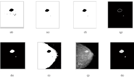
(d) (e) (f) (g)
(h) (i) (j) (k)
Figure 4: (d) Image after thresholding, (e) Dilated image, (f) Morphological opening, (g) Boundary extracted image, (h)
[image:5.595.59.522.74.358.2] [image:5.595.68.520.393.658.2]5. CONCLUSION
In this paper two algorithms, Local Thresholding and Mathematical Morphology, were successfully implemented on MATLAB 7.0. The algorithms were tested for approximately 50 digital mammogram images. It was observed that the results of mathematical morphology based breast tumor segmentation were better than that of local thresholding based segmentation for almost all of the mammographic images. Also segmentation of breast tumor from mammographic images using mathematical morphology was much faster than that of local thresholding method. Hence it can be concluded that mathematical morphology is a better segmentation algorithm for breast tumor demarcation than local thresholding. Segmented results can be further used for feature extraction and classification of breast tumors.
6. ACKNOWLEDGMENTS
We take this opportunity to express our gratitude to Dr. B. M. Patil for his valuable contribution in verifying the results.
7. REFERENCES
[1] Cancer[online]http://www.who.int/topics/cancer.
[2] Cancer [online]
http://www.mayoclinic.com/health/breast-cancer.
[3] Breast cancer [online]. Available:
http://en.wikipedia.org.
[4]Mammography[online].Available:http://www.radiologyinf o.org
[5]Cancer,Mammogram[online].Available:http://www.cancer. gov/cancertopics/factsheet/detection/mammograms.
[6] Sang Uk Lee, Seok Yoon Chung, Rae Hong Park, “A comparative performance study of several global thresholding techniques for segmentation”, Science Direct, Volume 52, Issue 2, November 1990, Pages 171– 190.
[7] P.K Sahoo, S Soltani, A.K.C Wong, “A survey of thresholding techniques”, Science direct, Volume 41, Issue 2, February 1988, Pages 233–260.
[8] P.K Sahoo, S Soltani, A.K.C Wong, “A survey of thresholding techniques”, Science direct, Volume 41, Issue 2, February 1988, Pages 233–260.
[9] S. Saheb Basha, Dr. K. Satya Prasad, “Automatic Detection Of Breast cancer mass in mammograms using morphological operators and Fuzzy C-Means”, Journal of Theoretical and Applied Information Technology, pp.:704-709, May 2005.
[10] American Cancer Society:Cancer Facts and Figures 2010.Altanta, Ga:American Cancer Society, 2010.Available:[online] (PDF-1543KB).
[11] Donald Mcrobbie. Medical Image Segmentation [Online]. Available: http://encyclopedia.jrank.org. [12] Basim Alhadidi, Mohammad H. Zu bi and hussam N
Suleiman, “Mammogram Breast Cancer Image Detection Using Image Processing Functions”, Information Technology Journal, Volume: 6, Issue: 2, pp.:217-221, December 2007.