Copyright © 2003, American Society for Microbiology. All Rights Reserved.
Broadly Reactive and Highly Sensitive Assay for Norwalk-Like Viruses
Based on Real-Time Quantitative Reverse Transcription-PCR
Tsutomu Kageyama,
1* Shigeyuki Kojima,
1Michiyo Shinohara,
2Kazue Uchida,
2Shuetsu Fukushi,
1Fuminori B. Hoshino,
1Naokazu Takeda,
3and Kazuhiko Katayama
3Section of Infectious Disease, R&D Center, BML, Kawagoe, Saitama 350-1101,
1Saitama Institute of Public Health, Saitama,
Saitama 338-0824,
2and Department of Virus Diseases and Vaccine Control, National Institute of
Infectious Diseases, Musashi-murayama, Tokyo 208-0011,
3Japan
Received 15 April 2002/Returned for modification 28 September 2002/Accepted 6 January 2003
We have developed an assay for the detection of Norwalk-like viruses (NLVs) based on reverse
transcription-PCR (RT-transcription-PCR) that is highly sensitive to a broad range of NLVs. We isolated virus from 71 NLV-positive stool
specimens from 37 outbreaks of nonbacterial acute gastroenteritis and sequenced the open reading frame 1
(ORF1)-ORF2 junction region, the most conserved region of the NLV genome. The data were subjected to
multiple-sequence alignment analysis and similarity plot analysis. We used the most conserved sequences that
react with diverse NLVs to design primers and TaqMan probes for the respective genogroups of NLV, GI and
GII, for use in a real-time quantitative RT-PCR assay. Our method detected NLV in 99% (80 of 81) of the stool
specimens that were positive by electron microscopy, a better detection rate than with the two available
RT-PCR methods. Furthermore, our new method also detected NLV in 20 of 28 stool specimens from the same
NLV-related outbreaks that were negative for virus by electron microscopy. Our new assay is free from
carryover DNA contamination and detects low copy numbers of NLV RNA. It can be used as a routine assay
for diagnosis as well as for elucidation of the epidemiology of NLV infections.
The Norwalk-like viruses (NLVs), members of the family
Caliciviridae
, are major causes of acute nonbacterial
gastroen-teritis and a major public health concern. NLV infections often
result from ingesting contaminated food (14, 30), such as
oys-ters (4, 8) and water (3, 33), or by person-to-person
transmis-sion in semiclosed communities, such as hospitals (35), schools
(20), nursing homes (18), and cruise ships (12).
The major obstacle in the laboratory diagnosis of NLV
in-fection is the lack of a tissue culture system for propagating the
viruses. Therefore, electron microscopy (EM) has been
rou-tinely used (5) to detect NLV particles in stool specimens.
However, the sensitivity of EM detection is low, requiring at
least 10
6viral particles per ml of stool.
Reverse transcription-PCR (RT-PCR) has been increasingly
used for detection of viruses and would be an attractive
alter-native for NLV detection. The key to an efficient RT-PCR
assay is finding conserved sequences to use as PCR primers.
The great sequence diversity of the NLV genome has made
this a challenging undertaking. Primers for previous assays
have been taken from open reading frame 1 (ORF1) of the
RNA-dependent RNA polymerase (RdRp) gene (1, 2, 9, 13,
15, 26, 27, 31, 37) or from ORF2 of the capsid protein gene (6,
10, 11, 13, 28, 38, 39).
Recently, we reported genogroup-specific primer sequences
in the capsid gene (21). With those primers, our assay had a
higher detection rate and allowed further differentiation of
strains when the PCR products were sequenced. However, not
all EM-positive stool specimens were positive by this RT-PCR.
In this study, we sought to improve the primers. We explored
sequences at the ORF1-ORF2 junction, the most conserved
region in the NLV genome (19), and established an NLV
detection assay for routine use with real-time quantitative
RT-PCR.
MATERIALS AND METHODS
Stool specimens and examination by EM.Stool specimens were obtained from 210 symptomatic individuals from 37 outbreaks of nonbacterial acute gastroen-teritis in Saitama Prefecture, Japan, between January 1997 and March 2000. The outbreaks occurred in a variety of settings, including restaurants, nursery schools, hotels, schools, dormitories, residences, and a nursing home. All stool specimens were examined by EM. NLVs were identified as round granular particles with a
diameter of⬃35 nm, a ragged edge, and an amorphous surface appearance,
according to described criteria (5). Screening by EM revealed NLV particles in 110 stool specimens from the 210 patients.
RNA extraction from stool specimens.A 10% (wt/vol) stool suspension was
prepared with distilled sterile water and clarified by centrifugation at 3,000⫻g
for 20 min. Viral RNA was extracted from 140l of a 10% stool suspension with
a QIAamp viral RNA kit (Qiagen, Valencia, Calif.) according to the
manufac-turer’s instructions. RNAs were eluted with 60l of diethyl
pyrocarbonate-treated water and kept at⫺80°C until used in RT.
PlotSimilarity analysis.We used the complete genome sequences of 14 NLVs. Five were previously described, including Nowalk/68 virus (16), Southampton virus (22), strain BS5 (32), Camberwell virus (34), and Lordsdale virus (7). Nine others resulted from our own work, including the NLV genogroup I (GI) strain SzUGI and the NLV GII strains Saitama U1, U3, U4, U16, U17, U18, U201, and U25 (19). These full-length sequences were analyzed to generate similarity plots with the PlotSimilarity program of the Wisconsin Sequence Analysis Package version 9 (Genetics Computer Group, Madison, Wis.).
Sequencing of the ORF1-ORF2 junction regions of NLVs.To amplify the NLV GI ORF1-ORF2 junction region, PCR was carried out with a mixture of three forward primers, G1FF (5⬘-ATHGAACGYCAAATYTTCTGGAC-3⬘, 5⬘-AT HGAAAGACAAATCTACTGGAC-3⬘, and 5⬘-ATHGARAGRCARCTNTGG TGGAC-3⬘, corresponding to nucleotides [nt] 5075 to 5097 in Norwalk/68), and a reverse primer, G1SKR (21). To amplify the NLV GII ORF1-ORF2 junction region, PCR amplification was also performed with a mixture of three forward primers, G2FB (5⬘-GGHCCMBMDTTYTACAGCAA-3⬘, 5⬘-GGHCCMBMDT TYTACAAGAA-3⬘, and 5⬘-GGHCCMBMDTTYTACARNAA-3⬘,
correspond-* Corresponding author. Mailing address: Section of Infectious
Dis-ease, R&D Center, BML, Matoba 1361-1, Kawagoe, Saitama
350-1101, Japan. Phone: 81-49-232-0440. Fax: 81-49-232-5480. E-mail:
[email protected].
1548
on May 15, 2020 by guest
http://jcm.asm.org/
ing to nt 4922 to 4941 in the Camberwell virus) and a reverse primer, G2SKR (21). The forward primers were designed by aligning the full-length NLV GI or GII sequences. RT was carried out as described previously (21). Ten microliters
of cDNA was added to 40l of PCR mixture containing 5l of 10⫻ExTaq
buffer; 2.5 mM MgCl2; 200M (each) dATP, dGTP, dTTP, and dCTP; 20 pmol
of primers; and 2.5 U of ExTaqDNA polymerase (Takara Shuzou, Kyoto,
Japan). Conditions for PCR on the GeneAmp PCR system 9600 (Perkin-Elmer, Wellesley, Mass.) were as follows: initial denaturation at 95°C for 10 min; 40 amplification cycles with denaturation at 95°C for 30 s, annealing at 48°C for 30 s, and extension at 72°C for 2 min; and a final incubation at 72°C for 7 min. The 597-bp NLV GI PCR product corresponded to nt 5075 to 5671 in Norwalk/68, and the 468-bp NLV GII PCR product corresponded to nt 4922 to 5389 in Camberwell. The PCR product was cloned into TA-cloning vector pT7 Blue (Novagen, Madison, Wis.). The DNA sequence was determined from at least three clones with the BigDye terminator cycle sequence kit and ABI 377A sequencer (Applied Biosystems, Foster City, Calif.).
Conventional RT-PCR assays.A part of the RdRp gene was amplified with primers MR3 and MR4 for the first PCR (23) and with Yuri22F and Yuri22R (31) for the nested PCR. The capsid N-terminal/shell (N/S) domain was ampli-fied with genogroup-specific primer pairs (21). RT-PCR was carried out as described previously (21). The PCR products were separated by electrophoresis in 3% agarose gels and visualized by ethidium bromide staining.
Real-time quantitative RT-PCR.To prevent carryover contamination by NLV cDNA and to reduce nonspecific amplification, viral RNA extracted with a QIAamp viral RNA kit was treated with DNase I before RT. Viral RNA (12.5 l) was added to a reaction mixture (2.5l) containing DNase I buffer (150 mM
Tris-HCl [pH 8.3], 225 mM KCl, 9 mM MgCl2) and 1 U of RQ1 DNase
(Promega Madison, Wis.). The reaction mixture was incubated at 37°C for 30 min to digest DNA and then at 75°C for 5 min to inactivate the enzyme. DNase
I-treated RNA (15l) was added to 15l of another mixture containing 100 mM
Tris-HCl (pH 8.3), 150 mM KCl, 6 mM MgCl2, a 1 mM concentration of each
deoxynucleoside triphosphate, 10 mM dithiothreitol, 75 pmol of random
hexam-ers (pdN6; Amhexam-ersham Pharmacia Biotech, Piscataway, N.J.), 30 U of RNasin (Promega), and 200 U of SuperScript II RNase H (⫺) reverse transcriptase (Gibco BRL, Gaithersburg, Md.). RT was performed at 42°C for 2 h, and the enzyme was inactivated at 70°C for 15 min. cDNA solutions were stored at ⫺20°C.
The real-time quantitative RT-PCR was carried out in 50l of a reaction
mixture containing 5l of cDNA, 25l of TaqMan Universal PCR Master Mix
(Applied Biosystems) containing dUTP and uracylN-glycosylase (UNG), a 400
nM concentration of each primer, and either 15 pmol of RING1(a)-TaqMan Probe (TP) and 5 pmol of RING1(b)-TP fluorogenic probes for NLV GI detec-tion or 5 pmol of RING2-TP fluorogenic probe for NLV GII detecdetec-tion. PCR amplification was performed with an ABI Prism 7700 sequence detector (Ap-plied Biosystems) under the following conditions: incubation at 50°C for 2 min to activate UNG, initial denaturation at 95°C for 10 min, and then 45 cycles of amplification with denaturation at 95°C for 15 s and annealing and extension at 56°C for 1 min. Amplification data were collected and analyzed with Sequence Detector software version 1.6 (Applied Biosystems).
In each operation, an NLV GI- or GII-specific standard curve was generated
by a 10-fold serial dilution (107to 101copies) of purified NLV GI or GII cDNA
plasmids. Plasmid standards containing PCR products of the ORF1-ORF2 junc-tion were prepared with strains SzUG1 and U201 with primer sets G1FF-G1SKR and G2FB-G2SKR, respectively.
Genome sequences.The following partial and complete genome sequences were also used in this study: Norwalk/68, GenBank accession no. M87661; Southampton, L07418; Camberwell, AF145896; Lordsdale, X86557; BS5, AF093797; KY-89, L23828; Desert Shield, U04469; Chiba, AB042808; Melk-sham, X81879; Hawaii, U07611; Toronto, U02030; OTH-25, L23830; Arg320, AF190817; Bristol, X76716; Mexico, U22498; Queensarms, AJ313030; and OC98008, AF315812.
[image:2.603.71.513.75.374.2]Nucleotide sequence accession numbers.The complete nucleotide sequences of the nine strains (19) were deposited in DDBJ with the following accession numbers: Saitama SzUG1 (SzUG1), AB039774; Saitama U1 (U1), AB039775;
FIG. 1. Nucleotide sequences of full-length NLV genomes were analyzed with the PlotSimilarity program of Genetics Computer Group
software. Similarity scores of a 150-nt sliding window were plotted. Four NLV GI strains (SzUGI, Norwalk/68, Southampton, and BS5) (A) and
10 NLV GII strains (U1, U3, U4, U16, U17, U18, U201, U25, Lordsdale, and Camberwell) (B) were compared. The locations of the ORFs are
noted. Thick lines depict the most conserved regions. The average similarity score within each genogroup is represented as a dotted line.
on May 15, 2020 by guest
http://jcm.asm.org/
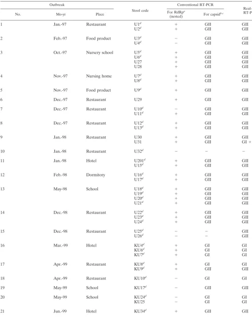
TABLE 1. Detection of NLVs by using conventional RT-PCRs and real-time quantitative RT-PCR with 81 EM-positive stool specimens from
36 outbreaks of nonbacterial acute gastroenteritis
Outbreak
Stool code
Conventional RT-PCR
Real-time
RT-PCRb
No. Mo-yr Place For RdRp(nested)a For capsidb,c
1
Jan.-97
Restaurant
U1
d⫹
GII
GII
U2
d⫹
GII
GII
2
Feb.-97
Food product
U3
d⫺
GII
GII
U4
d⫺
GII
GII
3
Oct.-97
Nursery school
U5
d⫹
GII
GII
U6
d⫹
GII
GII
U27
⫹
GII
GII
U28
⫹
GII
GII
4
Nov.-97
Nursing home
U7
d⫹
GII
GII
U8
d⫹
GII
GII
5
Nov.-97
Food product
U9
d⫹
GII
GII
6
Dec.-97
Restaurant
U29
⫹
GII
GII
7
Dec.-97
Restaurant
U10
d⫺
GII
GII
U11
d⫹
GII
GII
8
Dec.-97
Restaurant
U12
d⫹
GII
GII
U13
d⫹
GII
GII
9
Jan.-98
Restaurant
U30
⫹
GII
GII
U31
⫹
GII
GI
⫹
GII
10
Jan.-98
Restaurant
U32
d⫺
⫺
⫺
11
Jan.-98
Hotel
U201
d⫹
GII
GII
U15
d⫹
GII
GII
12
Feb.-98
Dormitory
U16
d⫹
GII
GII
U17
d⫹
GII
GII
13
May-98
School
U18
d⫹
GII
GII
U19
d⫹
GII
GII
U20
d⫹
GII
GII
U21
d⫹
GII
GII
14
Dec.-98
Restaurant
U22
d⫹
GII
GII
U23
d⫹
GII
GII
U24
d⫹
GII
GII
15
Dec.-98
Restaurant
U25
d⫺
⫺
GII
U26
d⫺
⫺
GII
16
Mar.-99
Hotel
KU4
d⫹
GI
GI
KU6
d⫹
GI
GI
KU7
d⫹
GI
GI
17
Apr.-99
Restaurant
KU8
d⫹
GI
GI
KU9
d⫹
GII
GII
18
Apr.-99
Restaurant
KU10
d⫺
GI
GI
19
May-99
School
KU17
d⫺
GII
GII
20
May-99
School
KU24
d⫺
GI
GI
KU25
⫺
GI
GI
21
Jun.-99
Hotel
KU34
d⫹
GII
GII
22
Jun.-99
Food product
KU44
d⫹
GII
GII
Continued on following page
on May 15, 2020 by guest
http://jcm.asm.org/
Saitama U3 (U3), AB039776; Saitama U4 (U4), AB039777; Saitama U16 (U16), AB039778; Saitama U17 (U17), AB039779; Saitama U18 (U18), AB039781; Saitama U201 (U201), AB039782; and Saitama U25 (U25), AB039780. The nucleotide sequences of the ORF1-ORF2 junctions were submitted to DDBJ; the accession numbers were AB058511 to AB058529 and AB058534 to AB058598.
RESULTS
PlotSimilarity analysis.
Our first goal was to identify a stable
region suitable for designing new primers and probes for
RT-PCR amplification. To identify the region with highest
similar-TABLE 1—
Continued
OutbreakStool code
Conventional RT-PCR
Real-time
RT-PCRc
No. Mo-yr Place For RdRp(nested)a For capsidb,c
23
Oct.-99
Restaurant
KU45
⫺
⫺
GII
24
Oct.-99
Nursery school
KU62
d⫹
⫺
GII
KU63
d⫺
⫺
GII
KU64
d⫺
⫺
GII
KU66
d⫹
⫺
GII
25
Nov.-99
School
KU68
d⫹
GII
GII
26
Nov.-99
Restaurant
eKU80
d⫹
GI
⫹
GII
GI
⫹
GII
KU82
d⫹
GI
⫹
GII
GI
⫹
GII
KU83
d⫹
GI
⫹
GII
GI
⫹
GII
KU84
d⫹
GII
GII
27
Dec.-99
School party
KU85
d⫺
GII
GII
KU88
d⫹
GII
GII
KU89
d⫹
⫺
GII
KU90
⫹
GII
GII
KU91
⫹
GII
GII
28
Dec.-99
School
KU93
d⫹
GII
GII
29
Dec.-99
Restaurant
KU98
d⫹
GII
GII
KU99
d⫺
GII
KU101
d⫹
GII
GII
30
Dec.-99
Restaurant
KU105
d⫹
GI
⫹
GII
GI
⫹
GII
KU106
⫹
GI
⫹
GII
GI
⫹
GII
KU109
d⫹
GI
⫹
GII
GI
⫹
GII
KU111
d⫺
GI
⫹
GII
GI
⫹
GII
KU112
d⫺
GI
GI
KU115
d⫺
GI
GI
31
Jan.-00
Food product
KU5
d⫹
⫺
GII
32
Jan.-00
Family
KU16
d⫹
GII
GII
33
Jan.-00
Restaurant
eKU18
d⫹
GII
GII
KU19
d⫹
GI
⫹
GII
GI
⫹
GII
KU26
d⫹
GII
GII
KU27
d⫺
GII
GII
34
Mar.-00
Restaurant
KU31
d⫹
⫺
GII
KU32
d⫹
⫺
GII
35
Mar.-00
Restaurant
eKU35
d⫹
GII
GII
KU36
d⫹
GI
⫹
GII
GI
⫹
GII
KU37
d⫹
⫺
GII
36
Mar.-00
Hotel
KU49
d⫹
GII
GII
KU53
d⫹
GII
GII
Total
81
62
67
80
Detection rate (%)
77
83
99
a⫹, positive;⫺, negative. Method described in reference 31.
bMethod described in reference 21.
cGI, GI detected; GII, GII detected;⫺, negative.
dUsed for sequence analysis.
eFrom shellfish-related outbreak.
on May 15, 2020 by guest
http://jcm.asm.org/
[image:4.603.45.542.82.609.2]on May 15, 2020 by guest
http://jcm.asm.org/
ity, PlotSimilarity was used to analyze full-length NLV GI
sequences, including those of SzUGI, Norwalk/68,
Southamp-ton, and BS5 (Fig. 1A), and NLV GII sequences, including
those of U1, U3, U4, U16, U17, U18, U201, U25, Lordsdale,
and Camberwell (Fig. 1B). The highest nucleotide similarity
score was found in the ORF1-ORF2 junction region (the
C-terminal RdRp to the N-C-terminal capsid) (19). We selected
this region, corresponding to nt 5250 to 5530 in Norwalk/68
(GI) and to nt 4980 to 5260 in Camberwell (GII), as a
prom-ising candidate for the design of real-time quantitative
RT-PCR primers and probes.
Nucleotide sequences of ORF1-ORF2 junction regions from
71 strains.
To confirm that it is highly conserved within each
genogroup, we sequenced the ORF1-ORF2 junction regions of
NLVs isolated from 71 stool specimens (Table 1). We used
RT-PCR and primer sets G1FF-G1SKR and G2FB-G2SKR to
amplify the NLV GI and GII ORF1-ORF2 junction regions,
respectively. All sequences of the ORF1-ORF2 junction region
were determined from at least three clones. The cDNA
se-quences for 70 samples were submitted to DDBJ (see
Materi-als and Methods). Only one stool specimen, stool code U32,
was not amplified by this RT-PCR.
Multiple alignment of the ORF1-ORF2 junction region was
performed on each genogroup and with available sequences in
the database (Fig. 2). Some stool specimens appeared to
con-tain both NLV GI and GII sequences (e.g., KU80 and KU19)
(Fig. 2). Four stool specimens (i.e., KU4, KU80, KU105, and
KU19) contained two or more NLVs with sequence variants
belonging to the same genogroup (Fig. 2). Multiple alignment
of the ORF1-ORF2 junction region revealed that the most
highly conserved nucleotide sequences were nt 5279 to 5381
(102 bases) with respect to Norwalk/68 virus (M87661) among
NLV GI and nt 4988 to 5108 (120 bases) with respect to
Camberwell virus (AF145896) among NLV GII. In this region,
the nucleotide identities within the genogroups were 88 to
100% and 86 to 100%, respectively. Interestingly, 20 nt (nt
5357 to 5376 in Norwalk/68) or 21 nt (nt 5080 to 5100 in
Camberwell) overlapped by ORF1 and ORF2 exhibited a
per-fect match within NLV GI or GII, respectively.
Development of real-time quantitative RT-PCR.
We
de-signed genogroup-specific primer sets and fluorescent probes
in the NLV ORF1-ORF2 junction region for use in real-time
quantitative RT-PCR. The primer pair COG1F-COG1R and a
mixture of fluorescent probes, RING1(a)-TP and
RING1(b)-TP, were used to detect NLV GI. The primer pair
COG2F-COG2R and the fluorescent probe RING2-TP were used to
detect GII NLVs. The locations of the primers and fluorescent
probes are illustrated in Fig. 2, and the nucleotide sequences
are shown in Table 2.
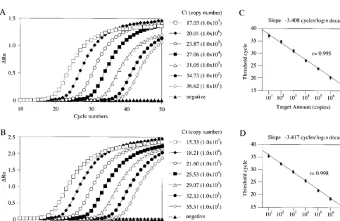
To determine the dynamic range of the real-time
quantita-tive RT-PCR, we generated standard curves with 10-fold serial
dilutions of NLV GI and GII standard plasmids (from 10
7to
10
1copies) (Fig. 3). The amplification curves shifted to the
right as the initial amount of the plasmid was reduced (Fig. 3A
and B). We constructed standard curves of the threshold cycle
(Ct) of the NLV GI and GII standard plasmid against the
amount of plasmid (Fig. 3C and D). The correlation between
the Ct and the amount of target template was good between
10
7and 10
1copies. Cross-reactivity between GI and GII was
not observed when the plasmid standards were used.
FIG.
2.
Alignment
of
nucleotide
sequences
and
partial
predicted
amino
acid
sequences
of
the
ORF1-ORF2
junctions
of
NLV
GI
(A)
and
GII
(B).
The
nucleoti
de
sequences
of
NLV
GI
were
aligned
from
nt
5254
to
5425
in
Norwalk/68,
and
those
of
NLV
GII
were
aligned
from
nt
4981
to
5152
in
Camberwell.
The
strain
name
and
accession
number
are
shown
beside
its
sequence;
a
lowercase
letter
in
the
strain
name
indicates
a
dif
ferent
clone
from
the
same
stool
specimen.
Some
strains
with
identical
sequence
are
omitted
from
the
figure.
The
conserved
amino
acid
sequences
of
NLV
GI
ORF1
are
indicated
above
the
sequences
between
nt
5279
and
5371
in
Norwalk/68,
and
those
of
ORF2
are
indicated
between
nt
5358
and
538
1.
The
conserved
amino
acid
sequences
of
NLV
GII
ORF1
are
indicated
above
the
nucleotide
sequences
between
nt
4988
and
5101
in
Camberwell,
and
those
of
ORF2
are
indicated
betw
een
nt
5085
and
5108.
The
asterisks
below
the
alignment
show
consensus
nucleotide
sequences.
Arrows
and
double
lines
show
locations
of
newly
designed
primers
and
probes,
res
pectively,
used
in
this
study.
on May 15, 2020 by guest
http://jcm.asm.org/
Quantitative detection of NLV RNA.
Real-time quantitative
RT-PCR was used to detect NLV RNAs from 81 EM-positive
stool specimens for evaluation (Table 1). NLV RNA was
de-tected in 80 (99%) of 81 stool specimens, whereas conventional
RT-PCR detected NLV in 62 of 81 stool specimens (77%) for
the RdRp and in 67 of 81 (83%) for the capsid N/S region
(Table 1). All three RT-PCR methods failed to detect NLV
RNA from stool code U32.
[image:7.603.45.543.81.174.2]Of 80 real-time quantitative RT-PCR-positive and
EM-pos-itive stool specimens, nine contained NLV GI viruses and 61
contained NLV GII viruses. Interestingly, 10 stool specimens
(U31, KU80, KU82, KU83, KU105, KU106, KU109, KU111,
[image:7.603.52.535.349.663.2]FIG. 3. Real-time RT-PCR quantification of NLV GI and GII standard plasmids. (A and B) Amplification plots of fluorescence intensities
(
⌬
Rn) versus the PCR cycle numbers are displayed for serial 10-fold dilutions of standard plasmids (10
7to 10
1copy equivalents per reaction) for
NLV GI (A) and GII (B). Each plot corresponds to a particular input target quantity marked by a corresponding symbol. Results are the average
of those from three reactions. (C and D) Relationship of known numbers of NLV GI (C) and GII (D) standard plasmids to the threshold cycle
(Ct). The Ct is directly proportional to the log of the input copy equivalents, as demonstrated by the standard curve. Results are the average of
those from three reactions, and error bars indicate standard deviations.
TABLE 2. Primer and probe oligonucleotides used for real-time quantitative RT-PCR
Genogroup Primer or probe Sequence (5⬘33⬘)a Polarityb Location
GI
Primer COG1F
CGYTGGATGCGNTTYCATGA
⫹
5291
ePrimer COG1R
CTTAGACGCCATCATCATTYAC
⫺
5375
eProbe
cRING1(a)-TP
FAM-AGATYGCGATCYCCTGTCCA-TAMRA
d⫺
5340
eRING1(b)-TP
FAM-AGATCGCGGTCTCCTGTCCA-TAMRA
⫺
5340
eGII
Primer COG2F
CARGARBCNATGTTYAGRTGGATGAG
⫹
5003
fPrimer COG2R
TCGACGCCATCTTCATTCACA
⫺
5100
fProbe RING2-TP
FAM-TGGGAGGGCGATCGCAATCT-TAMRA
⫹
5048
faMixed bases in degenerate primers and probes are as follows: Y, C or T; R, A or G; B, not A; N, any.
b⫹, virus sense;⫺, anti-virus sense.
cMixed probes are used for the GI NLVs.
d6-Carboxyfluorescein (FAM) as the reporter dye is coupled in the 5⬘end of the oligonucleotide, and 6-carboxy-tetramethylrhodamine (TAMRA) as the quencher
dye is coupled in the 3⬘end of the oligonucleotide.
eCorresponding nucleotide position of Norwalk/68 virus (accession no. M87661) of the 5⬘end.
fCorresponding nucleotide position of Camberwell virus (accession no. AF145896) of the 5⬘end.
on May 15, 2020 by guest
http://jcm.asm.org/
KU19, and KU36) from five EM-positive outbreaks (outbreaks
9, 26, 30, 33, and 35) contained a mixture of both NLV GI and
GII (Table 1).
The detection limits of NLV GI and GII RNA had Ct values
of 36.6 and 35.3, respectively, corresponding to 10 copies per
reaction tube. These Ct values corresponded to more than 2.0
⫻
10
4copies of NLV RNA per g of stool specimen. For each
gram of stool specimen in EM-positive specimens, Ct values of
17.7 to 30 and 14.1 to 23.8 corresponded to 1.4
⫻
10
10to 3.2
⫻
10
6and 6.3
⫻
10
10to 9.2
⫻
10
7copies/g of NLV GI and GII
RNAs, respectively.
We also used our method to test EM-negative stool
speci-mens from nine EM-positive outbreaks (outbreaks no. 8, 9, 14,
16, 19, 27, 28, 33 and 34) and one oyster-related but
EM-negative outbreak (outbreak 37). Of 28 samples tested, 20 were
NLV RNA positive by our method (Table 3). Ct values were
31.9 (9.6
⫻
10
5copies/g) for NLV GI (SzU) and 21.0 to 33.3
(6.1
⫻
10
8to 1.5
⫻
10
5copies/g) for NLV GII.
Since the real-time quantitative RT-PCR method was
de-signed to distinguish NLV GI and GII, we confirmed the
geno-group by sequencing the capsid N/S region of the PCR
prod-ucts from the 70 stool specimens. Using these sequence data,
we constructed the phylogenetic dendrogram and found that
the genogroups resulting from the phylogenetic analysis
matched completely with the results from the real-time
RT-PCR method (the dendrograms for the majority of the strains
sequenced were shown in our previous studies [19, 21]).
In this real-time quantitative RT-PCR, we observed no
cross-reactions in stool specimens containing other enteric
vi-ruses, such as rotavirus, enterovirus, adenovirus, and
poliovi-rus, or enteropathogenic bacteria, such as
Escherichia coli
,
Staphylococcus aureus
,
Campylobacter jejuni
, and
Vibrio
parah-aemolyticus
(data not shown).
DISCUSSION
We have established a sensitive and broadly reactive NLV
RNA detection system that uses real-time quantitative
RT-PCR. The primers were based on the ORF1-ORF2 junction
region, perhaps the most conserved region of the virus
ge-nome. Similarity plots (Fig. 1) and multiple alignment analysis
of sequences in this region (Fig. 2) indicated a high degree of
conservation in all isolates sequenced. Thus, it seemed an ideal
site for designing primers for an RT-PCR-based amplification
assay. The system does not require any other detection
proce-dures, such as electrophoresis, Southern hybridization, nested
PCR, or sequence analysis. Moreover, to prevent
false-posi-tives from carryover contamination, we treated all pre-PCR
samples with UNG to destroy any dUTP-containing PCR
products (24, 25, 29).
Because our method uses the TaqMan system, we could
quantitatively detect NLV cDNA. Linearity was obtained by
use of serially diluted standard DNA plasmids (10
7copies to
10
1copies) containing the ORF1-ORF2 junction sequence of
each genogroup (Fig. 3). With serially diluted standard DNA
plasmids, we found a detection limit of 10 copies of cDNA per
reaction tube with a cutoff value of 35 to 37 cycles, indicating
that our method is highly sensitive. For example, the slopes of
the curves of threshold cycle (Ct) values versus serial 10-fold
dilutions (range of 10
10to 10
1) for KU4 (GI), KU115 (GI),
KU98 (GII), and U201 (GII) suspended stool specimen were
⫺
3.381,
⫺
3.411,
⫺
3.422, and
⫺
3.412 cycles/log
10unit,
respec-tively. These slopes correlated well with the slopes of the
curves of Ct versus serially diluted NLV GI (
⫺
3.4075 cycles/
log
10unit) and GII (
⫺
3.4166 cycles/log
10unit) standard DNA
plasmid (
P
⬍
0.01). We estimated the number of copies of
NLV RNA in the original sample. Ct values of 17.0 (GI) and
15.3 (GII) are equivalent to 1
⫻
10
7copies of standard plasmid
in the reaction tube and correspond to about 2.0
⫻
10
10copies
of NLV RNA per g of stool specimen. Ct values for stool codes
U2, KU44, KU109, and KU111 of 20.1, 14.1, 16.7, and 23.8
corresponded to about 1.1
⫻
10
9, 6.3
⫻
10
10, 1.1
⫻
10
10, and
9.2
⫻
10
7copies of NLV RNA per g of stool specimen,
respec-tively.
The numbers of copies of NLV RNA in positive stool
spec-imens determined by the real-time quantitative RT-PCR were
considerably higher than previous estimates of 10
5to 10
6cop-ies by EM (17). The higher titers are probably due to the ability
of RT-PCR to detect NLV RNAs from additional sources
(e.g., from complete and incomplete virus particles and from
amplifiable RNA inside the infected intestinal epithelial cells).
Our findings are also consistent with those of others who found
higher numbers of NLV RNA by PCR methods (2, 13) than by
EM.
Given the greater sensitivity of our assay, we wanted to
examine more carefully EM-negative samples from
gastroen-teritis subjects within the same NLV-positive outbreaks. Of 28
EM-negative specimens, 20 were positive for NLV RNA
(Ta-ble 3). In our assay, EM-negative, RT-PCR-positive stool
spec-imens contained 3 to 30% as much NLV RNA as the
EM-positive stool specimens.
[image:8.603.43.284.90.227.2]The one outlier in this analysis was stool code U32 (outbreak
10), which was positive by EM (i.e., showed an image
resem-bling that of an NLV particle) but negative by all RT-PCR
methods. We speculate that the reason might be the presence
of an unidentified small round virus that is morphologically
similar to NLVs by EM. Conventional RT-PCR methods did
not detect other RNA viruses, such as hepatitis A virus,
hep-atitis E virus, poliovirus, Sapporo-like viruses, and Astro virus
(data not shown). Further investigation is needed to explain
the results for this sample.
TABLE 3. Detection of NLVs by real-time quantitative RT-PCR
against NLV EM-negative stool specimens
Outbreak no. Detection by TaqMan (no.positive/no. tested)a Genogroup
8
0/2
9
2/3
GII
14
3/3
GII
16
1/1
GI
19
7/8
GII
27
2/2
GII
28
1/3
GII
33
2/3
GII
34
1/2
GII
37
b1/1
cGI
⫹
GII
Total
20/28
aAll stool specimens were NLV negative by EM.
bFrom an oyster-related outbreak (February 1999) not included in Table 1.
cStool code SzU.
on May 15, 2020 by guest
http://jcm.asm.org/
Interestingly, 10 stool specimens from five EM-positive
out-breaks (outout-breaks 9, 26, 30, 33, and 35) (Table 1), and one
stool specimen from outbreak 37 that was negative by EM but
positive in our assay (Table 3), showed NLV GI and GII
strains. All of these outbreaks occurred in restaurants, and we
suspect that they have resulted from ingesting contaminated
foods. Four outbreaks (outbreaks 26, 33, 35 and 37) were
shellfish related. Similar results of coinfection have been
re-ported previously (36). Oysters, cockles, and mussels have
been implicated in food-borne outbreaks. Shellfish are
filter-feeding organisms and can concentrate NLV particles from a
contaminated habitat. Therefore, coinfection by multiple
strains is possible in shellfish-related NLV outbreaks.
The high sensitivity of our method allowed us to detect
NLVs that were undetectable by EM or other PCR methods.
Our method may have applications in the detection of NLV
pathogens in foods, such as oysters, as well as in the monitoring
of the levels and seasonal differences of NLV contamination in
sewage, river water, and seawater. Furthermore, our primer
and probe sequences may be used in other detection methods
such as the Light Cycler PCR technique. We believe that this
method will be useful for routine diagnosis as well as for
clarifying the epidemiology of NLV infections and thus for the
public health control of this disease in the future.
ACKNOWLEDGMENTS
We thank Sakae Inouye (Otsuma Women’s University, Tokyo,
Ja-pan) for critical review of the manuscript.
This work was supported by Research on Health Sciences focusing
on Drug Innovation grant KH51048 from the Japan Health Sciences
Foundation.
REFERENCES
1. Ando, T., S. S. Monroe, J. R. Gentsch, Q. Jin, D. C. Lewis, and R. I. Glass.
1995. Detection and differentiation of antigenically distinct small round-structured viruses (Norwalk-like viruses) by reverse transcription-PCR and
Southern hybridization. J. Clin. Microbiol.33:64–71.
2. Atmar, R. L., F. H. Neill, J. L. Romalde, F. Le Guyader, C. M. Woodley, T. G. Metcalf, and M. K. Estes.1995. Detection of Norwalk virus and hepatitis A
virus in shellfish tissues with the PCR. Appl. Environ. Microbiol.61:3014–
3018.
3. Beller, M., A. Ellis, S. H. Lee, M. A. Drebot, S. A. Jenkerson, E. Funk, M. D. Sobsey, O. D. Simmons, S. S. Monroe, T. Ando, J. Noel, M. Petric, J. P. Middaugh, and J. S. Spika.1997. Outbreak of viral gastroenteritis due to a
contaminated well. International consequences. JAMA278:563–568.
4. Berg, D. E., M. A. Kohn, T. A. Farley, and L. M. McFarland.2000. Multi-state outbreaks of acute gastroenteritis traced to fecal-contaminated oysters
harvested in Louisiana. J. Infect. Dis.181(Suppl. 2):S381–S386.
5. Caul, E. O., and H. Appleton.1982. The electron microscopical and physical characteristics of small round human fecal viruses: an interim scheme for
classification. J. Med. Virol.9:257–265.
6. De Leon, R., S. M. Matsui, R. S. Baric, J. E. Herrmann, N. R. Blacklow, H. B. Greenberg, and M. D. Sobsey.1992. Detection of Norwalk virus in stool specimens by reverse transcriptase-polymerase chain reaction and
nonradio-active oligoprobes. J. Clin. Microbiol.30:3151–3157.
7. Dingle, K. E., P. R. Lambden, E. O. Caul, and I. N. Clarke.1995. Human enteric Caliciviridae: the complete genome sequence and expression of virus-like particles from a genetic group II small round structured virus. J. Gen.
Virol.76:2349–2355.
8. Dowell, S. F., C. Groves, K. B. Kirkland, H. G. Cicirello, T. Ando, Q. Jin, J. R. Gentsch, S. S. Monroe, C. D. Humphrey, C. Slemp, et al.1995. A multistate outbreak of oyster-associated gastroenteritis: implications for
in-terstate tracing of contaminated shellfish. J. Infect. Dis.171:1497–1503.
9. Green, J., C. I. Gallimore, J. P. Norcott, D. Lewis, and D. W. Brown.1995. Broadly reactive reverse transcriptase polymerase chain reaction for the
diagnosis of SRSV-associated gastroenteritis. J. Med. Virol.47:392–398.
10. Green, S. M., K. E. Dingle, P. R. Lambden, E. O. Caul, C. R. Ashley, and I. N. Clarke.1994. Human enteric Caliciviridae: a new prevalent small round-structured virus group defined by RNA-dependent RNA polymerase
and capsid diversity. J. Gen. Virol.75:1883–1888.
11. Green, S. M., P. R. Lambden, E. O. Caul, and I. N. Clarke.1997. Capsid
sequence diversity in small round structured viruses from recent UK
out-breaks of gastroenteritis. J. Med. Virol.52:14–19.
12. Gunn, R. A., W. A. Terranova, H. B. Greenberg, J. Yashuk, G. W. Gary, J. G. Wells, P. R. Taylor, and R. A. Feldman.1980. Norwalk virus gastroenteritis aboard a cruise ship: an outbreak on five consecutive cruises. Am. J.
Epide-miol.112:820–827.
13. Hafliger, D., M. Gilgen, J. Luthy, and P. Hubner.1997. Seminested RT-PCR systems for small round structured viruses and detection of enteric viruses in
seafood. Int. J. Food Microbiol.37:27–36.
14. Inouye, S., K. Yamashita, S. Yamadera, M. Yoshikawa, N. Kato, and N. Okabe.2000. Surveillance of viral gastroenteritis in Japan: pediatric cases
and outbreak incidents. J. Infect. Dis.181(Suppl. 2):S270–S274.
15. Jiang, X., P. W. Huang, W. M. Zhong, T. Farkas, D. W. Cubitt, and D. O. Matson.1999. Design and evaluation of a primer pair that detects both
Norwalk- and Sapporo-like caliciviruses by RT-PCR. J. Virol. Methods83:
145–154.
16. Jiang, X., M. Wang, K. Wang, and M. K. Estes.1993. Sequence and genomic
organization of Norwalk virus. Virology195:51–61.
17. Kapikian, A. Z., and R. M. Chanock.1990. Norwalk group viruses, p. 671–
693.InB. N. Fields, D. M. Knipe, R. M. Chanock, M. S. Hirsch, J. L.
Melnick, T. P. Monath, and B. Roizman (ed.), Virology, 2nd ed., vol. 1. Raven Press, New York, N.Y.
18. Kaplan, J. E., L. B. Schonberger, G. Varano, N. Jackman, J. Bied, and G. W. Gary.1982. An outbreak of acute nonbacterial gastroenteritis in a nursing home. Demonstration of person-to-person transmission by temporal
cluster-ing of cases. Am. J. Epidemiol.116:940–948.
19. Katayama, K., H. Shirato-Horikoshi, S. Kojima, T. Kageyama, T. Oka, F. B. Hoshino, S. Fukushi, M. Shinohara, K. Uchida, Y. Suzuk, T. Gojobori, and N. Takeda.2002. Phylogenetic analysis of the complete genome of 18
Nor-walk-like viruses. Virology299:225–239.
20. Kobayashi, S., T. Morishita, T. Yamashita, K. Sakae, O. Nishio, T. Miyake, Y. Ishihara, and S. Isomura.1991. A large outbreak of gastroenteritis asso-ciated with a small round structured virus among schoolchildren and
teach-ers in Japan. Epidemiol. Infect.107:81–86.
21. Kojima, S., T. Kageyama, S. Fukushi, F. B. Hoshino, M. Shinohara, K. Uchida, K. Natori, N. Takeda, and K. Katayama.2002. Genogroup-specific
PCR primers for detection of Norwalk-like viruses. J. Virol. Methods100:
107–114.
22. Lambden, P. R., E. O. Caul, C. R. Ashley, and I. N. Clarke.1993. Sequence and genome organization of a human small round-structured (Norwalk-like)
virus. Science259:516–519.
23. Lew, J. F., M. Petric, A. Z. Kapikian, X. Jiang, M. K. Estes, and K. Y. Green.
1994. Identification of minireovirus as a Norwalk-like virus in pediatric
patients with gastroenteritis. J. Virol68:3391–3396.
24. Longo, M. C., M. S. Berninger, and J. L. Hartley.1990. Use of uracil DNA glycosylase to control carry-over contamination in polymerase chain
reac-tions. Gene93:125–128.
25. McGoldrick, A., E. Bensaude, G. Ibata, G. Sharp, and D. J. Paton.1999. Closed one-tube reverse transcription nested polymerase chain reaction for the detection of pestiviral RNA with fluorescent probes. J. Virol. Methods
79:85–95.
26. Moe, C. L., J. Gentsch, T. Ando, G. Grohmann, S. S. Monroe, X. Jiang, J. Wang, M. K. Estes, Y. Seto, C. Humphrey, et al.1994. Application of PCR to detect Norwalk virus in fecal specimens from outbreaks of gastroenteritis.
J. Clin. Microbiol.32:642–648.
27. Nakayama, M., Y. Ueda, H. Kawamoto, Y. Han-jun, K. Saito, O. Nishio, and H. Ushijima.1996. Detection and sequencing of Norwalk-like viruses from stool samples in Japan using reverse transcription-polymerase chain reaction
amplification. Microbiol. Immunol.40:317–320.
28. Noel, J. S., T. Ando, J. P. Leite, K. Y. Green, K. E. Dingle, M. K. Estes, Y. Seto, S. S. Monroe, and R. I. Glass.1997. Correlation of patient immune responses with genetically characterized small round-structured viruses in-volved in outbreaks of nonbacterial acute gastroenteritis in the United
States, 1990 to 1995. J. Med. Virol.53:372–383.
29. Pellett, P. E., T. J. Spira, O. Bagasra, C. Boshoff, L. Corey, L. de Lellis, M. L. Huang, J. C. Lin, S. Matthews, P. Monini, P. Rimessi, C. Sosa, C. Wood, and J. A. Stewart.1999. Multicenter comparison of PCR assays for detection of
human herpesvirus 8 DNA in semen. J. Clin. Microbiol.37:1298–1301.
30. Pether, J. V., and E. O. Caul.1983. An outbreak of food-borne
gastroen-teritis in two hospitals associated with a Norwalk-like virus. J. Hyg.91:343–
350.
31. Saito, H., S. Saito, K. Kamada, S. Harata, H. Sato, M. Morita, and Y. Miyajima.1998. Application of RT-PCR designed from the sequence of the local SRSV strain to the screening in viral gastroenteritis outbreaks.
Micro-biol. Immunol.42:439–446.
32. Schreier, E., F. Doring, and U. Kunkel.2000. Molecular epidemiology of outbreaks of gastroenteritis associated with small round structured viruses in
Germany in 1997/98. Arch. Virol.145:443–453.
33. Schvoerer, E., F. Bonnet, V. Dubois, A. M. Rogues, J. P. Gachie, M. E. Lafon, and H. J. Fleury.1999. A hospital outbreak of gastroenteritis possibly related to the contamination of tap water by a small round structured virus. J. Hosp.
Infect.43:149–154.
on May 15, 2020 by guest
http://jcm.asm.org/
34. Seah, E. L., J. A. Marshall, and P. J. Wright.1999. Open reading frame 1 of the Norwalk-like virus Camberwell: completion of sequence and expression
in mammalian cells. J. Virol73:10531–10535.
35. Stevenson, P., R. McCann, R. Duthie, E. Glew, and L. Ganguli.1994. A
hospital outbreak due to Norwalk virus. J. Hosp. Infect.26:261–272.
36. Sugieda, M., K. Nakajima, and S. Nakajima.1996. Outbreaks of Norwalk-like virus-associated gastroenteritis traced to shellfish: coexistence of two
genotypes in one specimen. Epidemiol. Infect.116:339–346.
37. Vinje, J., and M. P. Koopmans.1996. Molecular detection and epidemiology
of small round-structured viruses in outbreaks of gastroenteritis in the
Neth-erlands. J. Infect. Dis.174:610–615.
38. Wang, J., X. Jiang, H. P. Madore, J. Gray, U. Desselberger, T. Ando, Y. Seto, I. Oishi, J. F. Lew, K. Y. Green, et al.1994. Sequence diversity of small,
round-structured viruses in the Norwalk virus group. J. Virol.68:5982–5990.
39. Yamazaki, K., M. Oseto, Y. Seto, E. Utagawa, T. Kimoto, Y. Minekawa, S. Inouye, S. Yamazaki, Y. Okuno, and I. Oishi.1996. Reverse transcription-polymerase chain reaction detection and sequence analysis of small
round-structured viruses in Japan. Arch. Virol. Suppl.12:271–276.