EFFECT OF BRACKET TYPE AND WIRE DIMENSION ON ORTHODONTIC ALIGNMENT: AN ANALYSIS USING 3D IMAGING
Christian G. Piers
A thesis submitted to the faculty at the University of North Carolina at Chapel Hill in partial fulfillment of the requirements for the degree of Master of Science in the School of Dentistry
(Orthodontics).
Chapel Hill 2019
ABSTRACT
Christian G. Piers: Effect of Bracket Type and Wire Dimension on Orthodontic Alignment: An Analysis using 3D Imaging
(Under the direction of Ching-Chang Ko)
Many different brackets and archwires exist to align the teeth in orthodontics. We hypothesized that alignment efficiency is affected by wire dimension, bracket type and jaw. Accordingly, two clinical trials were conducted to compare: 1) customized to non-customized brackets; and 2) conventional brackets to self-ligating brackets in a customized system. Subjects were randomized to either .014” or .016” archwires. Intraoral scans were obtained at 3 time points and 3-way ANOVA was performed. In .016” wires, the mean reduction in malalignment in the customized group (mean=3.2mm, SD=1.9mm) was greater than that of the non-customized group (mean=2.2mm, SD=1.4mm) from 0-6 weeks (p=0.025). Conventional brackets were more efficient than self-ligating brackets from 6-12 weeks (p=.014). However, there were no
ACKNOWLEDGEMENTS
Thank you to my committee members, Dr. Ko, Dr. Wu, Dr. Lin, and Dr. Medland, for your expertise, guidance, and advice throughout my project. Thank you to Dr. Bryan
Whitecotton and Michael Touloupas for your research assistance. Thank you to Dr. Robert Selden for your assistance with recruitment and treatment in the private practice setting. Thank you to Dr. Haiping Zhang for your laboratory assistance. Thank you to Dr. Francisca Durán for your assistance with outcome assessment. Thank you to the Southern Association of
TABLE OF CONTENTS
LIST OF TABLES ... vii
LIST OF FIGURES ... viii
LIST OF ABBREVIATIONS ... ix
LIST OF SYMBOLS ...x
COMPARATIVE ASSESSMENT OF ALIGNMENT EFFICIENCY USING LITTLE INDEX BETWEEN .014” AND .016” INITIAL ARCHWIRES WITH CUSTOMIZED AND NON-CUSTOMIZED BRACKETS IN ADOLESCENTS AND ADULTS: A RANDOMIZED CONTROLLED TRIAL ...1
Introduction ...1
Materials and Methods ...5
Results ...9
Discussion ...10
Conclusions ...21
Tables ...23
Figures...27
COMPARATIVE ASSESSMENT OF ALIGNMENT EFFICIENCY USING LITTLE INDEX BETWEEN .014” AND .016” INITIAL ARCHWIRES WITH CUSTOMIZED
CONVENTIONAL BRACKETS AND CUSTOMIZED SELF-LIGATING BRACKETS IN
ADOLESCENTS AND ADULTS: A RANDOMIZED CONTROLLED TRIAL ...32
Introduction ...32
Materials and Methods ...36
Results ...41
Discussion ...42
Conclusions ...48
Tables ...50
Figures...51
LIST OF TABLES
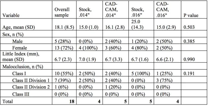
Table 1 – Demographic baseline characteristics of sample ...23
Table 2 – Overall Little Index at baseline (T0), 6 weeks (T1) and 12 weeks (T2) ...23
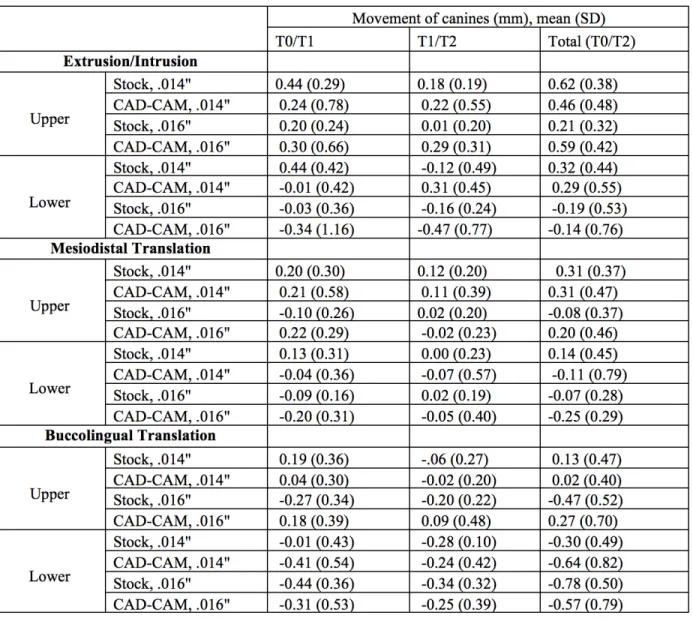
Table 3 – Linear movement of canines in three dimensions from baseline (T0) to 6 weeks (T1) and from 6 weeks to 12 weeks (T2) by archwire, bracket and jaw ...24
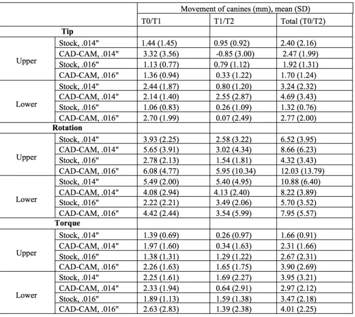
Table 4 – Rotational movement of canines in three dimensions from baseline (T0) to 6 weeks (T1) and from 6 weeks to 12 weeks (T2) by archwire, bracket and jaw ...25
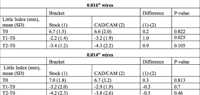
Table 5 – Comparison of baseline Little Index and change in Little Index with p-values during the first six weeks and total 12 weeks for stock and CAD/CAM bracket groups with archwire dimension held constant ...26
Table 6 – Demographic baseline characteristics of sample ...50
Table 7 – Number of patients with one de-bonded anterior bracket during study period ...50
LIST OF FIGURES
Figure 1 – Consort diagram of the flow of participants through the trial.
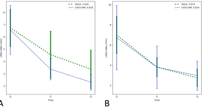
N, amount of patients ...27 Figure 2 – Mean + 1 SD of Little Index reduction based on archwire dimension ...28 Figure 3 – Mean + 1 SD of Little Index reduction based on bracket type for:
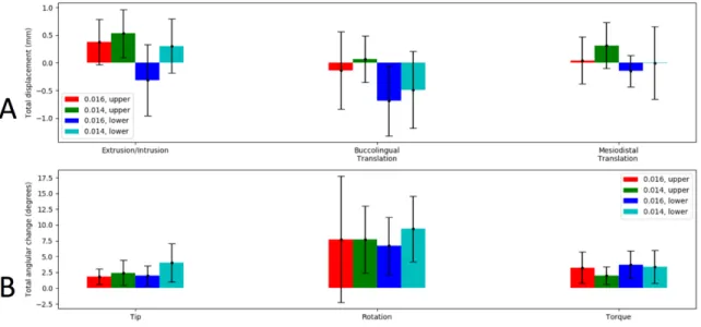
A) .016” archwires; and B) .014” archwires ...28 Figure 4 – Mean + 1 SD of total canine movement measurements
with 6 degrees of freedom based on archwire dimension and jaw, including: A) translational movement along 3 axes; and B) rotational
movement around 3 axes ...29 Figure 5 – Correlation between initial maximum malrotation
and rotational change for canines treated with: A) CAD/CAM
brackets and .016” archwires in the upper arch, and B) stock brackets
and .014” archwires in the lower arch ...21 Figure 6 – Consort diagram of the flow of participants through the trial.
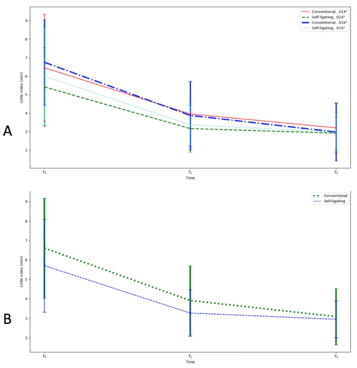
N, number of dental arches ...51 Figure 7 – Mean + 1 SD of Little Index reduction based on:
A) Bracket type and archwire dimension; and B) Bracket type alone ...52 Figure 8 – Mean + 1 SD of total upper right central incisor movements
with 6 degrees of freedom including translational movement along 3 axes and rotational movement around 3 axes based on: A) Bracket type
LIST OF ABBREVIATIONS
3D Three Dimensional
LIST OF SYMBOLS
© Copyright Symbol
® Registered Trademark
COMPARATIVE ASSESSMENT OF ALIGNMENT EFFICIENCY USING LITTLE INDEX BETWEEN .014” AND .016” INITIAL ARCHWIRES WITH CUSTOMIZED
AND NON-CUSTOMIZED BRACKETS IN ADOLESCENTS AND ADULTS: A RANDOMIZED CONTROLLED TRIAL
Introduction
when archwire diameter was increased from .014” to .016”.2 This implies a potential for clinical significance of both archwire dimension and bracket type.
Digital technologies including 3D scanning and CAD/CAM (computer-aided
design/computer aided manufacturing) have made bracket type—and specifically, customized orthodontic appliances— another possibility for increasing efficiency of treatment. Several systems now exist that use intraoral scans and digital tooth setups to correct malocclusions using computer software in order to streamline mechanics towards pre-established results.3 One such system is Insignia™ (Ormco©, Orange, Calif), which aims to reduce time spent finishing cases through the use of customized indirect-bonding jigs, customized brackets and customized archwires.4
To use this CAD/CAM system, intraoral scans are used by technicians to create
preliminary digital tooth setups. After orthodontists correct and approve these setups using the Insignia™ Approver software, twin brackets without slots are placed digitally on all of the teeth,
and slots are cut into the brackets based on their positions in the setup. Bracket bases are uniform,3 but the slots are individualized to each patient and cut to the necessary in-out, tip, and torque. These brackets are then placed using indirect-bonding jigs that are 3D-milled to fit the occlusal surfaces of the teeth.4
A follow-up retrospective study by Brown et al. evaluated 96 patients treated in a single practice with either direct-bonded stock brackets, bonded stock brackets, or indirect-bonded CAD/CAM brackets. There were no differences in ABO scores. While the total treatment time for the CAD/CAM group was 8 months shorter than the direct-bonded group, it was only 3 months shorter than the indirect-bonding group,6 which implies indirect-bonding may have provided some of the treatment effect.7
More recently, a prospective randomized clinical trial by Penning et al. compared 85 patients treated with customized CAD/CAM brackets to 89 patients treated with direct-bonded stock brackets and found no difference in either treatment time or PAR (Peer Assessment Rating) scores.8
No study has compared Insignia™ twin brackets to indirect-bonded stock twin brackets in order to control for the effect of indirect bonding. Further, all of these studies were performed with PVS impressions used to create digital tooth setups and bonding jigs for the CAD/CAM system, while today teeth are scanned directly using intraoral scanners. This could conceivably affect bracket placement and resultant efficiency.
These studies also all evaluated the total duration of treatment time without reporting data on specific stages of treatment. Measuring results at the end of treatment provides critical
information about overall treatment efficiency, but doesn’t track what was accomplished purely by the CAD/CAD system before bends or repositioned brackets were used to finish cases. Evaluating tooth position during Stage I (leveling and aligning) could provide information about how the system resolves malalignment solely through placement of an archwire with no
While crowding is the classical measurement of malalignment in orthodontics, there is subjectivity involved in identifying an “ideal archform” for each patient.9 The Little Index— which is defined as the sum of five measurements representing the linear distance between adjacent anatomic contact points of the mandibular anterior teeth—was developed to overcome this subjectivity.10 Studies have found that use of digital models obtained from intraoral scans can reduce both the subjectivity and variability associated with choosing contact points, increasing the reliability of the Little Index as a research tool.11,12 Subsequent studies have applied this measurement to maxillary teeth, as well.
Materials and Methods
This prospective, randomized controlled trial was approved by the Biomedical
Institutional Review Board at the University of North Carolina- Chapel Hill (IRB# 13-0924). All patients were recruited from the graduate orthodontic clinic at the University of North
Carolina- Chapel Hill between 2016 and 2017. Prior to initiating recruitment, the effect size in
the sense of Cohen’s f for the material effect was determined based on a previous study14 to be
about 0.4. It was determined that the current study would reach 93.7% power with a total sample
size of 80 dental arches, given that the type I error rate is 0.05 and the correlation among
repeated measurements is 0.5.
The protocol specified a subject age range of 10 years to 45 years, with more specific
inclusion criteria including non-extraction treatment, initial Little Index between 1 and 15mm
(with no spacing), and presence of all permanent anterior teeth. Specific exclusion criteria
included the following: systemic diseases such as diabetes, hypertension, temporomandibular
disorders (TMD) or craniofacial syndromes; periodontal pocketing of anterior teeth greater than
4mm; incisor mandibular plane angle (IMPA) ³ 100 degrees; or any anterior tooth completely
blocked from the arch form.
Preliminary assessment for inclusion in the study was performed by the investigators
using initial records obtained by the treating resident. Patients fulfilling the selection criteria
were recruited at their case presentation appointment, and consent/assent was obtained. The first
9 consecutive patients were enrolled in the “stock” group (Ormco© Mini Diamond® .022” twin
brackets), and the second 9 consecutive patients were enrolled in the “CAD/CAM” group
baseline (T0) 3D intraoral scan was obtained. Conventional indirect bonding setups were
completed for the stock group, and digital setups were completed for the CAD/CAM group. All
setups were verified by an ABO-certified orthodontist experienced in both techniques. For
subjects in each bracket group, randomization to either .014” or .016” initial archwires was
accomplished using a random number generator furnished by a third party to create a 4-arm
parallel study design. Allocations were concealed by third-party distribution of the archwires to
the treating residents in sealed packages that were labeled only with the numbers provided by the
random number generator. The trial was also initially planned to include a group of subjects
treated with archwires with a nano-crystalline ceramic coating. Due to problems with obtaining
the ceramic-coated archwires, this intervention was eliminated from the study.
On bonding day, indirect bonding trays or jigs were distributed to the treating residents
for completion of the bonding appointments. Each patient received the same dimension archwire
for both the maxillary and mandibular arches. Second molars were not engaged, and only
Ormco© silver elastomeric rings were used for engaging the archwires to the brackets. Patients
returned in 6 weeks for the first post-bonding appointment, at which time both archwires were
removed and a second intraoral scan (T1) was completed. The same archwires were then tied
back in. The patients returned in 6 weeks for the second post-bonding appointment. The
archwires were then collected, and the patients underwent a third and final intraoral scan (T2),
which concluded the patient’s participation in the study (Figure 1). No stopping guidelines were
specified, since subjects were receiving the same treatment materials as they would receive as
part of standard of care clinical treatment. Treating clinicians were given the authority to
Bracket type was not blinded in this study, since clear visual differences existed between
the two brackets. Investigators, participants, and outcome assessors were blinded to archwire
dimension for the duration of the study. The statistician was un-blinded to archwire dimension
only after data collection and outcome assessment was complete.
After obtaining all intraoral scans, the STL files generated from the scans were imported
into the Geomagic® Design X64 software (3D Systems©, Rock Hill, SC), where anterior tooth
contact points were precisely determined in order to obtain Little Index values (primary
outcome) for each time point in both arches. Scans were measured in serial sets to reduce
random error.15,16 Best-fit superimpositions were also performed in Geomagic® using palatal
rugae, which have been shown to be stable landmarks for the upper arch.17 In the lower arch,
second molars (which were not engaged with archwires) and gingival tissue lingual to the lower
incisors were used as stable landmarks. These superimpositions were used to assess Euclidean
rigid motion with 6 degrees of freedom for upper and lower canines in each subject (secondary
outcome). Canines were chosen for analysis because they connect the anterior segment of the
dental arch with the posterior segment and represent both a theoretical and literal turning point
within the arch. These 6 degrees of freedom involve translation in 3 planes of space, measured
as the linear distance between a specified point on the canine at each time interval, as well as 3
angular measurements generated from those same points via superimposition of the original and
final spatial orientation. For translational movements along the vertical, mesiodistal, and
buccolingual axes, positive values corresponded to extrusion, distal translation, and lingual
translation, respectively. Rotation around the vertical axis represented the traditional definition
of “rotation” in orthodontics, while the other 2 rotations represented tip and torque. The sizes of
so values corresponding to rotational movements are reported as absolute values without
directionality.
In order to measure initial maximum malrotation of canines, part of the incisor rotation
measurement method described by Charalampakis18was extended to canines. 2 points were
traced on the incisal edges of each lateral incisor, canine and first premolar directly above the
contact points, and the 2 points on each tooth were connected with a straight line. The angles
made between those lines on the mesial and distal of the canines was then measured on the
horizontal plane at baseline. Since these angles are not zero even in perfectly aligned teeth, a
standard angle derived from a perfectly aligned dentaform was subtracted from each measured
value to determine the initial mesial malrotation and initial distal malrotation. The larger of
these two angles was then used as the initial maximum malrotation for each canine.
To assess intraexaminer reliability, six scans were randomly selected from the study
cohort. After Little Index was determined, a wash-out period of three months was allowed
before Little Index was reassessed. The two measurements were highly correlated (Pearson’s
r=0.996, p<0.001), and the mean of the difference between the two measurements was -0.195mm
with 95% CI (-0.502, 0.113), which indicates there is no evidence that the difference was
different from 0.
The study consisted of 18 patients, including 36 dental arches used to evaluate the
primary outcome and 72 canines used to evaluate the secondary outcomes. Baseline
demographics were analyzed using chi-squared tests for categorical variables when comparing
percentages between groups and analysis of variance (ANOVA) for categorical variables when
comparing mean values. 3-way ANOVA was used to evaluate the effect of 3 factors: wire
repeated measure linear regression model was then used to evaluate any correlation between the
initial maximum malrotation and total rotation observed. For all measures, a p-value less than
0.05 was considered statistically significant.
Results
Demographic information regarding the study subjects is displayed in Table 1. 5 of the
subjects were male and 13 were female. The overall mean age was 18.1 y. No statistically
significant differences existed between the four groups at baseline with regard to age, gender,
Angle classification or pre-existing Little Index (p>0.05).
Changes in Little Index during the study are reported in Table 2, while translational and
rotational changes in canine position are reported in Tables 3 and 4. The difference in mean
Little Indices between subjects treated with .014” and .016” Copper NiTi™ wires (Figure 2) was
not statistically significant at 6 or 12 weeks (p>.05). However, when wire dimension was held
constant and the brackets were compared (Figure 3), there was a statistically significant
difference in the mean Little Indices in subjects treated with stock vs. CAD/CAM brackets in the
.016” archwire group at 6 weeks (p=0.025). Specifically, there was greater reduction in Little
Index with CAD/CAM brackets. This difference disappeared at 12 weeks (Table 5). There was
no statistically significant difference in Little Indices between the bracket types in the .014”
archwire group.
Based on 3-way ANOVA of the secondary outcomes—rigid motion of the canines (table
not shown)—there was a statistically significant difference in the mean vertical movement
(extrusion/intrusion) between .014” and .016” Copper NiTi™ archwires (p=.01) (Figure 4).
significant difference in the mean vertical change between the two jaws (p<.001). A tendency
for extrusion was seen in the maxillary canines, with a tendency for intrusion in the mandibular
canines. There was also a statistically significant interaction between wire diameter and jaw in
the vertical dimension (p=.029). The mandibular canines treated with .016” wires showed more
intrusion compared to the other 3 groups. The differences in translation in the buccolingual
plane were not found to be statistically significant (p>.05). However, a statistically significant
difference in mean mesiodistal translation existed between the two jaws (p=.024). Upper canines
had a tendency to move distally, while lower canines tended to move mesially. There was also a
statistically significant difference in the mean tip change between canines treated with .014” and
.016” Copper NiTi™ wires (p=.039). Specifically, canines treated with .014” wires experienced
greater tip (Figure 4). There was also a statistically significant interaction between bracket and
jaw for the mean rotational change (p=.045). CAD/CAM brackets were associated with more
rotation in upper canines than in lower canines, and canines treated with stock brackets had more
rotation in the lower arches than in the upper.
When a repeated measure linear regression model was used to examine the correlation
between initial maximum malrotation and the amount of derotation observed (Figure 5),
significant correlations were observed in the following bracket/wire/jaw combinations:
CAD/CAM, .016”, upper arch (p< .01) and stock, .014”, lower arch (p<.01).
No harms were detected during the study.
Discussion
The results of the current study suggest that when the impact of wire dimension alone is
difference in Little Index exists between arches treated with .014” and .016” Copper NiTi™
archwires at any time point.
It should be noted, however, that the different bracket types act as confounders for this
result, because different trends are seen when different bracket/wire groups are examined. When
wire diameter was increased from .014” to .016” in the CAD/CAM group, the correction
(improvement in Little Index) increased. When wire diameter was increased from .014” to .016”
in the stock group, the correction decreased. This is consistent with benchtop findings by
Montasser, who tested six different bracket systems with .014” and .016” wires and found that
increasing the wire diameter from .014” to .016” increased correction by up to 15% in some bracket/wire combinations and decreased correction by up to 25% in other combinations.2 These results thus reinforce in vitro findings which suggest that the increase in force levels brought
about by increased archwire diameter are not necessarily matched by a similar increase in
correction of malalignment, and further, that different bracket systems may have different ideal
initial wire dimensions for resolution of malalignment. Of the brackets and archwires tested in
this study, the stock brackets provided greater resolution of malalignment when paired with
.014” wires, and the CAD/CAM brackets provided greater resolution of malalignment when
paired with .016” wires. This relationship will be discussed further in a moment.
Upon closer examination, the data also show that when wire dimension is held constant,
the CAD/CAM twin brackets brought about a statistically significant improvement in Little
Index at 6 weeks when compared to stock twin brackets. This was only true in .016” archwires,
and the difference disappears at 12 weeks. One interpretation of this transient difference is that
although there is a “jump” in the resolution of malalignment at the beginning of treatment, this
It would follow to say that this evidence can’t justify the use of a particular bracket/wire
combination for more efficient alignment.
In clinical practice, however, a clinician who observes increased alignment at 6 weeks
doesn’t always re-tie the same wire as required by the study protocol. They may instead engage
a more rigid archwire to continue alignment. Although the difference in mean Little Indices at 6
weeks was small (1mm), even small differences in malalignment can determine whether a
clinician can progress in an archwire sequence or must retie the same wire. Montasser writes that
the decision to engage a particular archwire is an attempt to create an “optimum orthodontic
force that would produce maximum tooth movement, maximum biologic response, and
maximum patient comfort.”2 So it is reasonable to imagine that certain bracket/wire
combinations could create more efficient clinical alignment—even with small changes in Little
Index—due to the importance of small changes in malalignment in the scheme of archwire
progression.
The increased resolution of Little Index by the CAD/CAM brackets in the .016” group is
in agreement with studies by Weber5 and Brown6, which showed an overall increase in the
efficiency of the CAD/CAM system. It is unclear why differences were seen between the
brackets in the .016” group and not in the .014” group, but it is possible that because the .016”
wires more effectively filled the slot than the 014” wires, these wires may have been more
capable of expressing the customized aspects of the CAD/CAM brackets. This may have led to
more efficient resolution of malalignment when compared to the stock brackets in the .016”
group.
It should be noted, however, that a similar change in Little Index was observed during the
other words, the most effective bracket/wire pairing for each bracket type achieved the same
mean change of Little Index. So the difference in the .016” group is perhaps less accurately
described as increased alignment with CAD/CAM brackets, and more accurately described as
decreased alignment with stock brackets. The similarities in Little Index between the .014”
groups are in line with findings by Penning, who showed no difference in treatment efficiency
between CAD/CAM and stock brackets.8
An interesting trend in the data is that the Little Indices decreased a certain amount
during the first six weeks and appeared to decrease less during the second six weeks. This idea
of a lull or truncation in orthodontic movement is not a new discovery. It is known that
nickel-titanium wires experience force decay over time, so merit could lie in the idea that the forces
exerted by the wire may not be consistent. It is also known that while superelastic wires exert
light, continuous force over long ranges of deflection, their ranges are not infinite. The wires in
this study may thus have been less active from T1-T2 simply because of the reduction in
malalignment brought about during T0-T1. In other words, because the “initial condition” of the
brackets at T1 was not so severely malaligned as the initial condition of the brackets at T0, the
amount of tooth movement during that second interval decreased.
This could also suggest that the differences between the brackets disappear at 12 weeks
with .016” wires because the CAD/CAM brackets have “finished” resolving the malalignment
earlier. In other words, if the CAD/CAM brackets have already decreased the malalignment as
much as is possible without moving up to a larger archwire dimension (i.e. .018”), they will
remain largely inactive from T1-T2, and the stock brackets may catch up during that time simply
Limitations exist, however, in the measures of malalignment used in this study. Little
Index fails to account for vertical displacements, reciprocal rotations, or changes in torque or tip, and monitoring the movement of canines with 6 degrees of freedom provides only information about the canines and no information about changes in alignment. Further, while displacements in the horizontal plane tend to be an excellent indication of whether or not a patient in the retention phase will want to be retreated,19 Little Index is not a perfect tool for measuring alignment during orthodontic treatment. Vertical displacements, reciprocal rotations, and discrepancies in torque and tip are issues that, if not corrected during leveling and aligning, will need to be corrected later in treatment at the potential cost of elongating finishing and detailing.
Since the current study was limited to measuring alignment in terms of horizontal displacements, even more data could have been generated by progression to a rectangular superelastic archwire that was larger in the buccolingual dimension (.014”x.025” or larger) to evaluate how well the teeth aligned when the bracket slot was filled in that dimension. Of course, any archwire progression would have involved clinical judgement and introduced potential bias from un-blinded operators.
Some may argue that the CAD/CAM system analyzed in this study was not used
properly. Those familiar with the Insignia™ system will note that the use of “stock” arch forms in this study leaves out a key part of the system: customized archwires. These wires are pressed into arch arms carved from metal plates in the shape of the individualized, virtual arches by a 3D-milling process and then heated to set the arch forms. Since the bends in these wires are all in the first order (buccolingual) dimension,20 it could be argued that the current analysis wouldn’t observe all intended in-out corrections by using stock arch forms. However, since clinicians can only choose five customized archwires per case, it is not uncommon for doctors to “save” their customized arch forms for more rigid wires later in treatment. Many clinicians thus use stock initial round archwires before moving into a series of customized rectangular wires. Since the purpose of this study was to evaluate the effect of standard archwires and their interactions with different brackets during routine clinical use, the investigators elected not to introduce this confounder.
The repeated measure linear regression model examining the correlation between initial
maximum malrotation and observed derotation showed significant correlations in the upper arch
for CAD/CAM brackets and .016” wire and in the lower arch for stock brackets with .014”
wires. These correlations can be interpreted to denote “effective” or “predictable” combinations
of brackets and archwires, because with increasing amounts of initial malrotation, increasing
amounts of derotation (the expected result) was observed. Lack of significant correlation
between these numbers meant that the more rotated canines didn’t get correspondingly more
de-rotated. This represents unpredictability and would not be desired by clinicians.
It is surprising to see such unpredictability in 3 of the 4 “reasonable” bracket and wire
all indirect-bonding setups were verified by the same investigator, the stock brackets were placed
using traditional indirect-bonding setups, and the CAD/CAM setups were completed digitally
using the Insignia™ Approver software. Using this software, the investigator could only select
the final tooth position, which was then used by the software to reverse-engineer the bracket
position.
There could also be discrepancies in bonding accuracy between traditionally-fabricated
indirect-bonding trays and 3D-milled indirect bonding jigs. Several treating residents reported
that some of the CAD/CAM brackets ended up bonded in non-intuitive mesiodistal positions.
While this could be a reflection of operator error, a study superimposing scans of 15 patients
immediately after bonding over intended bracket positions found this bracket transfer to be
highly accurate, with error ranging between 0.1mm and 0.5mm.7 Further, in cases of severe
rotations, Insignia™ brackets are sometimes placed by the software in non-ideal mesiodistal
positions with the slots purposefully cut at an angle to derotate teeth without the need for
repositioning.4
It is also possible that the differences seen between the two bracket types are due to
bracket dimension and not the CAD/CAM nature of the system. The CAD/CAM canine brackets
were about 0.7mm wider in both the upper and lower arches. This could mean that the
CAD/CAM brackets and .016” wires were a predictable combination in the upper arch simply
because the decreased interbracket distances brought about by the wider CAD/CAM brackets
made the .016” wires act more rigid. If all four of the wire-bracket combinations were able to
adapt quite fully to the bracket slots due to the generally increased interbracket distances in the
born out in the Little Index measurements: CAD/CAM brackets and .016” wires were
significantly more effective in reducing Little Index for that first six weeks.
This same line of reasoning could explain why .014” wires and stock brackets had
predictable effects in the lower arch. If all of the combinations tested had trouble adapting to the
brackets due to the shorter bracket spans, a bracket that allowed slightly greater
inter-bracket span could make the wire less rigid and thus able to adapt more fully to the inter-bracket for
fuller expression. The relationship of stiffness to inter bracket span is cubic, so even though the
difference in span is small, the difference in stiffness could be large. This reasoning is supported
by the statistically significant interaction between bracket and jaw in the rotation measurements,
which showed that canines bonded with CAD/CAM brackets experienced more rotation in the
upper arches than in the lower, while canines bonded with stock brackets rotated more in the
lower arches than the upper. This could potentially support the idea of selecting a particular
bracket type based on the arch that requires more derotation. Interestingly, while the derotations
were predictable for one combination of wires and brackets in the lower arch, there was no
significant difference in the resolution of malalignment between .014” stock brackets and .014”
CAD/CAM brackets at any time point.
With respect to the secondary outcome measures, the six degrees of freedom results
showed that .014” archwires generally caused greater extrusion and that .016” generally brought
about greater intrusion. This was surprising. When pitting extrusion against intrusion,
conventional wisdom suggests extrusion will “win out”—especially when heavier forces are
used. Intrusion requires a very light force (~10-20g) directed down the long axis of the tooth to a
pinpoint region of the alveolar socket at the apex. The ideal force for extrusion is 3 times as
archwires, and the adjusted model showed the CAD/CAM .016” group had the greatest intrusion.
This result is difficult to explain. Another perplexing finding is the increased tipping in .014”
compared to .016” wires. Intuitively, larger diameter round wire should exert more force in the
tip dimension. Finally, the CAD/CAM .016” group had significantly greater torque changes than
the stock, .014” group in the upper arch. This makes sense because a larger round wire should
create more crown torque than a smaller round wire. Since the round wires used in this study
don’t express torque, torque should theoretically be the only rotation that is influenced solely by
the archwire and experiences no effects from the customized nature of the CAD/CAM bracket.
Interestingly, the stock, .016” group was the only bracket/wire combination in the upper arch that
expanded the canines, while all combinations expanded canines in the lower arch. The
implication of these findings is that different combinations of brackets and archwire dimensions
may be more effective than others at accomplishing specific directional movements for the
canines in each jaw.
These data help answer the question, “Does it really matter what wires and brackets are
used to align the teeth, or will the teeth just end up the in the same position no matter how one
approaches leveling and aligning?” It seems intuitive that there is really only one 3D orientation
in which crowded teeth can be aligned into a dental arch with proper bonding and no IPR or
extractions before bends or auxiliaries or heavy expanded or constricted archwires are used to
accomplish further changes. Based on the data in the current study, however, it seems possible
that even after all of the horizontal displacements are worked out, some arches will have more
extrusion, intrusion, torqueing or expansion, at least in the canines, depending on which archwire
or bracket was used. It thus seems possible that orthodontists could use initial archwire and
movements during leveling and aligning. While archwire mechanics can always be used to
intrude or extrude these same teeth later in treatment to undo unintended movements during
leveling and aligning, it could be more efficient to avoid or reduce the duration of that step by
using targeted archwire and bracket selection to accomplish needed movements during
alignment.
These rigid motion findings should be interpreted with caution. With a sample of only 36
dental arches from 18 distinct subjects, the sample size may not be large enough to mask existing
differences between the groups, even when the baseline demographics show no differences. This
is especially true when only canines are considered. For example, a greater number of ectopic,
infra-erupted buccal canines in one group could bring about greater extrusion measures in that
group simply because more canines in that group were infra-erupted relative to adjacent teeth at
baseline. This same sort of sampling error has the potential to affect tooth measurements in all 6
degrees of freedom, and more research with larger sample sizes will be needed to decrease this
potential for sampling error.
Unfortunately, superimposition is a challenging prospect in the lower arch. While the
median palatal rugae have been shown to be a stable landmark in the upper arch,17 similar
landmarks are difficult to find in the mandible. Previous researchers have superimposed upon
the mandibular teeth by selecting points in the central pits of both first and second molars and
using regions of interest on the occlusal surfaces of those teeth as a basis for superimposition.18
This wasn’t practical in the current study because first molars were engaged and thus may have
moved. When superimpositions were performed using the second molars alone, however, the
adaptations of the scans were very poor. This may be partially explained by the fact that several
period. However, adaptation was poor even in subjects without bracketed second molars. When
tissue gingival to the mandibular incisors was used as an additional area of interest for
superimposition, adaptation improved. While Tables 3 and 4 report all translational and rotation
data collected for the canines, the investigators feel this technique is less reliable than
superimposing on palatal rugae and suggest viewing the rigid motion results in the lower arch
with greater caution.
Limitations also exist in the fact that it was impossible to blind the principal investigator
to bracket type during the indirect-bonding/digital tooth setup stage. While operator bias was
likely reduced by limiting the study period to 12 weeks with a protocol that didn’t allow for
bracket repositioning or archwire progression, more blinding could have been achieved by
digitally removing the brackets from the teeth in each scan to blind outcome assessors during
digital model evaluation.
It should also be noted that these findings can only be generalized to patients aged 10-45
with cases of mild-moderate crowding and no teeth blocked-out from the dental arch who are
treated without extractions. Some severe cases of crowding simply will not allow engagement of
an .016” archwire without debonding brackets, and these malocclusions all require an initial
archwire of .014” or smaller.
This study disputes the idea that clinicians should simply use the biggest wire that can fit
into the brackets without de-bonding them. Based on the current data, the influence of archwire
dimension on tooth movement is nuanced and should also include assessments of need for
vertical movement of teeth in each arch, need for tip correction, and whether rotations are a
greater concern in the upper or lower arch. The latter question must also consider the bracket
for resolving rotations in this study. In the future these values could allow clinicians to
individualize treatment for specific types and degrees of malalignment (not just rotation, but also
other types of “crookedness”) by measuring the interbracket distances after bonding and using a
formula to select the initial archwire dimension that would generate the stiffness values shown
by research to be most predictable for that bracket type and jaw.
Conclusions
In the leveling and aligning of orthodontic cases with mild-moderate crowding treated
without extractions:
1. CAD/CAM brackets initially resolve malalignment more efficiently than stock brackets
when .016” archwires are used. The difference in mean change in Little Index was small
(1mm), and taking advantage of it would require appointing the patient 6 weeks
post-bonding for archwire progression.
2. CAD/CAM brackets and stock brackets may be more effective at rotating anterior teeth
in different arches. This could support selection of bracket type for each case based on
the arch that requires more derotation.
3. If leveling of a deep bite is required, clinicians may consider .016” initial wires in the
lower arch to intrude mandibular canines. In cases with open bite tendency, .014” wires
may be preferred.
5. If expansion of upper canines is desired, .016” wires and stock brackets were the most
effective of the combinations tested in this study. All combinations expanded canines in
Table 1. Demographic baseline characteristics of sample
Figure 2. Mean + 1 SD of Little Index reduction based on archwire dimension
Figure 4. Mean + 1 SD of total canine movement measurements with 6 degrees of freedom based on archwire dimension and jaw, including: A) translational movement along 3 axes; and B) rotational movement around 3 axes
REFERENCES
1. Montasser MA, Keilig L, El-Bialy T, Reimann S, Jäger A, Bourauel C. Effect of archwire cross-section changes on force changes during complex tooth alignment with conventional and self-ligating brackets. Am J Orthod Dentofacial Orthop 2015;147(4):S101-S108. 2. Montasser MA, Keilig L, Bourauel C. Archwire diameter effect on tooth alignment with
different bracket-archwire combinations. Am J Orthod Dentofacial Orthop 2016;149(1):76-83.
3. Grauer D, Wiechmann D, Heymann GC, Swift EJ. Computer-aided design/computer-aided manufacturing technology in customized orthodontic appliances. J. Esthet. Restor. Dent. 2012;24(1):3-9.
4. Scholz RP, Sarver DM. Interview with an Insignia doctor: David M. Sarver. Am J Orthod Dentofacial Orthop 2009;136(6):853-856.
5. Weber DJ, Koroluk LD, Phillips C, Nguyen T, Proffit WR. Clinical effectiveness and efficiency of customized vs. conventional preadjusted bracket systems. J. Clin. Orthod. 2013;47(4):261-6.
6. Brown MW, Koroluk L, Ko C-C, Zhang K, Chen M, Nguyen T. Effectiveness and efficiency of a CAD/CAM orthodontic bracket system. Am J Orthod Dentofacial Orthop 2015;148(6):1067-1074.
7. Nguyen T, Jackson T. 3D technologies for precision in orthodontics. Semin Orthod 2018;24(4):386-392.
8. Penning EW, Peerlings RHJ, Govers JDM, et al. Orthodontics with Customized versus Noncustomized Appliances: A Randomized Controlled Clinical Trial. J. Dent. Res. 2017;96(13):1498-1504.
9. Howe RP, McNamara JA, O’Connor KA. An examination of dental crowding and its relationship to tooth size and arch dimension. Am J Orthod 1983;83(5):363-373.
10. Little RM. The irregularity index: a quantitative score of mandibular anterior alignment. Am J Orthod 1975;68(5):554-563.
11. Dowling AH, Burns A, Macauley D, Garvey TM, Fleming GJP. Can the intra-examiner variability of Little’s Irregularity Index be improved using 3D digital models of study casts? J. Dent. 2013;41(12):1271-1280.
13. Thurston WP, Levy S. Three-Dimensional Geometry and Topology. Princeton, NJ: Princeton University Press; 1997.
14. Cobb NW, Kula KS, Phillips C, Proffit WR. Efficiency of multi-strand steel, superelastic Ni-Ti and ion-implanted Ni-Ti archwires for initial alignment. Clin Orthod Res
1998;1(1):12-19.
15. Houston WJB. The analysis of errors in orthodontic measurements. Am J Orthod 1983;83(5):382-390.
16. Jones ML. A Comparison of Orthodontic Treatment Changes as Measured from Study Casts and Cephalometric Radiographs. Br J Orthod 1991;18(2):99-103.
17. Almeida MA, Phillips C, Kula K, Tulloch C. Stability of the palatal rugae as landmarks for analysis of dental casts. Angle Orthod 1995;65(1):43-48.
18. Charalampakis O, Iliadi A, Ueno H, Oliver DR, Kim KB. Accuracy of clear aligners: A retrospective study of patients who needed refinement. Am J Orthod Dentofacial Orthop 2018;154(1):47-54.
19. Kearney M-K, Pandis N, Fleming PS. Mixed-methods assessment of perceptions of mandibular anterior malalignment and need for orthodontic retreatment. Am J Orthod Dentofacial Orthop 2016;150(4):592-600.
20. Gracco A, Tracey S. The insignia system of customized orthodontics. J. Clin. Orthod. 2011;45(8):442-51; quiz 467.
COMPARATIVE ASSESSMENT OF ALIGNMENT EFFICIENCY USING LITTLE INDEX BETWEEN .014” AND .016” INITIAL ARCHWIRES WITH CUSTOMIZED CONVENTIONAL BRACKETS AND CUSTOMIZED SELF-LIGATING BRACKETS IN
ADOLESCENTS AND ADULTS: A RANDOMIZED CONTROLLED TRIAL
Introduction
Digital orthodontics has become a common focus of conversations about treatment efficiency, yet basic questions about the efficiency of both initial archwire dimension and bracket type remain unanswered. The answers to these questions are important because they will have a bearing on how best to optimize emerging digital orthodontic technologies.
increases alignment for some brackets, and decreases it for others. This implies a potential for clinical significance of both archwire dimension and bracket type.
Although the first self-ligating bracket was introduced in 1935,5 there has been a resurgence of interest in these brackets as a means of improving treatment efficiency. Two early retrospective studies showed a significant decrease in both total treatment duration and number of visits when self-ligating brackets were compared to conventional brackets.6,7 Badawi concluded that self-ligation produced a more accurate force system with fewer undesirable forces and moments when compared with conventional elastic ligation in an in vitro high-canine scenario.8 Self-ligating brackets have also been purported to reduce friction between the bracket and archwire, thus improving efficiency by reducing the number of visits and overall duration of treatment.9 A systematic review of 19 in vitro studies determined that self-ligating brackets produced less friction than conventional brackets when small round wires were used when tipping or malalignment was not present,10 but these conclusions may not be generalizable to the “crooked” teeth which round wires are typically used to align.
The majority of prospective controlled studies comparing alignment efficiency between conventional and self-ligating brackets have also found no overall differences between the groups.14–19 However, one randomized controlled trial found conventional brackets to be significantly more efficient.20 These alignment efficiency trials have been conducted in many scenarios: with extractions;16,20,21 without extractions;14,15 with ceramic brackets;19 and with both passive and active self-ligating brackets.20 The authors are aware of no comparisons between self-ligating and conventional brackets in customized CAD/CAM systems.
This raises an interesting question, because customized brackets are also alleged to increase treatment efficiency. Customized CAD/CAM systems use digital tooth setups to correct malocclusions with computer software in order to streamline mechanics towards pre-established results.22 One such system is Insignia™ (Ormco©, Orange, Calif), which aims to improve treatment efficiency through the use of customized indirect-bonding jigs, customized brackets and customized archwires.23 To use this system, technicians create preliminary digital tooth setups from intraoral scans, and orthodontists correct and verify the setups using the Approver software. If conventional metal brackets are selected, the bracket bases are uniform, but the slots are individually-milled based on the position of each bracket in the setup22 to requisite tip, in-out and torque values.23. If self-ligating brackets are selected, the thickness and angulation of the bracket bases are varied for each tooth to achieve customization.24 All brackets are placed using indirect-bonding jigs that are 3D-milled to fit the occlusal surfaces of the teeth.23
customized and non-customized groups.27 Again, none of these trials compared conventional and self-ligating brackets.
Fleming and O’Brien have noted the results of clinical trials comparing twin and self-ligating brackets may be unintentionally biased by inaccurate bracket placement.28 This bias could be controlled using customized CAD-CAM systems that allow brackets to be digitally placed on perfectly-aligned teeth and delivered with highly-accurate indirect-bonding jigs. A study showed the error in bracket placement for the Insignia™ system ranged from 0.1mm to 0.5mm, with the highest error associated with lower second molars and high canines.24 Reduction of this confounder could be aided by excluding patients with blocked-out canines, since second molars are not routinely measured in alignment efficiency studies.
Penning noted that the orthodontist was the most important factor in the primary outcome of his trial comparing customized and non-customized appliances,27 and similar results have been reported in a retrospective comparison of self-ligating and conventional brackets.29 Evaluating efficiency of alignment brought about by a single initial arch wire could allow for greater control of operator-related confounders such as decisions to progress in arch wire or reposition brackets.
jaw (upper vs. lower). Secondary outcomes measured included movements of upper right central incisors, which were measured using Euclidean rigid motion concepts with 6 degrees of freedom (3 translations and 3 rotations). These measurements, along with Little Index and contact point displacement, were determined using digital models generated directly from intraoral scans.30 The university setting allowed protocols to be tightly controlled and monitored directly. This study has the potential to demonstrate the impact of both wire dimension and bracket type upon the efficiency of alignment in a customized CAD-CAM system, which could prove to be valuable clinical information for the orthodontist as intraoral scans and digital tooth setups become more commonplace.
Materials and Methods
This prospective, randomized controlled trial was approved by the Biomedical Institutional Review Board at the University of North Carolina- Chapel Hill (IRB# 17-1446). All patients were recruited from the graduate orthodontic clinic at the University of North Carolina- Chapel Hill or the private practice of a single UNC orthodontic faculty member between 2017 and 2019. Prior to initiating recruitment, the effect size in the sense of Cohen’s f for the primary outcome was determined based on a study by a previous study to be about 0.4mm.31 It was determined that the current study would reach 93.7% power with a total sample size of 80 dental arches, given that the type I error rate is 0.05 and the correlation among repeated measurements is 0.5.
included systemic diseases such as diabetes, hypertension, temporomandibular disorders (TMD) or craniofacial syndromes, periodontal pocketing of anterior teeth greater than 4mm, incisor mandibular plane angle (IMPA) ³ 100 degrees, or any anterior tooth completely blocked from the arch form.
Preliminary assessment for inclusion in the study was performed by the investigators using initial records obtained by the treating clinician. Patients fulfilling the selection criteria were recruited at their case presentation appointment, and consent/assent was obtained. A baseline (T0) 3D intraoral scan was obtained, and digital tooth setups were completed using the Approver software. All setups were verified by an ABO-certified orthodontist experienced with Insignia™ and submitted for custom bracket fabrication. Subjects were assigned to a bracket group (conventional vs. self-ligating) according to the preference of their treating clinician and randomly assigned to an arch wire dimension group. Block randomization with size 8 was accomplished using a random number generator furnished by a third party to create a 4-arm parallel study design. Study groups were as follows: (G1) .014” wire with conventional brackets; (G2) .016” wire with conventional brackets; (G3) .014” wire with self-ligating brackets; and (G4) .016” wire with self-ligating brackets. Allocations were concealed by third-party distribution of the archwires to the treating residents in sealed packages that were labeled only with the numbers provided by the random number generator.
brackets. Second molars were not engaged. Arch wires for patients allocated to the self-ligating groups were secured into the bracket slots by full closure of the bracket doors, while arch wires for patients allocated to the twin groups were secured with silver Ormco© elastomeric modules with standard ligation at the bonding appointment and figure-8 ligation permissible at the first arch wire adjustment. Patients returned in 6 weeks for the first post-bonding appointment, at which time both archwires were removed and a second intraoral scan (T1) was completed. The same archwires were then tied back in. The patients returned in 6 weeks for the second post-bonding appointment. The archwires were then retrieved by research personnel, and the patients underwent a third and final intraoral scan (T2), which concluded the patient’s participation in the study (Figure 1). No stopping guidelines were specified since subjects were receiving the same treatment materials they would receive as part of standard of care clinical treatment. Treating clinicians were given the authority to withdraw subjects from the study if the wire dimensions or bracket positions were deemed inappropriate.
Bracket type was not blinded in this study, since clear visual differences existed between the two brackets. However, the investigator performing the digital tooth setups was blinded to each subject’s bracket assignment during the setup stage. Investigators, participants, and outcome assessors were blinded to archwire dimension for the duration of the study. The statistician was un-blinded to archwire dimension only after data collection and outcome assessment was complete.
defined as the sum of five measurements representing the linear distance between adjacent anatomic contact points of the mandibular anterior teeth in the horizontal plane.32 It was developed to overcome the subjectivity in determining an ideal arch form to measure “crowding.”33 Studies have found that the use of digital models obtained from intraoral scans can reduce both the subjectivity and variability associated with choosing contact points, increasing the reliability of the Little Index as a research tool.34,35 Scans were measured in serial sets to reduce random error.36,37 Contact point displacement is based on Little Index but measures the 3-dimensional linear distance between anatomic contact points of adjacent anterior teeth to allow for detection of vertical and tip discrepancies. A similar measure has been used in previous studies.14,38
Rotation around the vertical axis represented the traditional definition of “rotation” in orthodontics, while the other 2 rotations represented tip and torque. The sizes of these rotations were of greater interest to the investigators than the directionality of the rotations, so values corresponding to rotational movements are reported as absolute values without directionality.
To assess intraexaminer reliability, six scans were randomly selected from the study cohort. After anterior contact points were determined, a wash-out period of three months was allowed before contact points were reassessed. The two measurements were highly correlated both in Little Index and contact point displacement (Pearson’s r=0.996, p<0.001; and Pearson’s r=0.994, p<0.001, respectively), and the mean of the difference between the two measurements was -0.195mm with 95% CI (-0.502, 0.113) for Little Index and 0.228mm with 95% CI (-0.149, 0.605) for contact point displacement, which indicates in both cases there is no evidence that the difference was different from 0.
Results
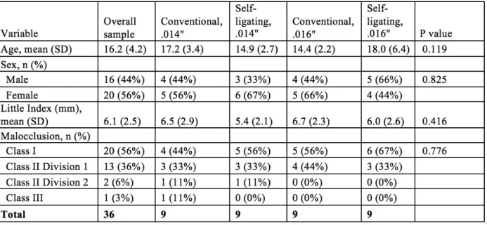
Demographic information regarding the study subjects is displayed in Table 1. 16 of the subjects were male and 20 were female. The overall mean age was 16.2 y. No statistically significant differences existed between the four groups at baseline with regard to age, gender, Angle classification or pre-existing Little Index (p>0.05). 5 patients (14%) experienced anterior bracket de-bonds during the study period involving a single bracket in each case (Table 2). Brackets were re-bonded at repair appointments using 3D-milled single tooth indirect bonding jigs per study protocol. There were no statistically significant differences in the number of anterior bracket debonds between the study groups (p>0.05).
compared over any interval (p>.05), and no statistically significant difference in mean reduction of either Little Index or contact point displacement when jaws (upper vs. lower) were compared (p>.05).
Based on results of 2-way ANOVA considering bracket type and archwire dimension for the secondary outcome—movement of upper right central incisors with six degrees of freedom— there was a statistically significant difference in the mean vertical movement (extrusion/intrusion) of incisors treated with conventional and self-ligating brackets during both T0-T1 and T0-T2 (p=.039 and p=.007, respectively). Specifically, self-ligating brackets showed a greater tendency towards intrusion (Figure 3). From T1-T2 a significant difference in buccolingual translation of central incisors was observed between .014” and .016” Copper NiTi™ wires (p=.043), with a tendency towards buccal movement observed with .014” wires. No other statistically significant differences in movement of upper right central incisors were observed in any of the six degrees of freedom over any interval (p>.05).
Discussion
The results of the current study suggest that when the impact of wire dimension alone is considered in cases of mild-moderate crowding treated without extractions in a customized CAD-CAM system, no statistically significant difference in efficiency of alignment exists between arches treated with .014” and .016” Copper NiTi™ archwires at any time point. There is also no significant difference in alignment between arches treated with conventional and self-ligating brackets.
in reduction of Little Index and contact point displacement. In other words, while there is a trend in both .014” and .016” wires towards greater mean reduction of both Little Index and contact point displacement in conventional brackets for the first six weeks, this trend intensifies to statistical significance during the second six weeks with no change in the archwire.
In vitro tests may provide an explanation for this. Gibson et al. has described the
frictional forces that constrain wire sliding in the initial phase of treatment as “constraining forces.” His group showed in benchtop tests that as malalignment increases in severity, there is a severity turning point—marked by a sudden increase in constraining force—where the forces of classical friction are likely joined by those of elastic binding (elucidated by Kusy and Whitley41) as well as plastic binding. They hypothesized that these forces between the wire and bracket could create the expansion necessary to allow alignment of partially blocked-out teeth. In a lingually blocked-out lower incisor, for example, the large constraining forces at each bracket would prevent the wire from sliding through the brackets, causing the excess wire between the three brackets to act like an opening coil as the wire assumed its initial shape.42
These clinical and benchtop findings contradict the findings of Pandis et al., who found patients with moderate crowding experienced alignment 2.7x faster in self-ligating brackets compared to conventional brackets. The authors reasoned this effect came from the decreased friction in a moderately-crowded ligating group when compared to a severely-crowded self-ligating group and conventional groups.18 In other words, the previous study reasoned that friction decreased alignment, a claim that has been insinuated by Damon,43 while the current study supports the hypothesis of Gibson et al. that the frictional forces that constrain wire sliding may increase alignment.
These contradictory findings may be explained by the fact that the previous study measured the time to total alignment in days, while the current study evaluated only the first 12 weeks of alignment. The forces at work in larger arch wires could be different from those in small, round aligning wires. Further, the previous study used different archwire sequences for the two bracket groups, while archwire dimension was controlled in the current study.
the self-ligating bracket relative to the twin group in the current study occurred during the second interval, at a point when rotations had already been partially corrected (see Figure 2). This means that in both studies, the conventional brackets displayed greater alignment efficiency during the interval when the wires in the self-ligating brackets had more rotational “play,” and theoretically were experiencing less friction, than those in the conventional brackets. If increased dimension of an aligning wire in a rotated tooth has a similar effect on constraining force as an increased rotation, the findings by Miles could thus also conceivably be explained by increased constraining forces allowing the self-ligating group to “catch up” with the alignment of the conventional brackets during the second interval.
It’s important to note that even with these differentials in alignment efficiency, no significant difference in the overall efficiency of alignment existed during either the 20-week previous study period or 12-week current study period. The small differences during the first 6-week interval in the current study diluted the differences observed during the second six 6-weeks, so that the overall increase in mean resolution of Little Index brought about by the conventional brackets was 0.7mm in the .014” groups and 0.8mm in the .016” groups. These differences are neither statistically nor clinically significant.
This is only the second alignment efficiency study comparing conventional and self-ligating brackets to allow for detection of vertical displacements and changes in tip by incorporating a 3-dimensional measure of contact point displacement. Alignment is often measured in the horizontal dimension alone using Little Index. A prior study used a coordinate measuring machine to analyze plaster models,14 while the current study used digital models to select contact points, which may have allowed for increased accuracy. Differences in resolution of contact point displacement mirrored those of the changes in Little Index during all intervals in this study. Over the 12 weeks of the study, conventional brackets showed a 0.9mm greater mean resolution of contact point displacement and 0.8mm greater mean resolution of Little Index compared to self-ligating brackets. Neither of these differences were statistically significant.
At the end of the day, self-ligating brackets are more expensive. The current study adds to the body of evidence that suggests there is no rationale to justify the increased cost of self-ligating brackets for efficiency of initial alignment, and further suggests that this relationship is not altered by the customized CAD/CAM nature of conventional and self-ligating brackets.
It is possible that there are efficiencies to customized CAD/CAM systems that manifest later in treatment. Perhaps as archwire dimension is increased, fuller expression of the customized bracket prescriptions will be observed, potentially increasing the efficiency and/or quality of full treatment. Further studies will be needed to determine whether any differences exist between conventional and self-ligating brackets in a customized system when full treatment is concerned.
0.2mm and 0.4mm. These numbers are small compared to the resolution achieved by these same brackets during the first 6 weeks, and small enough to be considered clinically insignificant. Unless there is some reason other than reduction of malalignment to use these longer treatment intervals, it is possible these longer appointment intervals should be reexamined.
One may also speculate that the difference in alignment efficiency between the bracket types during T1-T2 was not created by a decrease in the frictional forces that had constrained the movement of the archwires through the self-ligating brackets, but rather by the “fresh” elastomeric modules applied to the conventional brackets at 6 weeks. It is unknown whether this increased efficiency would have been seen during that interval without replacing the modules, but future research could evaluate this question.
The secondary outcome measurements of upper right central incisor movements were difficult to explain. It is unknown why the self-ligating groups showed an increased tendency towards intrusion. Movements in the vertical dimension should be largely governed by the incisal and gingival aspects of the bracket slot. These surfaces should be functionally similar between the two brackets. Perhaps more perplexing is the fact that .014” archwires tended to move incisors more buccally than .016” arch wires. Conventional wisdom might suggest that the greater force of .016” archwires would create a greater risk of incisor flaring.
two groups. More lingually-displaced central incisors in the .014” group, for example, could have led to a tendency to more buccal movements during alignment.
Another limitation of this study is the fact that some of the treating clinicians were unwilling to participate in full randomization of bracket type. This means that only archwire dimension was truly randomized, and bracket type was quasi-randomized. There is always a risk of selection bias due to the nature of allocation in quasi-randomized trials. However, since the operators did not choose between conventional or self-ligating brackets based on the presentation of each case, but instead requested that all of their cases be set up with the bracket type they used in their own clinical practice, the effect of allocation bias was decreased. While the possibility of sampling bias remains, no statistically significant difference in age, gender, Angle classification or pre-existing Little Index existed between the four groups at baseline.
Finally, the data presented as clinical evidence for the constraining force are only in reference to one type of malalignment (rotation), while Gibson et al. describes constraining force for what they describe as the four major malaligment types that limit alignment.42 Further clinical research will be required to either support or refute the existence of this force not only in rotations but also in the other three factors that limit alignment: tipping, in-outs and the vertical steps.
It should also be noted that these findings can only be generalized to patients aged 10-45 with cases of mild-moderate crowding and no teeth blocked-out from the dental arch who are treated without extractions. Further research will be required to determine whether these findings apply to other groups.
In the alignment of orthodontic cases with mild-moderate crowding treated without extractions in a customized CAD-CAM system:
1. There is no overall difference in the alignment efficiency between conventional and self-ligating brackets.
2. There is no overall difference in the alignment efficiency between .014” and .016” archwires.
3. The frictional forces that constrain the sliding of archwires through brackets may actually increase the efficiency of alignment.
Table 6. Demographic baseline characteristics of sample
Table 7. Number of patients with one de-bonded anterior bracket during study period