Genetic Engineering to Investigate Driver Mutations of Gastrointestinal Neuroendocrine Tumors
By Sanjna Iyengar
Senior Honors Thesis
Department of Biomedical Engineering University of North Carolina at Chapel Hill
Abstract
In the intestine, rare neuroendocrine epithelial cells secrete hormones that aid the digestive process and modulate the immune response. Gastrointestinal neuroendocrine tumors (GI-NETs) are slow growing and comprised of cells that look similar to neuroendocrine cells. The Magness Lab has found that a subset of transit-amplifying (TA) cells found in the crypts of the small intestine have a gene expression signature with features of both stem and
Background and Significance Intestinal Biology
The small intestine is responsible for most digestion and absorption of nutrients and minerals. The organ can be divided into three sections, the duodenum, jejunum, and ileum. The first segment of the small intestine, the duodenum, is about 25 cm long, and can be further
divided into the superior, descending, horizontal and ascending duodenum. The duodenum starts at the pyloric sphincter and moves behind the perineum before curving around the pancreas and moving back into the peritoneal cavity to connect to the jejunum. The jejunum is
approximately 90 cm long and connects the duodenum to the ileum, the longest segment of the small intestine. This 1.8 meter segment is thicker and more vascular than the jejunum, and connects to the cecum of the large intestine at the ileocecal sphincter. The mesentery connects both the jejunum and ileum to the posterior wall of the abdomen1.
Four distinct tissue layers constitute the small intestine: the serosa, the muscularis, the submucosa, and the mucosa. The innermost layer of the intestine, the mucosa, is a mucous membrane that consists of villi and crypts. Villi are finger-like projections into the lumen of the intestine. They are semi-permeable and serve to increase the surface area available for
cells, and are responsible for the absorption of nutrients. They contain a “brush border” comprised of microvilli that further increase the surface area of the intestine. Goblet cells produce mucins for protection as well as growth and repair of the epithelium3. Finally, enteroendocrine cells are responsible for secreting peptide hormones to regulate glucose homeostasis, food intake, and gastric emptying4.
Between the villi are intestinal glands, known as crypts, that extend away from the lumen. Crypts consist of stem and progenitor
cells, as well as Paneth cells. The
undifferentiated stem and progenitor cells help to restore the differentiated pool of epithelial cells. As they differentiate, they migrate upwards into the villi, with the exception of Paneth cells. Paneth cells, which secrete antimicrobial peptides, digestive enzymes and growth factors, remain at the base of the crypt3.
Neuroendocrine Tumors
Neuroendocrine cells receive signals from the nervous system and synthesize and release hormones in response. They are diffusely distributed throughout the body. The enteroendocrine cells of the gastrointestinal tract are a type of neuroendocrine cell. Gastrointestinal
diagnosed (or misdiagnosed) before developing into a rather aggressive metastasis. Symptoms of GI-NETs, if present, may include diarrhea, constipation, abdominal pain, heart palpitations and others. Over 12,000 people are diagnosed with neuroendocrine cancer each year, and this number is increasing annually by more than five percent16. The five-year survival rate for GI-NETs is only about 52-77%17. This thesis aims to understand the biological mechanisms behind the onset of neuroendocrine tumors in the small intestine to aid the development of treatments for GI-NETs.
Intestinal Stem Cells (ISCs)
The stem cells of the small intestine are able to self-renew or differentiate into the many types of mature cells in the intestinal epithelium. ISCs reside in stem cell niches, specific
Currently, there is little experimental evidence on the specific set of mutations that induce GI-NET formation. However, two genes, MEN1 (Multiple Endocrine Neoplasia Type 1) and SOX4 (Sry-Box 4) are known to have properties that make them likely candidates of promoting GI-NET formation when mutated.
MEN1
MEN1 encodes a protein known as menin. Menin serves as a tumor suppressor; it aids in copying and repairing DNA as well as regulating apoptosis6. A loss of function in MEN1 has been observed in human GI-NETs.
SOX4 is a member of the Sox-family of transcription factors that play powerful roles in early cell fate decisions18. The protein encoded by this gene may serve as a transcriptional regulator as well as play a role in the apoptosis pathway. SOX4 marks a progenitor cell type and is known to promote endocrine cell differentiation in the small intestine5. SOX4 has been found to be highly upregulated in human GI-NETs.
Experimental Goal
The purpose of this experiment was to induce a loss of function mutation in MEN1 and test the resulting cells for signs of GI-NET formation, including increased proliferation and increased incidence in Chromogranin A (ChgA), a GI-NET marker. The effects of SOX4 overexpression will be explored as a part of future experiments.
CRISPR-Cas9
Once the Cas9/gRNA complex is bound to the DNA, Cas9 generates a double stranded break at the target site that is then repaired using either non-homologous end joining (NHEJ) or homology-directed repair (HDR) if a DNA template is provided. NHEJ is the predominant repair pathway and is prone to insertion and deletion (indel) mutations13. Therefore, this mechanism can be used to induce a loss of function mutation in a gene of interest.
Materials and Methods
Retrieval of Human Intestinal Stem Cells
Intestinal stem cells were retrieved from the duodenum of an anonymous human donor. Segments of the donor intestine were cut longitudinally and transversely to expose the
The intestinal pieces were added to conical tubes containing 30 mL of colon isolation buffer. The tube was manually shaken to remove additional mucus and debris. This process was repeated until the isolation buffer appeared clear after shaking. The tissue was then added to a 30 mL solution containing colon isolation buffer, EDTA and DTT for thirty minutes at room
temperature. The tubes were shaken by hand with moderate force for two minutes and placed on ice immediately after. This process served to extract the villi into the solution. The tissue was then transferred to a new tube containing colon isolation buffer, EDTA and DTT. The tube was placed on a rocker at room temperature for ten minutes and then shaken for two minutes to extract the crypts into the solution. The tissue was transferred once again to a new tube containing isolation buffer, EDTA and DTT and rocked at room temperature for ten minutes. The solution was shaken a third time to release more crypts, transferred to a new tube and shaken one last time to release any remaining crypts. Isolated crypts (from shakes 2 and 3) were pooled into a new tube and centrifuged at 600 g for one minute.
The crypts were resuspended in expansion media (EM) and Y-27632 (or Y-27). Media containing about 15,000 crypts was transferred to a new tube and centrifuged at 600 g for one minute. The cells were then resuspended in 12 mL of EM and 10 µM of Y-27. Finally, the cells
were plated in a 6-well expansion plate (4 mL per well).
Cell Culture Medium
The expansion medium used to culture the human intestinal stem cells (hISCs) consists of the following components: basal culture medium, EGF, noggin, R-spondin, Wnt3A,
coiled-coil containing protein kinase (ROCK). It serves to prevent anoikis, or dissociation-induced apoptosis, when cells are broken down into single cells, thereby enhancing the survival of the hISC monolayers12.
Selection of gRNA Target Sites for MEN1 Knockout
A loss of function was induced in the MEN1 gene using CRISPR-Cas9. The process began with selection of the gRNA target sites that Cas9 would cleave at in the MEN1 gene. CCTop (CRISPR-Cas9 target online predictor) was
used to select the gRNA target sites. The MEN1 exon sequence was input into the program, along with the desired PAM sequence, NGG. A list of possible target sequences was generated, along with a list of potential off-target sites. To reduce the possibility that Cas9 would cleave at an incorrect locus in the genome, target sequences were chosen based on the criteria that the off-target sequences should have at least three mismatches from the target sequence. Three gRNA targets (denoted as T1, T2 and T10) were selected.
Transfection of hISCs with CRISPR-Cas9 Tools
(tracRNA), gRNAs and tracRNAs from IDT were first resuspended in nuclease-free annealing buffer. 5 µL of each of the three gRNAs was mixed with 5 µL of the tracRNA and annealed.
Cas9/gRNA mixes were prepared by adding 1 µL of the IDT Cas9 nuclease with 1 µL of each of
the annealed gRNA/tracRNA mixes. The Cas9/gRNA mixes were incubated at room temperature for five minutes and placed on ice. For the positive control, pMaxGFP was used as it
constitutively expresses GFP. The annealing buffer served as the negative control. Nucleofection was performed using the Lonza 4D Nucleofection system. The
nucleofection solution was prepared by mixing 16.4 µL of buffer P1 with 3.6 µL of supplement 1
per reaction. The cells were then dissociated. Collagenase IV (500 units/mL final concentration) was first used to digest the collagen in the wells containing the wild type hISC populations. Conditioned media (containing old media from the wells) was set aside as well. The solution containing the cells and collagenase was spun down at 1000 g for five minutes at room temperature. The media was removed, and the cells were resuspended in PBS. Once the cells were repelleted and the PBS was removed, the cells were resuspended in 500 µL of prewarmed
TrypLE with Collagenase IV (500 units/mL final concentration) and incubated for five minutes. The cells were passed through an insulin syringe to be dissociated into single cells. TrypLE was then neutralized with media and the solution was spun down at the conditions listed previously. The cells were rinsed with PBS once again and then resuspended in nucleofection solution P1 and supplement. The solution was divided into aliquots to allow for multiple experimental conditions and incubated for five minutes at room temperature. The cells, except for the negative control, were then nucleofected (EN150 nucleofector program) and incubated for ten minutes at room temperature. Finally, the cells were resuspended in 3 mL of conditioned medium and 3 µL
Assaying for CRISPR activity
Transfection of the Cas9 protein and gRNAs into the hISCs was confirmed using a T7 endonuclease assay. T7 endonuclease is an enzyme that cleaves heteroduplexes, or bulges in the DNA that form due to mismatches in the nucleotides.
The genomic DNA was first extracted from the cells. This was done by dissociating and pelleting the cells using the standard single cell dissociation procedure (mentioned in the “Single Cell Dissociaton” section). The cells were resuspended in 500 µL of proteinase K solution
(containing 10 mg/mL proteinase K diluted 1:10 in buffer TE) and incubated overnight at 55 °C.
The DNA was then purified with a 1:1 phenol/chloroform extraction.
The purified DNA was then PCR amplified. A sample of each PCR product (10 µL) was
combined with NEB Buffer (2 µL) and molecular grade water (7 µL). Each solution was added
to the thermal cycler, and the temperature was ramped down from 95 °C to room temperature to
denature and slow cool the DNA. This process would allow the positive and negative DNA strands to separate and re-ligate with arbitrary complementary strands in the mixture. Since the mutations made during NHEJ are distinct among the different DNA molecules, ligation between the DNA strands would result in mismatches at the target site, therefore leading to the formation of heteroduplexes. Ten units of T7 endonuclease (1 µL) were added to each solution, allowing
the enzyme to recognize and cleave the heteroduplex DNA. The samples were then incubated at 37 °C for an hour. Next, the samples underwent gel electrophoresis; the presence of two bands
was also completed using the chromatograms for the control and test samples to quantify the percentage of MEN1 knockout for each of the targeted hISC populations.
Single Cell Dissociation
The T1 and T10 cell populations, which were found to contain editing based on the T7 endonuclease assay, were dissociated into single cells and allowed to grow into individual colonies consisting of genetically identical cells. This allows for isolation of the colonies and subsequent sequencing of the cells to determine their genotype.
The single cell dissociation was performed by digesting the collagen scaffold in culture medium containing 500 units/mL Collagenase IV. The solution was spun down at 1000 g for five minutes at room temperature to obtain a cell pellet. Following an intermediate PBS wash and an additional spin at the same conditions, a 500 uL solution containing TrypLE and 500 units/mL Collagenase IV was used to digest intracellular adhesion molecules in the cells and remove any remaining collagen. The cells were passed through a 28-gauge insulin needle six times to mechanically dissociate the cells into single cells. TrypLE was then neutralized with 4.5 mL of EM and the solution was spun down at 600 g for five minutes at room temperature. The cells were then resuspended in 1 mL EM. A hemocytometer was used to determine the total number of cells in the 1 mL sample, and the cells were then plated at 500 cells per well for the T1 population and 1500 cells per well for the T10 population. The cells were plated at different densities in order to determine the optimum density for clonal isolation.
confluency, the T10 cell population was passaged 1:3 in a 6-well plate. The cells were permitted to grow to about 70% confluency once again. Two of the wells were frozen, while one of the wells was dissociated into single cells using the procedure mentioned above. The well was found to contain greater than one million cells which were then plated at densities ranging from
approximately 63,000 cells per well to over 500,000 cells per well in a 6-well plate.
The cells were monitored and fed over the next few days. The well containing more than 500,000 cells was observed to have too high of a density for picking individual colonies. Four days after plating, the single cell dissociation procedure was carried out again on the well containing more than 500,000 cells. This time, the cells were plated at the following densities in a 6-well plate: 5000, 10000, 25000, 50000, 75000, and 100,000 cells per well.
Isolation of Clones
The clones were ready to be isolated when the wells contained around 10+ clones with clonal widths ranging between 300 - 400 µm. To isolate the clones, 300 µL of 5000 units/mL
Analysis of Isolated Clones
The colonies were allowed to grow to about 30% confluency and then passaged to a 24-well plate. This procedure first involved adding a 50 µL 1:1 collagenase (2500 units/mL final
outside of the collagen patty from the plate. The cells were then incubated at 37 °C for ten
minutes. The media and partially digested collagen were then transferred to a separate tube, and a pipette was used to break up the collagen. The tubes were incubated in a 37 °C water bath for
five minutes to digest the remaining collagen. The tubes were centrifuged at approximately 1000 g for five minutes, and the supernatant was discarded. The cell pellet was resuspended in 50 µL
of TrypLE and incubated at 37 °C for five minutes. The cells were then broken up by pipetting
the solution about fifteen times.
A small sample each cell suspension (5 µL) was transferred to a PCR tube, along with 1 µL of 10 mg/mL proteinase K and 4 µL of Buffer TE. These 10 µL solutions were run in a
thermal cycler at 50 °C for ten minutes, 94 °C for ten minutes, and held at 15 °C overnight.
The remaining cells in the TrypLE suspensions were neutralized with 450 µL of media
per suspension. The tubes were centrifuged at approximately 1000 g for five minutes, and the supernatant was discarded. The cells were then resuspended in 1 mL EM and 1 µL Y27 per well
in a 24-well pre-rinsed collagen plate.
The 10 µL solutions containing the extracted DNA were then prepared to be sequenced in
order to determine the type of MEN1 mutation in the clonal population. Overall, 8 T1 clones and 34 T10 clones were tested. A sample of each solution (4 µL) was mixed with 1 µL of each of the
forward and reverse MEN1 primers diluted 1:10, 25 µL of PCR mastermix (Apex Hot Start Taq
BLUE Mastermix) and 19 µL of molecular grade water to bring the total sample volume to 50 µL. The DNA samples were then amplified and loaded on to the gel to check for the presence of
bands. The samples were sequenced to determine the type of MEN1 mutation.
Chromogranin A (ChgA) is a protein secreted by neuroendocrine cells. Since stem cells are not expected to secrete ChgA, the protein can serve as a GI-NET marker. In this experiment, a wild type and MEN1+/- colony was therefore each stained for the presence of ChgA, with the wild type population representing the control.
The staining process involved the use of a rabbit-derived primary antibody that is specific to ChgA, and a goat-derived, anti-rabbit secondary antibody that is specific to any rabbit protein. The secondary antibody also contained a fluorophore to allow for visualization. To begin the staining process, the cells were washed with PBS. Paraformaldehyde (PFA) was then added to the cells, and the cells were incubated at room temperature for twenty minutes. The PFA was removed, and the cells were washed twice with 3% BSA. The cells were then washed with PBS for fifteen minutes, and subsequently washed with 0.1% PBST for ten minutes. 0.3% PBST was then used to permeabilize the cells for fifteen minutes. A 1X Cell Signaling Blocking reagent (3 mL) was then added to the cells for thirty minutes. The blocking solution would serve to reduce nonspecific binding of the antibody14. The 1X blocking solution was then removed from the cells, and the primary antibody, diluted in 1X blocking solution 1:200, was added to the cells. The MEN1+/- colony was then incubated at room temperature for two hours, while the wild type colony was incubated at 4 °C overnight.
The cells were then washed with 0.1% PBST for fifteen minutes. The PBST was
was then used to stain the cells for five minutes. Finally, the cells were washed twice with PBS, with five minutes in between washes.
Staining for EdU
EdU (5-ethynyl-2’-deoxyuridine) is a thymidine analog and can serve as an S-phase marker to quantify cell proliferation. The molecule is incorporated into DNA during the DNA replication stage of the cell cycle. A fluorescent azide that covalently cross-links to the EdU molecule must be added to the sample to allow for visualization of the EdU marker15. In this experiment, a wild type and MEN1+/- cell population was each stained for EdU to measure and compare the amount of cell proliferation between the two genotypes.
Approximately three hours prior to the start of the procedure, fresh EM (3 mL)
containing EdU (3 µL) was added to the cells. After the three hours, the media was replaced with
4% PFA. The cells were then incubated at room temperature for twenty minutes. The PFA solution was then removed, and the cells were washed twice with 3% BSA in PBS for five minutes per wash. This was followed by the addition of 0.5% Triton X-100 in PBS to the cells. The cells were incubated for another twenty minutes. The cells were then washed twice with 3% BSA for five minutes per wash. An EdU staining solution containing 76% EdU reaction buffer, 4% CuSO4, 2.5% Sulfo-CY5 azide and 20% EdU reaction buffer additive (L-aspartic acid) was then added to the cells. The cells were incubated at room temperature for an hour, away from any light. After the hour, the EdU staining solution was removed, and the cells were rinsed with PBS for five minutes.
minutes, away from any light. Finally, the cells were washed twice with PBS for five minutes per wash. The cells were covered and stored at 4 °C for four days.
Results
Analysis of Transfected Cell Populations
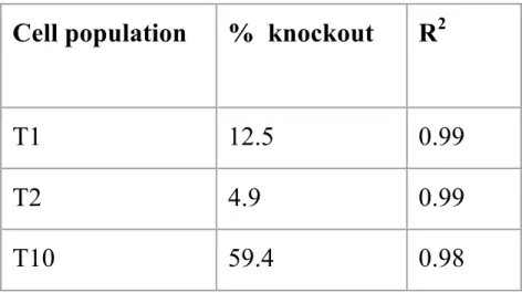
A T7 endonuclease assay was used to verify that the transfection of Cas9 and the MEN1-targeting gRNAs was successful. The gel electrophoresis results from this assay showed two bands for the T1 and T10 populations. No bands were visible for the T2 population. This
Cell population
% knockout
R
2T1
12.5
0.99
T2
4.9
0.99
T10
59.4
0.98
Analysis of Isolated Clones
Sanger sequencing was used to sequence the MEN1 gene in the DNA samples of each of the isolated clones. The nucleotide sequence for each clone was compared to the wild type sequence. Manual comparison between the wild type and test sample sequences allowed for the determination of the number of wild type, MEN1+/- and MEN1-/- colonies. Out of the 34 T10 test samples, 6 were wild type (two wild type alleles), 17 were heterozygous for the MEN1 mutation (one wild type allele and one mutant allele), and 1 was homozygous for the MEN1 mutation (two mutant alleles). The remaining samples either contained multiple clones or had unclear
sequencing results.
ChgA Staining Results
EdU Staining Results
Discussion
Until recently, culturing primary intestinal epithelial cells was a challenge due to cells’ lack of proliferation when they were placed in culture. Researchers alternatively needed to use cancer cell lines as these cells were able to effectively proliferate as confluent monolayers. However, the cell lines varied greatly in phenotype from normal epithelium, therefore introducing uncertainties in their ability to accurately represent healthy intestinal form and function. To combat this problem, 3D organoid culture was recently introduced. This culture system allows for the maintenance of a stem cell niche as well as differentiation of the stem cells into mature cell types, mimicking the native villus-crypt structure present in the normal human intestine24. However, studies on intestinal stem cells are a challenge with 3D organoids because the organoid’s enclosed architecture limits access to the lumen of the organoid.
Wang et. al determined culture conditions that allowed for 2D culture of primary human rectal epithelial cell monolayers on collagen hydrogel.19 2D cultures are beneficial because the cells are exposed and easily accessible for experimental manipulation. For this reason, our experiment utilized 2D culture techniques to effectively culture stem and progenitor cells from the small intestine. Additionally, our experiment utilized novel methods of single cell
In this experiment, Cas9 and the gRNAs were delivered by directly transfecting the hISCs with the Cas9 protein in complex with the gRNA22. This is a higher-throughput approach than transfecting a plasmid, which results in delayed expression and continuous generation of the Cas9 protein, therefore increasing the chance of off-target mutations and lowering editing
efficiency23. Therefore, direct transfection of the Cas9 protein and gRNAs was the preferred method as it increased overall transfection efficiency, as indicated by the 60% transfection efficiency achieved in the T10 population (which is twice as high as the highest recorded transfection efficiency prior to this experiment).20
Single cell dissociation of the hISCs provided an effective way to isolate single colonies consisting of genetically identical cells. The process was done three times, each at different ranges of densities, in order to determine the ideal density for isolation of colonies. When the cells were plated at too low of a density, the colonies grew very slowly and could not be picked until several weeks later. On the other hand, colonies plated at too high of a density could not be distinguished from one other at the time of isolation. The wells containing 50,000, 75,000, and 100,000 cells were able to be picked after the fewest number of days (9 days), while the well with 25,000 cells produced the most number of colonies (22 colonies). As a result, we concluded that the optimum density for plating hISCs in a 6-well plate for clonal isolation is between 25,000 to 50,000 cells per well.
300 µL of 5000 units/mL Collagenase IV to the cells and resuspending them in PBS before
attempting to isolate colonies served to be a quick and simple method for isolation since the cells flowed freely in the PBS without dissociating. The optimum clonal size was found to be at least 300 to 400 um in width, and was determined by testing different colonies of varying sizes. Colonies below this size range were observed to grow more poorly.
The results from the ChgA and EdU staining procedures revealed that there was no significant difference in the amount of ChgA or EdU between the wild type and MEN1+/- cells. This indicates that a heterozygous mutation in MEN1 is insufficient for increased incidence of enteroendocrine differentiation or increased cell proliferation, both of which are features of GI-NETs. Experimental evidence suggests that these results were expected. According to a 2008 study by Marini et al, mice with a heterozygous mutation in MEN1 developed endocrine tumors late in life while mice with a homozygous mutation in MEN1 died while still in utero. This leads us to believe that a homozygous MEN1 mutation (which leads to a complete loss in menin production) may be necessary for significant alterations in proliferative capacity or
differentiation of the mutated cell25.
MEN1-/- clones to overexpress the gene. These populations will be compared to the wild type cells and cells containing a MEN1 knockout alone.
Additional genes that could potential serve as driver genes for GI-NETs will be explored as well. FOXA1 is one such gene with characteristics favorable for further investigation. FOXA1 is a transcription factor that plays a role in the establishment and regulation of tissue-specific gene expression in differentiated tissues. It has been found to bind to compacted chromatin through its interactions with histones in order to open the chromatin for other proteins. FOXA1 is also involved in modulation of nuclear hormone receptor transcriptional activity, cell cycle regulation and glucose homeostasis7. FOXA1 has been prominently expressed in esophageal cancer, lung cancer, and 12T-10 invasive undifferentiated neuroendocrine carcinomas8. One study showed that miR-1290, a microRNA with high expression in colon cancer, “promotes gastric tumor cells proliferation and metastasis through FOXA19.” It therefore may be beneficial to test the overexpression of FOXA1 independently and in combination with mutations in the MEN1 and SOX4 genes.
Conclusion
function in MEN1 and an overexpression of SOX4 have been found to be present in human GI-NETs. Therefore, this experiment first tested the effects of a MEN1+/- mutation on the
proliferation rate and incidence of the GI-NET marker, ChgA. The procedure involved using CRISPR-Cas9 to induce a loss of function mutation in the MEN1 gene. This was followed by single cell dissociation of the targeted population, clonal isolation, and sequencing of the isolated clones to obtain a clonal population containing a MEN+/- mutation and a clonal population
containing a MEN-/- mutation. The MEN+/- cells were stained for ChgA and EdU. Visual comparison of the staining images between the wild type and MEN+/- colonies indicated that there was no significant difference in the amount of either ChgA or EdU between the two colonies. Therefore, this indicates that a heterozygous mutation in MEN1 is insufficient for increased proliferation or increased incidence of endocrine differentiation.
The next step will be to test the MEN-/- colony for signs of GI-NET formation.
Overexpression of SOX4 as well as mutations in other potential candidates of GI-NET driver genes, such as FOXA1, will be explored as well.
By identifying driver mutations of GI-NETs, we hope to better understand the biological and genetic basis behind this disease, and thereby improve our ability to diagnose and treat this aggressive form of cancer.
Acknowledgments
References
1. OpenStax. Anatomy and Physiology. 2013 Mar 6.
2. The Small Intestine. Lumen.
3. Ensari A, Marsh MN. Exploring the villus. Gastroenterology and hepatology from bed to bench. 2018.
4. Mellitzer G, Beucher A, Lobstein V, Michel P, Robine S, Kedinger M, Gradwohl G. Loss
of enteroendocrine cells in mice alters lipid absorption and glucose homeostasis and
impairs postnatal survival. The Journal of Clinical Investigation. 2010 May 3.
5. SOX4 Gene. genecards.org.
6. MEN1 gene - Genetics Home Reference - NIH. U.S. National Library of Medicine.
7. FOXA1 Gene. genecards.org.
8. FOXA1. WikiGenes.
9. Lin M, Shi C, Lin X, Pan J, Shen S, Xu Z, Chen Q. sMicroRNA-1290 inhibits cells proliferation and migration by targeting FOXA1 in gastric cancer cells. ScienceDirect. 2016 Feb 3.
10. Tissue-engineered Small Intestine - Oley Foundation.
11. Sato T, Stange DE, Ferrante M, Vries RGJ, Van Es JH, Van den Brink S, Van Houdt WJ, Pronk A, Van Gorp J, Siersema PD, et al. Long-term expansion of epithelial organoids
from human colon, adenoma, adenocarcinoma, and Barrett's epithelium.
Gastroenterology. 2011 Nov.
13. CRISPR/Cas9 & Targeted Genome Editing: New Era in Molecular Biology. New England Biolabs.
14. Preparing Fixed Cells for Labeling. Thermo Fisher Scientific - US.
15. EdU Assay / EdU Staining Proliferation Kit (iFluor 488) (ab219801). Abcam.
16. Neuroendocrine Tumor. Cancer.Net. 2014 Jun 17.
17. Small bowel carcinoids. What is the prognosis for small intestinal neuroendocrine
tumors?
18. Gracz AD, Samsa LA, Fordham MJ, Trotier DC, Bailey Zwarycz Y-HL, Bao K, Starmer
J, Shroyer NF, Reinhardt RL, Magness ST. Sox4 drives Atoh1-independent intestinal
secretory differentiation toward tuft and enteroendocrine fates. bioRxiv. 2018 Jan 1.
19. Wang Y, DiSalvo M, Gunasekara DB, Dutton J, Proctor A, Lebhar MS, Williamson IA, Speer J, Howard RL, Smiddy NM, et al. Self-renewing Monolayer of Primary Colonic or
Rectal Epithelial Cells. Cellular and molecular gastroenterology and hepatology. 2017
Mar 6.
20. Fujii M, Matano M, Nanki K, Sato T. Efficient genetic engineering of human intestinal
organoids using electroporation. Nature News. 2015 Sep 3.
21. Matano M, Date S, Shimokawa M, Takano A, Fujii M, Ohta Y, Watanabe T, Kanai T, Sato T. Modeling colorectal cancer using CRISPR-Cas9-mediated engineering of human
intestinal organoids. Nature medicine. 2015 Mar.
22. Hempstead A. CRISPR 101: Ribonucleoprotein (RNP) delivery. addgene blog. 2018 Sep
23. Farboud B, Jarvis E, Roth TL, Shin J, Corn JE, Marson A, Meyer BJ, Patel NH, Hochstrasser ML. Enhanced Genome Editing with Cas9 Ribonucleoprotein in Diverse
Cells and Organisms. Journal of visualized experiments : JoVE. 2018 May 25.
24. Sato T, Vries RG, Snippert HJ, van de Wetering M, Barker N, Stange DE, van Es JH,
Abo A, Kujala P, Peters PJ, et al. Single Lgr5 stem cells build crypt-villus structures in
vitro without a mesenchymal niche. Nature. 2009 May 14.
25. Marini F. Multiple Endocrine Neoplasia Type 1 (MEN1) Syndrome. NCBI. 2008 Aug 9. 26. Zheng T, Hou Y, Zhang P, Zhang Z, Xu Y, Zhang L, Niu L, Yang Y, Liang D, Yi F, et
al. Profiling single-guide RNA specificity reveals a mismatch sensitive core sequence.
Scientific reports. 2017 Jan 18.
27. GI tract. AboutKidsHealth.
28. Sancho R, Cremona1 CA. Rocio Sancho. Embo reports . 2015 May 1