1 Biopsy in Renal Cell Carcinoma
Surveillance Epidemiology and End Results (SEER) estimates that 58,240 people will be diagnosed with cancer of the kidney and renal pelvis, while 13,040 will die in 20101. Renal cell cancer accounts for about two percent of all adult cancers2. More than 80% of cancers of the kidney arise in the renal parenchyma, while the rest arise in the pelvis3. Historically, renal cell carcinoma (RCC) has been treated with nephrectomy, often with no tissue diagnosis, because of the adverse effects and possibility of false negatives associated with renal mass biopsy. With the advancement of imaging modalities in the last decades, smaller tumors are now being discovered that pose therapeutic dilemmas that did not exist in the past. Growing numbers of studies are showing a substantial proportion of these masses is benign on nephrectomy pathology leading urologists to question the current paradigm and reconsider biopsy4, 5. Due to improved sampling techniques and advances in immunohistochemistry, recent studies have reported higher levels of sensitivity and specificity for biopsy than were previously reported in the literature6-8. If biopsy could be proven to be dependable, options such as active surveillance and ablative therapies could provide physicians and patients with better choices if patients are not good surgical candidates.
Etiology
2 significant relationship to cancer. This may be due to the fact that initial studies considered phenacetin, which has now been off the market for over 25 years9. Studies have failed to show a significant association between diet and RCC9.
Tumor Characteristics
In 2002, the International Union Against Cancer (UICC) updated the TNM staging system for renal cell carcinoma by further classifying the T1 group into T1a and T1b; T1a refers to tumors within the kidney that are less than or equal to four centimeters, while T1b refers to tumors within the kidney that are between four and seven centimeters11. Stage II cancer is larger than 7 centimeters but is still limited to the kidney. Stage III refers to tumors that have spread to one lymph node or into the fatty tissue or veins around the kidney, but not to any distant organs. Lastly, Stage IV cancer involves spread to more than one lymph node, a lymph node not near the kidney, or to distant sites12. With regards to RCC grading, Fuhrman et al. presented a system based on 100 patients after nephrectomy in which four nuclear grades were defined based on size, irregularity and nucleolar prominence. Though many alternate grading schemes have been presented, the Fuhrman grading system remains the most widely used system in the United States12.
Changes in Epidemiology
3 RCC at Yale New Haven Hospital from 1989 and 1993 and found that more than 60% were discovered incidentally on imaging and showed this to be an increasing trend compared to previous decades2. Of the 135 pathology specimens collected from the lab, 131 had complete medical records accessible. The authors referred to masses as incidental if they lacked signs or symptoms of RCC, such as flank mass, flank pain, or hematuria, or if the physician had no suspicion of RCC as noted in the medical record. Findings such as these have been attributed to increasing detection of asymptomatic tumors by imaging procedures such as ultrasound,
computed tomography and magnetic resonance imaging. Unpublished data from the Health Care Financing Administration, which reported a 73% rise in abdominal/pelvic CT scans or MRIs to Medicare beneficiaries, supports this hypothesis3. The increased detection is coupled with a decrease in the average size of masses at diagnosis due to the advancing precision of imaging modalities; a recent study reported a decrease in mean tumor size from 4.1 to 3.6 cm between 1993 and 200414.
4 rates rose from 1.2 to 3.2 deaths per 100,000 people, with an increase seen in all tumor size groups. Although five-year survival for RCC has improved in that time period, the overall mortality has increased, which the authors attribute to lead and length time bias. Because smaller tumors are being detected, the five-year survival percentages are increasing, but overall survival is not. The authors argue that despite earlier detection of masses and increased rates of surgery, population data do not show a decrease in mortality, which argues for “a reassessment of the current treatment paradigm.” As the authors admit, the study does have limitations. Among them, SEER does not collect patient comorbidity data and thus it is impossible to know the causes of mortality in all patients. Additionally, over 15% of patients were missing tumor size information and were excluded from the study16.
Imaging
5 on angiography have been shown to be neither sensitive nor specific19. Although recent advances in imaging allow accurate differentiation between cystic and solid lesions, differentiating benign and malignant renal masses remains largely unreliable20, 21.
Imaging can be diagnostic, however, in one specific type of renal mass: angiomyolipomas, also known as renal hamartomas. According to studies by Bosniak,
attenuation of fat on CT scans in angiomyolipomas can be diagnostic in more than 90% of these masses2223. However, when angiomyolipomas have no fat present on imaging, they are
indistinguishable from renal cell carcinoma and merit further investigation24.
Treatment
Despite substantial advances in oncologic treatment, renal cancer still remains resistant to standard chemotherapeutic agents. However, low stage RCC can be successfully treated with extirpative surgery, with better prognosis for those with lower stage disease25. Historically, radical nephrectomy, with wide excision of the kidney outside of Gerota’s fascia to include the adrenal gland and perirenal fat, was preferred because of concern that extrarenal involvement lead to surgical failure26. However, nephron-sparing surgery, initially performed in those with a solitary kidney or pre-existing renal insufficiency with the hopes of preserving renal function, has become increasingly popular in recent decades.
6 were similar rates of contralateral recurrence and metastasis. The cumulative incidence of
chronic renal insufficiency (defined as greater than 2.0 mg/dL at least 30 days after surgery) was 22.4% and 11.6% in the radical nephrectomy group and nephron-sparing surgery group,
respectively. The authors suggest that nephron-sparing surgery is as effective as radical nephrectomy from an oncologic standpoint and may potentially have better outcomes with respect to complications. Compared to earlier studies in which nephron-sparing surgery was only done in those with solitary kidneys or pre-existing renal insufficiency, the study enrolled patients with single unilateral RCC and a normal contralateral kidney, which strengthens generalizability. These findings coincided with those from a multi-center Austrian study as well as a single
institution German study showing comparable cancer-free survival in nephron-sparing surgery versus radical nephrectomy27,28.
However, due to its non-randomized and retrospective design, the conclusions from this study are limited. Despite matching on tumor size, the average size in the radical nephrectomy group was 3.7 cm compared with 3.3 cm in the nephron-sparing surgery group, with the difference being statistically significant. It is likely that the smaller size of tumors in the
7 The intention-to-treat analysis showed a 10-year overall survival rate of 75.7% for NSS and 81.1% for RN, with an estimated hazard ratio of 1.50 (95% CI 1.03-2.16). However, this number became insignificant when the analysis was limited to those with actual renal cell carcinoma rather than surgery performed for benign masses. The hazard ratio of death from renal cancer was not significantly higher in the NSS group, nor was progression of cancer. A major weakness of the study was that the authors reported recruiting only 541 patients due to poor accrual despite calculating a necessary sample size of 1300 patients to detect a proposed 3% difference in 5-year survival. Additionally, the authors reported that 5.9% of the patients randomized to radical nephrectomy underwent nephron sparing surgery and 14.6% of patients randomized to nephron sparing surgery underwent radical nephrectomy29. The finding that overall survival is slightly better in RN than NSS is in contrast with many previously published studies on this topic. However, those studies are limited by their retrospective design, heterogeneous stage of tumors between groups, and lack of renal function outcomes. As far as we know, this is the only randomized trial to date on this subject and more studies like this need to be done in order to definitively answer this question.
8 Elective indications are for those patients with unilateral renal cell carcinoma less than 4 cm and a normal contralateral kidney26.
Benign Findings on Nephrectomy
For tumors smaller than 4 cm, the gold standard for treatment is radical or nephron sparing surgery30. However, a paper from 1987 showed that roughly 15% of renal masses detected on CT scan were benign31. More recent papers have shown even higher rates of benign findings on nephrectomy pathology. In a paper published in 2003, Lane and Gill retrospectively examined patients who underwent laparoscopic partial nephrectomies at the Cleveland Clinic and found 56 patients with at least five years of follow-up. Though the purpose of the study was to report oncologic outcomes after partial nephrectomy, the data showed a benign diagnosis on final pathology in 19 of the 56 patients (38%)32. These numbers may not provide an accurate representation of the proportion of benign masses because some of the masses were
symptomatic; in a population of asymptomatic incidental renal masses, it is possible that the proportion of benign masses would be even higher. A similar paper in 2006 found that 38 of 123 (31%) masses removed by laparoscopic partial nephrectomy were found to be benign on final pathology. The average size of the masses in the study was 2.6 cm, which is promising for generalizability to small renal masses, which are generally considered to be less than 4 cm. The indications for nephrectomy, however, were difficult to ascertain as the authors only reported performing surgery for “enhancing renal masses33.” In 2007, Gill et al. reported their findings of
9 The proportion of nephrectomies for benign masses is even more disconcerting when stratified by size, as shown in a case series by Frank et al. that examined 2770 nephrectomies due to solid renal masses and found that those less than 3 cm were 25% benign, those less than 2 cm were 30% benign and those less than 1 cm were 44% benign34. With the average age of RCC being 65 years old, many patients undergoing procedures are at higher risk for post-operative complications or mortality due to various comorbidities30. Because of this increased risk, active surveillance and modern ablative therapies are likely a better option in this population. Biopsy, if proven to be accurate and dependable, could assist the physician and the patient in making a more informed decision. Even young patients may have chronic renal insufficiency, solitary kidneys, or other medical conditions that seriously complicate surgery; thus, watchful waiting would be a welcome option to many patients if it could be proven as a safe alternative35.
The resurgence of biopsy
Benign findings on nephrectomy and the known indolence of small malignancies coupled with the emergence of newer treatments such as radio-frequency ablation and cryoablation, which require pre-procedure biopsy, have caused physicians to reconsider the role of renal mass biopsy19. Supporters of biopsy claim that it is safe, that the majority of biopsied lesions are benign, and that biopsy can decrease unnecessary surgery34. Opponents argue that biopsy carries too high a risk of false negatives36, can lead to biopsy track seeding with cancer cells, and leads to complications37.
10 after biopsy to check for complications, the authors reported no patients in whom a hematoma was found or any patients requiring intervention related to biopsy complications7. In a meta-analysis by Lane et al., the authors observed complications in renal mass biopsies in seven case series done after 2001. Out of a total of 362 biopsies, one series in the review showed one major complication from biopsy while the remaining six showed zero major complications. There were 17 minor complications reported in the review19. An important consideration is that these are case series reported from academic centers, and that their characteristics may not be applicable to all types of hospitals or urology practices. The authors did not report skills levels of surgeons or the volume of cases that these centers are exposed to. Additionally, the papers did not adjust for baseline status of the patients and thus conclusions are limited.
A case report of biopsy needle track seeding with malignant cells from a study in 1991 raised alarms about the risk of percutaneous biopsy39. Recently, however, reports of tumor seeding have been exceedingly rare, in part because of the use of the coaxial system. This approach uses a larger gauge introducer which then remains in place while a smaller needle can make multiple passes to obtain tissue20. This approach has been reported to reduce the chance of tumor seeding because it limits the number of times the renal capsule is penetrated40. Numerous reports with follow-up periods ranging from one to five years have shown no evidence of needle track seeding38, 40-42. The main argument against these findings is that the follow-up period may not be long enough to detect possible tumor seeding.
11 biopsy. For example, in an American Urology Association Update Series from 2000, the authors report sensitivity for core biopsy of renal masses as low as 70% and state that “false negatives are most often due to insufficient specimens which are often bloody aspirates.” Based on these reports, they conclude that biopsy is not indicated for a solitary renal lesion that looks like RCC on imaging43. However, taking these biopsies out of the denominator can portray a drastically different situation depending on the frequency of biopsy failure. In more recent studies, where inconclusive biopsies, i.e. those showing normal renal parenchyma, necrotic tissue, or hemorrhage are excluded from the analysis, the accuracy of biopsy in deciphering benign versus malignant masses has been reported between 89% and 98%6, 7, 44. Because false negatives based on an adequate sample have been shown to be less common38 45, 46, it is possible that the rising sensitivity values seen in recent studies are attributed not to improvements in techniques, but to improvements in denominators.
12 Methods
This systematic review included published studies describing different measures of performance for renal mass biopsy in imaging-detected small renal masses. Biopsy of renal masses is
currently not routine practice and thus the studies were limited to academic medical centers participating in research. Because of recent advances in immunohistochemistry and pathology techniques, we sought to perform a review of recent articles and chose 2005 as a cutoff. In March 2011, we identified prospective or retrospective cohort studies and case series by searching the MEDLINE and EMBASE databases from January 1, 2005 to March 1, 2011. We also hand searched reference lists. A research librarian at the University of North Carolina Health Sciences Library assisted in creation of the search strategy.
We searched the following terms in MEDLINE:
("Kidney Neoplasms"[Majr] AND "Biopsy"[Mesh] AND ("Predictive Value of Tests"[mh] OR "Sensitivity and Specificity"[mh] OR "Reproducibility of Results"[mh] OR "Retrospective Studies"[mh] OR "Risk Assessment"[mh] OR "Prognosis"[mh] OR "Incidental Findings"[mh] OR "diagnosis"[Subheading] OR "classification"[Subheading] OR "pathology"[Subheading])) (English[lang] AND "adult"[MeSH Terms].
[majr] = Major MeSH term [mh] = MeSH term
[sh] = Subheading
13 “kidney tumor'/exp/mj AND biopsy'/exp/mj AND ('predictive value of tests' OR 'sensitivity and specificity' OR 'reproducibility of results' OR 'retrospective studies' OR 'risk assessment' OR 'prognosis' OR 'incidental findings' OR 'diagnosis' OR 'classification' OR 'pathology')
Limits: Humans; English”
Two readers independently first reviewed all titles identified by both searches excluding those about non-renal malignancies, editorial or “letter” articles, studies not discussing biopsy, and individual patient case reports. The readers then read the remaining abstracts excluding those in which renal masses over 5 cm were reported, articles reported ex-vivo biopsies, articles
reported laparoscopic biopsies, and articles that reported biopsy before radiofrequency ablation. All remaining articles were fully reviewed.
14 Population Adults over age 40 with incidentally detected renal mass found on
imaging
Intervention Image-guided biopsy
Comparison If nephrectomy specimen available, compare biopsy findings to nephrectomy pathology
If no nephrectomy, consider 2 year symptom-free surveillance of a benign biopsy correct
Outcome Benign versus malignant biopsy result
Time At least a two-year follow-up of cases under surveillance Setting Academic medical centers
Study Type Randomized control trials, cohort studies, case series Table 1. USPSTF criteria for cohort studies.
Quality Assessment Tool
The United States Preventive Services Taskforce provides the following criteria for randomized control trials and cohort studies:
Initial assembly of comparable groups:
o For RCTs: adequate randomization, including first concealment and whether potential confounders were distributed equally among groups.
o For cohort studies: consideration of potential confounders with either restriction or measurement for adjustment in the analysis; consideration of inception cohorts. Maintenance of comparable groups (includes attrition, cross-overs, adherence,
contamination).
Important differential loss to follow-up or overall high loss to follow-up.
Measurements: equal, reliable, and valid (includes masking of outcome assessment). Clear definition of interventions.
All important outcomes considered.
15 Based on these criteria, the USPSTF offers the following three categories:
Good: Meets all criteria: Comparable groups are assembled initially and maintained throughout the study (follow-up at least 80 percent); reliable and valid measurement instruments are used and applied equally to the groups; interventions are spelled out clearly; all important outcomes are considered; and appropriate attention to confounders in analysis. In addition, for RCTs, intention to treat analysis is used.
Fair: Studies will be graded "fair" if any or all of the following problems occur, without the fatal flaws noted in the "poor" category below: Generally comparable groups are assembled initially but some question remains whether some (although not major) differences occurred with follow-up; measurement instruments are acceptable (although not the best) and generally applied equally; some but not all important outcomes are considered; and some but not all potential confounders are accounted for. Intention to treat analysis is done for RCTs.
16 Figure 1. Search strategy and outcomes.
Full review led to exclusions 8 duplicate studies 7 masses over 5 cm 4 no nephrectomy group 3 ex vivo
1 endoscopic US Original Search: 572 articles
369 MEDLINE 203 EMBASE
Identified for Full Review: 37 articles 20 MEDLINE
17 EMBASE
Included in Final Analysis: 11 articles 8 MEDLINE
3 EMBASE Hand searched
reference lists
Title review led to exclusions: Non-renal malignancies Editorials/letters Studies not discussing biopsy
17 Results
Study Selection
The PUBMED search resulted in 369 articles, which we narrowed down to 123 based on the titles of the articles. We subsequently reviewed 123 abstracts and from these, chose 9 papers to include in the study. The EMBASE search yielded 203 articles, which we narrowed down to 39 articles based on titles. We subsequently reviewed 39 abstracts, and chose 3 papers to include in the study. There was substantial overlap between the two databases despite efforts to prevent duplication. Because we searched EMBASE after PUBMED many of the papers from the EMBASE search were already included because they arose in the PUBMED database.
FNA versus core
Eight of the studies used needle core biopsy rather than fine needle aspiration, however three studies did use both methods. In the study by Veltri et al., interventional radiologists only used FNA technique when a pathologist was present in the room.
CT/US guided
While many studies included both computed tomography and ultrasound-guided biopsy, very few presented details on the performance of one compared with the other. Nine of the studies reported use of both CT and ultrasound, while one study reported ultrasound-guided only, and another reported CT only. Only Volpe et al. reported an analysis of one method versus the other and found that there was no statistical significance between the accuracy of ultrasound-guided biopsy versus CT-guided biopsy. There was also no statistically significant improvement when using both methods30.
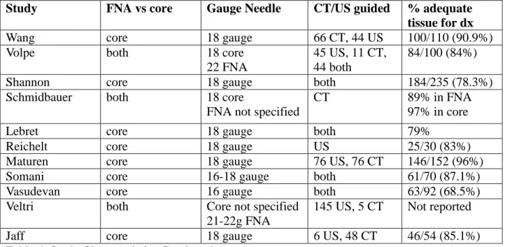
18 One of the central arguments against biopsy has been that it often yields inadequate tissue for diagnosis. While we did not search for this specifically, all of the studies included in the analysis provided this information. The values ranged from as low as 68.5% adequate biopsy in the Vasudevan et al. study to 100% adequate biopsy in the Lebret et al. study. All of the studies used 18 gauge needles to perform core biopsy, with the exception of the Somani et al. study using 16 and 18 gauge needles and the Vasudevan study using 16 gauge needles only. No stratification was provided based on needle size in the Somani study, but it is interesting that the lowest rate of successful biopsies was reported in the study using exclusively the largest gauge needle48. In the studies using FNA technique, one did not report needle size49, one used 22 gauge needles30 and another reported 21-22 gauge use50.
Many studies described normal renal parenchyma (i.e. not the mass), necrotic or hemorrhagic areas, inflammation, fibrosis, or specimens in which the tissue origin could not be determined. Depending on the study, the numbers are derived from either the first biopsy taken, with subsequent re-biopsy success rates reported, or in many studies, patients went on to repeat biopsy if the first sample was inadequate and these numbers are included in the total percentage of adequate biopsies. The Schmidbauer et al. study reported two different rates of adequate biopsy, 89% FNA, and 97% for core biopsy. For FNA, failed biopsies reported no cells in one sample and blood cells only in the remaining samples, while core biopsy showed normal renal parenchyma. This study eventually stopped using FNA after the first 44 patients.
Sensitivity for Benign versus Malignant
19 benign versus malignant pathology ranges from 92% to 100% in those with adequate biopsy. The Veltri et al. study reported accuracy for FNA alone as 88.4%, core biopsy alone as 94.8%, and in those undergoing both FNA and core biopsy, they reported an accuracy of 94.8%. Maturen et al. reported a sensitivity of 85/87 (97.7%) and a specificity of 100% for diagnosing benign versus malignant pathology. The two incorrect diagnoses in the sensitivity calculation were not “benign” but rather “nondiagnostic” biopsies that turned out to be malignant on nephrectomy. In all of the studies, substantially fewer patients underwent nephrectomy than had a diagnostic biopsy. There were very few nephrectomy specimens for those with a benign biopsy. In all but one paper, accuracy was only reported for the subset of patients with a diagnostic biopsy that went on to nephrectomy, because the nephrectomy specimen could serve as a gold standard. However, Maturen et al. created criteria under which they considered an observed patient with a benign biopsy correctly diagnosed after two years of no mass growth or symptoms.
RCC subtype
20 Adverse Events
Though definitions of adverse events were not standardized, the majority of studies dichotomized adverse events into significant versus insignificant, or major versus minor groups. In most cases, major adverse events required intervention, an emergency room visit, or hospital admission. Minor events were those that could be monitored without any action. Studies differed in their post-procedure protocols, with some centers performing ultrasound on all patients who underwent biopsy, while others only took action if complications arose. No studies reported needle track seeding, a major source of concern in early studies. The Wang et al. study reported the highest rate of complications requiring intervention, at 7.3%. Six studies reported either no complications or only minor events requiring no intervention. The Vasudevan study reported one complication in which a patient needed two units of red blood cells, while Maturen et al. reported one patient who needed four units. The total number of biopsies in all the studies considered is 1242 and the total number of serious complications (emergency room visit, admission, or medical intervention) reported was 14, resulting in a rate of 1.1%.
Management of Benign Biopsy
21 studies reported surveillance regimens using CT or MRI every six months, with some spacing out imaging to once a year after a certain amount of time with no growth. The Shannon et al. study reported growth of monitored lesions, but reported no symptoms or subsequent surgery for the masses.
Change in Management
Because eight of the eleven studies were retrospective, the authors reported results as if they would have changed management. These changes referred to patients avoiding surgery based on
biopsy diagnosis of benign lesions, diagnosis of low grade lesions, or diagnosis of metastases that required treatment of the initial cancer rather than nephrectomy. In other cases, the biopsy results affected the type of surgery (i.e. nephron sparing surgery versus radical nephrectomy) or led physicians to use radio frequency ablation techniques rather than extirpative surgery. Even in cases where surveillance was planned initially, a benign biopsy led to less strict surveillance protocols. Eight of the studies reported the percentage of patients with a change in a
22 Study Initial
Assembly of Comparable Groups
Differential or high loss to follow-up
Measurement Clear definition of intervention s Important outcomes considered Adjustment for potential confounders Grade
Wang 52 no 3% benign biopsies
lost to follow-up. 0% of malignant
Details of IHC-unknown. Biopsy pathology compared to nephrectomy pathology
yes yes no poor
Volpe 30 no No reported loss to
follow-up Details of IHC-yes. Biopsy pathology compared to nephrectomy pathology
yes yes no poor
Shannon53 no 7/28 patients with
no diagnosis lost to follow-up. 0 patients with diagnostic biopsy lost to follow-up
Details of IHC-unknown. Biopsy pathology compared to nephrectomy pathology
yes yes no poor
Schmidbauer49 no No report of loss to
follow-up Details of IHC- unknown. Biopsy pathology compared to nephrectomy pathology
yes yes no poor
Lebret 51 no No report of loss to
follow-up Details of IHC-yes. Biopsy pathology compared to nephrectomy pathology
yes yes no poor
Reichelt 54 no No report of loss to
follow-up Details of IHC-yes. Biopsy pathology compared to nephrectomy pathology
yes yes no poor
Maturen 20 no No report of loss to
follow-up Details of IHC-unknown. Biopsy pathology compared to nephrectomy pathology
yes yes no poor
23 nephrectomy group. 14/149 in nonbiopsy nephrectomy group IHC-yes. Biopsy pathology compared to nephrectomy pathology
Vasudevan 48 no No report of loss to
follow-up Details of IHC-unknown. Biopsy pathology compared to nephrectomy pathology
yes yes no poor
Veltri 50 no 12/150 biopsies lost
to follow-up, no further details provided Details of IHC-unknown. Biopsy pathology compared to nephrectomy pathology
yes yes no poor
Jaff 55 no No report of loss to
follow-up Details of IHC-unknown. Biopsy pathology compared to nephrectomy pathology
yes yes no poor
Table 2. Quality criteria adapted from USPSTF cohort study quality criteria.
*important outcomes: findings on biopsy, findings on nephrectomy (if available), adverse events, tumor seeding, death
24 Study # biopsies Mean tumor size Mean Age
(years)
% Male
Wang 110 2.7 cm 60.4 68%
Volpe 100 2.4 cm 60 (median) unknown
Shannon 235 2.9 cm (median) 64 unknown
Schmidbauer 122 4 cm 63 80%
Lebret 119 3.3 cm 66.5 60%
Reichelt 30 2.9 cm 63 60%
Maturen 152 4.1 cm 60 46%
Somani 70 unknown 63.8 61%
Vasudevan 100 unkown 62 unknown
Veltri 150 3.4 cm 64.5 63%
Jaff 54 3.3 cm 72 (median) 60%
Table 3. Study Characteristics
Study FNA vs core Gauge Needle CT/US guided % adequate tissue for dx
Wang core 18 gauge 66 CT, 44 US 100/110 (90.9%)
Volpe both 18 core
22 FNA
45 US, 11 CT, 44 both
84/100 (84%)
Shannon core 18 gauge both 184/235 (78.3%)
Schmidbauer both 18 core
FNA not specified
CT 89% in FNA
97% in core
Lebret core 18 gauge both 79%
Reichelt core 18 gauge US 25/30 (83%)
Maturen core 18 gauge 76 US, 76 CT 146/152 (96%)
Somani core 16-18 gauge both 61/70 (87.1%)
Vasudevan core 16 gauge both 63/92 (68.5%)
Veltri both Core not specified
21-22g FNA
145 US, 5 CT Not reported
Jaff core 18 gauge 6 US, 48 CT 46/54 (85.1%)
25
Study Accuracy
Benign vs Malig Accuracy RCC subtype Accuracy for Fuhrman grade Adverse Events Change in Management
Wang 34/34 (100%) 28/29
(96.6%)
- 8/110= 7% Not reported
Volpe 20/20 (100%) 20/20
(100%)
8/12 (66.7%) none 43/100 (43%) Shannon 108/108 (100%) 106/108
(98%)
- 2/108 (0.9%) 62/235 (26%) Schmidbauer 95.2% (core
sens) 100% (core spec) 90.6% (FNA sens) 100% (FNA spec) 91% core 86% FNA 28% FNA 76% core None requiring intervention 19/122 (15.6%)
Lebret Not presented 86% 46%
76% if high vs low
none 31/102 (30%)
Reichelt 17/17 (100%) - - 1 renal
hematoma
Not reported
Maturen 97.7%
(sensitivity) 100% (specificity)
- - 2
post-procedure hematomas, 1 needing 4 units pRBC
90/152 (60.5%)
Somani 32/32 (100%) - - 1 requiring
admission
17/70 (24%)
Vasudevan 38/38 (100%) - - 1 pt needed 2
units blood
Not reported
Veltri 119/129 (92.2%) - - 8/150 (5.3%) 89/150
(68.9%)
Jaff 100% 100% 75% none 32/46 (69.6%)
Table 5. Review findings for benign vs. malignant, RCC subtype, Fuhrman grade, adverse events and change in management.
26 Study Study Design Inclusion
Criteria
Blinding of pathologists?
Details of
immunohistochemistry
Wang retrospective case
series
SRM ≤ 4 cm. Adults over 18, excluded cystic masses
unknown unknown
Volpe retrospective case
series
Incidental, isolated
SRM ≤4 cm. unknown yes
Shannon retrospective case
series
Incidental,
asymptomatic mass under 5 cm
unknown unknown
Schmidbauer prospective case series
Solid renal masses, cystic excluded
unknown unknown
Lebret retrospective case
series
SRM ≤ 4 cm or those missing radiologic criteria
unknown yes
Reichelt prospective case
series
Noncystic,
homogenous masse found on US
unknown yes
Maturen retrospective case
series
- unknown unknown
Somani prospective case
series
Biopsied those in which "it was not possible to characterize renal masses as either malignant or benign on imaging
characteristics alone"
unknown yes
Vasudevan retrospective case
series
Asymptomatic, incidental masses under 5 cm
unknown unknown
Veltri retrospective case
series
Radiology did not provided sufficient diagnosis
unknown unknown
Jaff retrospective case
series
Solitary kidney, RFA candidate, suspect oncoytoma, lymphoma or mets, high risk for surgery
unknown unknown
27 Discussion
The papers we reviewed claim promising results for the use of biopsy in lesions smaller than four centimeters. In these papers, biopsy was effective at distinguishing benign versus malignant masses; in the papers that reported it, biopsy could predict RCC subtype with accuracy in the 80-100% range. None of the papers reported needle track seeding, and major
complications were rare. These findings coincide with other recent literature reviews on the subject that found improved sensitivity and specificity of renal mass biopsy and reported low rates of complications and no needle track seeding19, 56. Two recent studies found that Fuhrman grade was correctly interpreted in 70%44 and 83% 7 of cases, which also coincides with our findings.
However, an important distinction must be made. As proposed in reviews by Lane et. al in 2008 and Samplaski et al. in 2010, the idea that biopsy failures should not be counted as false negatives is a departure from past literature46, 57-62. Changing these denominators can give substantially higher values for sensitivity depending on the rate of biopsy failure, which may not be an entirely accurate representation of the data. By not counting these patients, some argue that the numbers ignore the non-trivial process of going through biopsy after which there is no
additional information. While this is true, the rate of complications in our study is very low. Additionally, we argue that clinicians and pathologists know that a failed biopsy does not
28 due in part to this method of reporting; a true comparison to previous studies is difficult because of this change in the denominator.
Even if renal mass biopsy is safer and more accurate than previously thought, questions have arisen about the costs incurred by this approach. A cost-effectiveness analysis by
Pandharipande, et al. showed that the risks that have kept physicians from using renal biopsy are “at least equaled by those risks incurred by performing empiric surgery in all patients.” The Markov model developed in the paper used a base-case of a 65 year old man with an incidentally detected tumor under four centimeters who could undergo empiric surgery or tumor surveillance. The base-case sensitivity and specificity were 0.9 and 1, respectively, but sensitivity analyses for both values ranged from 0.5 to 1. By using a quasi-societal model, the authors did not factor in time costs to the patient. Using the base-case model, the biopsy strategy yielded a four day longer life expectancy with a $3466 lower lifetime cost than empiric nephron sparing surgery. The biopsy strategy also dominated across the majority of sensitivity ranges during a one-way sensitivity analysis. Even when the mortality of nephron sparing surgery was assumed to be 0%, the empiric surgery model did not dominate the biopsy model. However, when biopsy sensitivity fell to less than 0.78, the surgical model dominated the biopsy model. Based on findings from the studies that we reviewed, the biopsy model should dominate in the current environment.
Limitations of the study should not nullify its findings, but they are worth considering. For example, because there is limited data on surveillance studies, the authors concede that it was difficult to model biopsy negative tumor outcomes. The authors also note that there are no accepted guidelines for surveillance, which creates difficulty in making assumptions for the model63.
29 Despite increased interest in the area of renal mass biopsy, the current research still has many limitations. Based on USPSTF quality criteria, all of the papers in our study earned a poor quality rating. The majority of the studies are retrospective case series from major academic medical centers, in which patient outcomes are reported from medical records that are often lacking demographic information and long-term follow-up. All of the studies lacked
randomization and control groups, and no study adjusted for baseline status of patients or other possible confounding variables. Additionally, many studies do not elaborate on inclusion criteria for biopsy. For the purposes of our research question, it is important that patients undergo biopsy for an asymptomatic incidental renal mass, because this is the clinical scenario that perplexes urologists most.
30 raised regarding tumor seeding, noting that studies may not have followed patients long enough to detect seeding if it occured later on.
The behavior of supposedly benign masses under surveillance is an important aspect of the conclusions of the studies. Because of a lack of nephrectomy follow-up in benign biopsy, authors were forced to use growth-free or symptom-free surveillance periods as the gold standard for an accurate benign biopsy. This is problematic if, as some studies have suggested, growth-free or symptom-growth-free masses can still go on to be malignant. In a 2004 study by Wehle et. al, out of 29 patients enrolled in a surveillance program, the authors reported three cases that went on to nephrectomy because of patient wishes (i.e. not symptoms or substantial growth) and were found to be RCC. For two of these, they reported no growth in 12 months and 38 months of follow-up, respectively. For the third case, they reported a 0.2 cm/year growth rate during 43 months of follow-up35. Though this study did have a relatively small sample size, and the majority of patients continued surveillance with no deaths or reports of metastases, the study casts doubt on the use of growth-free surveillance as a true marker of benignity. However, growth-free
malignant masses may not be clinically significant if they remain growth-free or grow at a very slow pace. For patients with a limited life expectancy, a mass growing at 0.2 cm/year could be inconsequential even if it is histologically malignant. A longer term study could provide information about the growth of such masses; unfortunately, because of the apprehension of urologists to observe renal masses, there are few surveillance studies in the literature.
31 more training and are exposed to higher case volumes than those practicing in community
settings. For these reasons, reported outcomes and complications may not accurately portray results in other settings.
Future Studies
Here, we propose the design of future studies that would address many of the logistic and epidemiologic shortfalls of the current research. All of the studies in our review were case series reported from major academic medical centers, where patients are more rigorously selected and can introduce unpredictable bias into conclusions. To strengthen generalizability, patients in future studies should be enrolled from population-based registries such as the VA Hospital system or Kaiser because these are more representative of populations across the country. We propose that patients who pose a therapeutic dilemma be pre-specified in some way. For
example, those with tumors under 4 cm, age over 65 years old, or those with a pre-defined set of comorbidities that make surgery a less than ideal option. From this point, these patients could be randomized to renal mass biopsy or watchful waiting. In the biopsy group, further treatment would be dictated by biopsy results; type of surgery, biopsy technique, and
32 generalizable than case series reporting surgical outcomes of highly selected patients at academic medical centers.
Future Direction
Renal epithelial cancers are a diverse group with differing natural histories and prognoses due to their own cytogenetic and molecular aberrations65. A series of studies have reported improvements in biopsy results with the addition of cytogenetic studies. In a paper by Barocas65 et al., the authors combined histopathology with a molecular diagnostic algorithm presented in a previous study66 to improve diagnostic accuracy of core biopsy. This algorithm is based on high CA9 expression in clear cell carcinoma, AMACR in papillary RCC, and CLCNKB in
chromophobe RCC and oncocytoma66. The study reported the diagnostic accuracy of biopsy improved from 83.3% to 95% (57 of 60 biopsies) when histology was combined with the molecular diagnostic algorithm compared to histology alone. The combination increased sensitivity from 87.1% to 100% and improved the negative predictive value from 87.5% to 100%65. However, this study has limited generalizability because of the ex-vivo sampling of kidney masses. Direct visualization and palpation of masses in the study make extrapolation to a true biopsy scenario difficult. Additionally, the authors report using a 14 gauge needle for biopsy which is substantially larger than the more routinely used 18 gauge needle found in the studies we reviewed. The authors note that there is currently no molecular marker than can distinguish between chromophobe renal cell carcinoma from oncocytoma, which is an often a source of incorrect diagnoses.
33 chromosome 3p in clear cell RCC, trisomy 7 and 17 in papillary RCC, and losses of parts of chromosomes 10, 13, 17 or 21 in chromophobe RCC67. The paper showed that adding FISH led to the correct identification of four additional tumors out of 36 total masses, improving the accuracy of biopsy from 75% to 86%. The study shares weaknesses with the aforementioned study in its ex-vivo design and use of 14 gauge needles. Additionally, FISH is accurate for detecting losses and translocations, but can miss mutations, which can be relevant in clear cell carcinoma67. If costs can kept under control, molecular studies could be the future of renal mass management as they will increase diagnostic capabilities and tailor treatment to specific
individuals.
34 REFERENCES
1. Altekruse SF, Kosary CL, Krapcho M, Neyman N, Aminou R, Waldron W, Ruhl J, Howlader N, Tatalovich Z, Cho H, Mariotto A, Eisner MP, Lewis DR, Cronin K, Chen HS, Feuer EJ, Stinchcomb DG, Edwards BK (eds). SEER cancer statistics review, 1975-2007.
http://seer.cancer.gov/csr/1975_2007/.
2. Jayson M, Sanders H. Increased incidence of serendipitously discovered renal cell carcinoma. Urology. 1998;51(2):203-205.
3. Chow WH, Devesa SS, Warren JL, Fraumeni Jr JF. Rising incidence of renal cell cancer in the united states. JAMA. 1999;281(17):1628.
4. Gill IS, Matin SF, Desai MM, et al. Comparative analysis of laparoscopic versus open partial nephrectomy for renal tumors in 200 patients. J Urol. 2003;170(1):64-68.
5. Gill IS, Kavoussi LR, Lane BR, et al. Comparison of 1,800 laparoscopic and open partial nephrectomies for single renal tumors. J Urol. 2007;178(1):41-46.
6. Lechevallier E, André M, Barriol D, et al. Fine-needle percutaneous biopsy of renal masses with helical CT Guidance1. Radiology. 2000;216(2):506.
7. Neuzillet Y, Lechevallier E, Andre M, Daniel L, Coulange C. Accuracy and clinical role of fine needle percutaneous biopsy with computerized tomography guidance of small (less than 4.0 cm) renal masses. J Urol. 2004;171(5):1802-1805.
8. Zhou M, Roma A, Magi-Galluzzi C. The usefulness of immunohistochemical markers in the differential diagnosis of renal neoplasms. Clin Lab Med. 2005;25(2):247-257.
9. Lipworth L, Tarone RE, McLaughlin JK. The epidemiology of renal cell carcinoma. J Urol. 2006;176(6):2353-2358.
10. Bjørge T, Tretli S, Engeland A. Relation of height and body mass index to renal cell carcinoma in two million norwegian men and women. Am J Epidemiol. 2004;160(12):1168. 11. Ficarra V, Schips L, Guillè F, et al. Multiinstitutional european validation of the 2002 TNM staging system in conventional and papillary localized renal cell carcinoma. Cancer.
2005;104(5):968-974.
12. Belldegrun A, MD, Blute MM, Chow GKM, et al. Guidelines for the management of a clinical stage 1 renal mass. American Urologic Association. 2009.
13. Miller DC, Ruterbusch J, Colt JS, et al. Contemporary clinical epidemiology of renal cell carcinoma: Insight from a population based case-control study. J Urol. 2010.
14. Cooperberg MR, Mallin K, Ritchey J, Villalta JD, Carroll PR, Kane CJ. Decreasing size at diagnosis of stage 1 renal cell carcinoma: Analysis from the national cancer data base, 1993 to 2004. J Urol. 2008;179(6):2131-2135.
15. Hollenbeck BK, Taub DA, Miller DC, Dunn RL, Wei JT. National utilization trends of partial nephrectomy for renal cell carcinoma: A case of underutilization? Urology.
2006;67(2):254-259.
16. Hollingsworth JM, Miller DC, Daignault S, Hollenbeck BK. Rising incidence of small renal masses: A need to reassess treatment effect. J Natl Cancer Inst. 2006;98(18):1331.
17. Silverman S, Lee B, Seltzer S, Bloom D, Corless C, Adams D. Small (< or= 3 cm) renal masses: Correlation of spiral CT features and pathologic findings. Am J Roentgenol.
1994;163(3):597.
18. Li G, Cuilleron M, GENTIL‐PERRET A, Tostain J. Characteristics of image‐detected solid renal masses: Implication for optimal treatment. International Journal of Urology.
35 19. Lane BR, Samplaski MK, Herts BR, Zhou M, Novick AC, Campbell SC. Renal mass biopsy--A renaissance? J Urol. 2008;179(1):20-27.
20. Maturen KE, Nghiem HV, Caoili EM, Higgins EG, Wolf Jr JS, Wood Jr DP. Renal mass core biopsy: Accuracy and impact on clinical management. Am J Roentgenol. 2007;188(2):563. 21. Silverman SG, Gan YU, Mortele KJ, Tuncali K, Cibas ES. Renal masses in the adult patient: The role of percutaneous Biopsy1. Radiology. 2006;240(1):6.
22. Bosniak MA, Megibow AJ, Hulnick DH, Horii S, Raghavendra BN. CT diagnosis of renal angiomyolipoma: The importance of detecting small amounts of fat. Am J Roentgenol.
1988;151(3):497.
23. Bosniak MA. Angiomyolipoma (hamartoma) of the kidney: A preoperative diagnosis is possible in virtually every case. Urol Radiol. 1981;3(3):135-142.
24. Zagoria RJ. Imaging of small renal masses: A medical success story. Am J Roentgenol. 2000;175(4):945.
25. Motzer RJ, Bander NH, Nanus DM. Renal-cell carcinoma. N Engl J Med. 1996;335(12):865-875.
26. Novick AC, Derweesh I. Open partial nephrectomy for renal tumours: Current status. BJU Int. 2005;95 Suppl 2:35-40.
27. Steinbach F, Stockle M, Hohenfellner R. Clinical experience with nephron-sparing surgery in the presence of a normal contralateral kidney. Semin Urol Oncol. 1995;13(4):288-291.
28. Petritsch PH, Rauchenwald M, Zechner O, et al. Results after organ-preserving surgery for renal cell carcinoma. an austrian multicenter study. Eur Urol. 1990;18(2):84-87.
29. Van Poppel H, Da Pozzo L, Albrecht W, et al. A prospective, randomised EORTC intergroup phase 3 study comparing the oncologic outcome of elective nephron-sparing surgery and radical nephrectomy for low-stage renal cell carcinoma. Eur Urol. 2010.
30. Volpe A, Mattar K, Finelli A, et al. Contemporary results of percutaneous biopsy of 100 small renal masses: A single center experience. J Urol. 2008;180(6):2333-2337.
31. Davis CJ,Jr. Pathology of renal neoplasms. Semin Roentgenol. 1987;22(4):233-240. 32. Lane BR, Gill IS. 5-year outcomes of laparoscopic partial nephrectomy. J Urol. 2007;177(1):70-74.
33. Venkatesh R, Weld K, Ames CD, et al. Laparoscopic partial nephrectomy for renal masses: Effect of tumor location. Urology. 2006;67(6):1169-1174.
34. Frank I, Blute ML, Cheville JC, Lohse CM, Weaver AL, Zincke H. Solid renal tumors: An analysis of pathological features related to tumor size. J Urol. 2003;170(6):2217-2220.
35. Wehle MJ, Thiel DD, Petrou SP, Young PR, Frank I, Karsteadt N. Conservative management of incidental contrast-enhancing renal masses as safe alternative to invasive therapy. Urology. 2004;64(1):49-52.
36. Campbell SC, Novick AC, Herts B, et al. Prospective evaluation of fine needle aspiration of small, solid renal masses: Accuracy and morbidity. Urology. 1997;50(1):25-29.
37. Smith EH. Complications of percutaneous abdominal fine-needle biopsy. review. Radiology. 1991;178(1):253.
38. WOOD BJ, KHAN MA, McGOVERN F, HARISINGHANI M, HAHN PF, MUELLER PR. Imaging guided biopsy of renal masses: Indications, accuracy and impact on clinical
management. J Urol. 1999;161(5):1470-1474.
36 40. Eshed I, Elias S, Sidi A. Diagnostic value of CT-guided biopsy of indeterminate renal
masses. Clin Radiol. 2004;59(3):262-267.
41. Wehle MJ, Grabstald H. Contraindications to needle aspiration of a solid renal mass: Tumor dissemination by renal needle aspiration. J Urol. 1986;136(2):446-448.
42. Richter F, Kasabian N, IrwinJr R, Watson R, Lang E. Accuracy of diagnosis by guided biopsy of renal mass lesions classified indeterminate by imaging studies. Urology.
2000;55(3):348-352.
43. Herts BR, Remer E. The role of percutaneous biopsy in the evaluation of renal and adrenal mass. AUA Update Series. 2000;19:282-287.
44. Wunderlich H, Hindermann W, Mustafa AMA, Reichelt O, Junker K, Schubert J. The accuracy of 250 fine needle biopsies of renal tumors. J Urol. 2005;174(1):44-46.
45. Truong LD, Todd TD, Dhurandhar B, Ramzy I. Fine-needle aspiration of renal masses in adults: Analysis of results and diagnostic. Diagn Cytopathol. 1999;20(6):339.
46. Murphy WM, Zambroni BR, Emerson LD, Moinuddin S, Lee LH. Aspiration biopsy of the kidney. simultaneous collection of cytologic and histologic specimens. Cancer. 1985;56(1):200-205.
47. Somani BK, Nabi G, Thorpe P, N'Dow J, Swami S, McClinton S. Image-guided biopsy-diagnosed renal cell carcinoma: Critical appraisal of technique and long-term follow-up. Eur Urol. 2007;51(5):1289-1297.
48. Vasudevan A, Davies RJ, Shannon BA, Cohen RJ. Incidental renal tumours: The frequency of benign lesions and the role of preoperative core biopsy. BJU Int. 2006;97(5):946-949. 49. Schmidbauer J, Remzi M, Memarsadeghi M, et al. Diagnostic accuracy of computed tomography-guided percutaneous biopsy of renal masses. Eur Urol. 2008;53(5):1003-1012. 50. Veltri A, Garetto I, Tosetti I, et al. Diagnostic accuracy and clinical impact of imaging-guided needle biopsy of renal masses. retrospective analysis on 150 cases. Eur Radiol. 2010:1-9. 51. Lebret T, Poulain JE, Molinie V, et al. Percutaneous core biopsy for renal masses:
Indications, accuracy and results. J Urol. 2007;178(4):1184-1188.
52. Wang R, Wolf Jr JS, Wood Jr DP, Higgins EJ, Hafez KS. Accuracy of percutaneous core biopsy in management of small renal masses. Urology. 2009;73(3):586-590.
53. Shannon BA, Cohen RJ, de Bruto H, Davies RJ. The value of preoperative needle core biopsy for diagnosing benign lesions among small, incidentally detected renal masses. J Urol. 2008;180(4):1257-1261.
54. Reichelt O, Gajda M, Chyhrai A, Wunderlich H, Junker K, Schubert J. Ultrasound-guided biopsy of homogenous solid renal masses. Eur Urol. 2007;52(5):1421-1427.
55. Jaff A, Molinié V, Mellot F, Guth A, Lebret T, Scherrer A. Evaluation of imaging-guided fine-needle percutaneous biopsy of renal masses. Eur Radiol. 2005;15(8):1721-1726.
56. Samplaski MK, Zhou M, Lane BR, Herts B, Campbell SC. Renal mass sampling: An enlightened perspective. International Journal of Urology.
57. Holm HH, Pedersen JF, Kristensen JK, Rasmussen SN, Hancke S, Jensen F. Ultrasonically guided percutaneous puncture. Radiol Clin North Am. 1975;13(3):493-503.
58. Nosher JL, Amorosa JK, Leiman S, Plafker J. Fine needle aspiration of the kidney and adrenal gland. J Urol. 1982;128(5):895-899.
59. Juul N, Torp-Pedersen S, Grønvall S, Holm H, Koch F, Larsen S. Ultrasonically guided fine needle aspiration biopsy of renal masses. J Urol. 1985;133(4):579-581.
37 61. Leiman G. Audit of fine needle aspiration cytology of 120 renal lesions. Cytopathology. 1990;1(2):65-72.
62. Niceforo J, Coughlin B. Diagnosis of renal cell carcinoma: Value of fine-needle aspiration cytology in patients with metastases or contraindications to nephrectomy. Am J Roentgenol. 1993;161(6):1303.
63. Pandharipande PV, Gervais DA, Hartman RI, et al. Renal mass biopsy to guide treatment decisions for small incidental renal tumors: A cost-effectiveness Analysis1. Radiology. 2010;256(3):836.
64. Chawla SN, Crispen PL, Hanlon AL, Greenberg RE, Chen DYT, Uzzo RG. The natural history of observed enhancing renal masses: Meta-analysis and review of the world literature. J Urol. 2006;175(2):425-431.
65. Barocas D, Rohan S, Kao J, et al. Diagnosis of renal tumors on needle biopsy specimens by histological and molecular analysis. J Urol. 2006;176(5):1957-1962.
66. Chen YT, Tu JJ, Kao J, Zhou XK, Mazumdar M. Messenger RNA expression ratios among four genes predict subtypes of renal cell carcinoma and distinguish oncocytoma from carcinoma. Clinical cancer research. 2005;11(18):6558.
67. Barocas DA, Mathew S, DelPizzo JJ, et al. Renal cell carcinoma sub‐typing by