EVALUATION OF AKR1B10 AND SORD MRNA EXPRESSION IN IN VIVO AND
IN VITRO MODELS
Anita R. Gandhi
Department of Nutrition
University of North Carolina at Chapel Hill
2016
Approved by:
Dr. Ramon Bataller
ACKNOWLEDGEMENTS
I would like to thank my research mentor, Dr. Bataller, for allowing me to conduct
research in his lab for the past two years. In addition, I wanted to acknowledge my
research project advisor, Veronica Massey, for her diligence, genuine care, and for being
there every step of the way. I am also thankful for the unwavering and constant support
TABLE OF CONTENTS CHAPTER
I. ABSTRACT___________________________________________________ 4
II. INTRODUCTION______________________________________________ 5
III. METHODS___________________________________________________ 10
IV. RESULTS____________________________________________________ 14
V. DISCUSSION_________________________________________________19
VI. CONCLUSION________________________________________________24
I. ABSTRACT Author: Anita R. Gandhi
Advisors: Ramon Bataller, Veronica Massey
Background: Alcoholic Hepatitis (AH) is the most severe form of alcoholic liver disease. To identify targets that may be dysregulated in AH, AH patient tissue samples
were deep-sequenced, identifying that AKR1B10 is the most up-regulated and SORD is
down-regulated.
Aims: To use both in vivo and in vitro models to study the induction of AKR1B10 and SORD mRNA in response to exposure to ethanol or signaling factors.
Methods: AKR1B10 and SORD mRNA expression were analyzed in patients with various liver diseases, HepG2 cells, VL-17A cells, primary human hepatocytes, and
ethanol-fed mice. RNA was extracted from all samples and gene expression was both
measured and analyzed using real-time qPCR.
Results: AKR1B10 mRNA expression is inducible by EGF in HepG2 cells and primary human hepatocytes. It is also up-regulated in the presence of ethanol in VL-17A cells.
SORD mRNA expression was not significantly changed in any of the in vitro models
studied, but is up regulated by ethanol exposure in the in vivo mouse model.
Conclusion: AKR1B10 mRNA induction by ethanol and EGF indicates that ethanol or EGF signaling may play a role in the up-regulation of AKR1B10 in patients with AH. In
addition, due to the lack of significant changes in SORD mRNA expression in the in vitro
models studied, it is important to identify other signaling factors that may dysregulate
AKR1B10 or SORD expression, perform co-cultures, perform additional ethanol
II. INTRODUCTION Alcoholic Hepatitis
Alcohol is a widely consumed substance around the world. Given that the liver is the
primarily site of alcohol metabolism, over-consumption of alcohol can lead to many
negative effects on the liver. Indeed, heavy alcohol drinkers are at a greater risk of
developing a multitude of liver-specific adverse health conditions, such as alcoholic liver
disease (ALD). ALD is a spectrum of disease states, which include steatosis, cirrhosis,
fibrosis, and alcoholic hepatitis (AH), which is the most severe form of ALD (1)
. Though
AH presents after the onset of the first three diseases, not all patients reach this state. It is
unknown as to why some patients develop alcoholic hepatitis and others do not despite a
similar history of alcohol use. However AH has become a worldwide problem.
Apparently, liver cirrhosis has become one of the top causes of death in the United States,
and almost half of them were alcohol related (1)
. Despite the prevalence of this condition,
more research needs to be conducted on the pathogenesis or patient susceptibility to AH,
as both are still largely unknown. Additionally, the current therapy for AH,
corticosteroids, is ineffective in many patients with AH (1)
. Therefore, a major goal of our
research group is to explore the potential mechanisms underlying AH and find targets for
therapy. In order to do so, our group has previously deep-sequenced genes in individuals
diagnosed with alcoholic hepatitis and compared gene expression to normal liver tissue.
The deep sequencing analysis identified aldo-keto reductase family 1 member B10
(AKR1B10) as the most up regulated gene in the liver of patients with AH. The analysis
setting of AH. Therefore, we aim to determine the effects of ethanol and signaling
factors on the mRNA expression of AKR1B10 and SORD in vitro and in vivo models.
AKR1B10:
AKR1B10 is a known gene that is located on chromosome 7 (2)
. The expression of
AKR1B10 is highly induced in the cells of various cancers such as lung non-small-cell
carcinoma and hepatocellular carcinoma (3)
. Since the enzyme exhibits broad substrate
specificities toward various xenobiotics such as anti-tumor drugs or various endogenous
compounds such as retinaldehyde, AKR1B10 may play an important role in tumor
progression or drug resistance (3)
. It also has an important role in the sorbitol polyol
pathway, an alternative pathway for glucose metabolism (4)
. Since AKR1B10 was up
regulated in patients with AH, as the deep-sequencing identified, the expression was
induced and studied in various cell models. These models include HepG2, VL-17a, mice,
and Human hepatocytes, especially when exposed to tumor necrosis factor α-(TNFα),
epidermal growth factor (EGF), and insulin. EGF and insulin have been previously
shown to induce AKR1B10 expression (5). TNFα was explored, as it is highly involved in
severe liver diseases, including alcoholic hepatitis. It is important to note that in mice, the
mouse ortholog for AKR1B10 is AKR1B8(6)
SORD
SORD, a gene located on chromosome 15, codes for an enzyme that converts polyols into
their corresponding ketoses (7)
. Together, SORD and aldose reductases constitute the
fructose(7) .
The polyol pathway is hypothesized to play an important role in the
progression and development of diabetes (7)
. Since SORD was down regulated in patients
with AH, the expression was induced and studied in various cell lines such as HepG2,
VL-17a, primary human hepatocytes, and in a mouse model. Specifically, the effect of
TNFα, EGF, and insulin, and ethanol exposure on AKR1B10 and SORD mRNA
expression was studied.
Lieber-DeCarli Diet
The Lieber-DeCarli diet was developed in 1982 as a method of ethanol exposure in
rodent models. It is currently the most commonly used model for alcoholic liver injury (8)
.
The administration of this diet in rodent models can result in mild steatosis, slight
elevation of serum alanine transaminase (ALT) but little or no inflammation (8)
. This diet
is suitable for exposing mice to ethanol in order to investigate the effects of AKR1B10
up-regulation on hepatocytes.
Sorbitol Polyol Pathway
AKR1B10 and SORD are both genes that code for enzymes involved in the polyol
pathway. In the polyol pathway, glucose is metabolized into sorbitol via AKR1B10, and
SORD then converts sorbitol to fructose (9)
. When produced inside the cell, sorbitol is
sequestered and cannot leave. In contrast, fructose is able to leave the cell via a fructose
transporter (9) .
Since AKR1B10 is up regulated and SORD is down regulated in patients
with AH, we hypothesized that these patients may experience sorbitol accumulation in
inside the cell while decreased SORD expression may prevent sorbitol degradation
leading to intracellular sorbitol accumulation. Since hepatocytes, unlike other cells in the
body, do not have known sorbitol transporters, sorbitol accumulation could cause
hepatocellular damage including osmotic and oxidative stress.
In renal cells and erythrocytes, there is an enzyme called “sorbitol permease” which has
the ability to remove excess sorbitol from the cell (10)
. Before the cell is lysed due to
osmotic pressure, the sorbitol permease allows release of the sorbitol into the
bloodstream. It is in response linearly to sorbitol concentration and is not affected by
substances that normally block other sugar transporters (10)
. However, there has been no
such mechanism found in the liver.
Since it has been determined that AKR1B10 and SORD mRNA expression are altered in
patients who present with alcoholic hepatitis, a major goal of the experiments detailed in
this paper was to study the potential mechanisms underlying AKR1B10 and SORD
mRNA expression in hepatocytes. In order to do so, AKR1B10 and SORD mRNA
expression are studied in in vivo and in vitro models. The in vitro models used included
HepG2 cells, primary human hepatocytes, and VL-17A cells. Both the primary human
hepatocytes and HepG2 cells were used to study AKR1B10 and SORD mRNA induction
in hepatocytes. Human hepatocytes possess many of the functions lost in immortalized
hepatocellular carcinoma lines and are a useful model for studying hepatocytes10 . As
insulin and EGF have proven to induce AKR1B10 expression6
, and TNFα is involved in
the progression of many hepatic disorders, human hepatocytes are to be exposed to each
a liver biopsy of a 15-year-old Caucasian male11
and are commonly used to study
hepatocytes and the progression of liver diseases. HepG2 cells are to be exposed to EGF,
which will theoretically induce the expression of SORD and AKR1B10. Unlike HepG2,
VL-17A cells possess enzymes that metabolize alcohol, such as alcohol dehydrogenase
and CYP2E19
. Thus, VL-17A cells are a useful for studying the effects of ethanol and
ethanol metabolism in vitro. For these studies, VL-17A cells will be used to determine
changes in mRNA expression of AKR1B10 and SORD caused by ethanol exposure. An
in vivo model of ethanol exposure in mice was used to compliment the in vitro models used for these studies. For the in vivo studies, mice were fed and ethanol containing
Lieber DeCarli diet over the course of four weeks.
Given the goals of our experiments, our main hypotheses were that 1) EGF, insulin, and
TNFα can induce AKR1B10 and SORD mRNA expression in primary human
hepatocytes 2) EGF can induce AKR1B10 and SORD mRNA expression in HepG2 cells,
and 3) AKR1B10 mRNA expression will be up-regulated and SORD will be
down-regulated after ethanol exposure in VL-17A cells and in the liver of mice. A proposed
mechanism for hepatocyte damage in AH is dysregulation of AKR1B10 and SORD
III. METHODS Patient Samples:
Seventeen patients admitted to the Liver Unit of the Hospital Clinic in Barcelona, Spain
from 2007 to 2009 with features of alcoholic hepatitis were included as patient samples
for this study (1)
. In addition, seven patients that were diagnosed with compensated liver
disease, indicating that they had non-alcohol mediated cirrhosis, were included for
analysis. For control purposes, samples from nine patients diagnosed with chronic
hepatitis-C induced liver disease were also analyzed. These patients had HCV genotype 1
and had not received previous antiviral treatment. The inclusion criterion has been
described in previous studies (1)
. Liver biopsies from these patients were obtained using a
trans-jugular approach. For all patients, an expert liver pathologist examined each
specimen to confirm diagnosis. Portions of the biopsy were submerged into an RNA
stabilization solution (Austin, Texas, USA) and subsequent mRNA isolation. All patients
involved in the study gave informed consent and the protocol was conformed to the
ethical guidelines of the 1975 Declaration of Helsinki and was approved by the Ethics
Committee of the Hospital Clinic of Barcelona (1) .
Mouse Models:
The mice used for the study were a generous gift from the laboratory of Scott Magness
(UNC Chapel Hill). All experimental procedures were reviewed and approved by the
University of North Carolina at Chapel Hill Institutional Animal Care and Use
Committee. Eight-week old C57BL6/J female mice were exposed to an
an in vivo model of alcohol exposure, as described previously (8)
. Briefly, ethanol fed
mice were given a liquid diet (Dyets, Bethlehem, PA) that contained increasing
concentrations of ethanol over a period of four weeks until a concentration of six percent
ethanol was reached(8).
Control animals were pair fed an isocaloric control diet (8)
. Mice
were fed once per day between the hours of 4:30 and 5:30 PM and consumed 12 mL per
mouse per day. Mice were kept on a 12-hour light/dark cycle and housed in temperature
and humidity controlled rooms. At the time of sacrifice, mice were anesthetized and liver
samples were collected for the analysis of AKR1B10 and SORD mRNA expression. A
fragment of liver tissue was immediately submerged in Trizol (ThermoFisher, Carlsbad,
CA, USA) for RNA isolation.
Cell Cultures
All cells were maintained at 37 degrees C in a 5% CO2 environment. HepG2 cells
(ATCC, Rockville Maryland) and VL-17A cells (a generous gift from the Zeisel Lab,
Kannapolis, NC) were cultured as previously described (11). Specifically, cells were
maintained in appropriate media and were passaged twice per week with Trypsin (Gibco,
Grand Island, USA). Primary human hepatocytes (Triangle Research Labs, North
Carolina) were grown following the supplier’s protocol(12) .
HepG2 cells and primary human hepatocytes were exposed to EGF (50 ng/mL) for 24
hours as described by others (5)
. VL-17A were exposed to 100 mM ethanol for 24 hours
and Human hepatocytes, were exposed to EGF (50 ng/mL), insulin (10 mM), or TNFα 50
Cells were passaged using PBS (Gibco, Grand Island, USA) and trypsin. RNA was
isolated from the cells using Trizol (ThermoFisher, Carlsbad, CA, USA) following
manufacturer’s protocol.
RNA isolation and qPCR
RNA was isolated from both animal and human cells using Trizol (ThermoFisher,
Carlsbad, CA, USA) following manufacturer’s protocol. To isolate the mRNA, phenol:
chloroform extraction was used. To determine the concentration and purity of RNA
present in the samples, absorbance was measured at 260 and 280 nm, respectively, using
a NanoDrop 1000. A cDNA reverse transcription kit (Applied Biosystems, Foster City,
CA, USA) was used to reverse-transcribe mRNA. For quantitative PCR analysis, cDNA
was amplified for forty cycles using a StepOne PlusTM
Real-Time PCR System (Applied
Biosystems). Both mouse and human primers were used in analysis and were purchased
from Applied Biosystems as kits (Foster City, CA). Mouse probes included:
Mm03928990_g1 for 18S, Mm03048764 for AKR1B10, and Mm00455377_g1 for
SORD. These probes were used for the in vivo model of ethanol exposure. Human probes
included: Hs03928985_g1 for 18S, Hs01546975_gH for ARK1B10, and
Hs00162091_m1 for SORD and were used for the in vitro models. Results were
normalized to 18S expression for animal and human samples. Gene expression was
calculated using the ΔCT method. This method determines the amount of target gene,
normalized to 18S, an endogenous reference gene, and relative to a calibrator (2-ΔΔCT
Statistics
Results of quantitative variables are expressed as the mean ± standard error of the mean.
To analyze results, Prism by Graphpad software was used. Two-sample t-tests were
performed and statistical significance was determined as p < 0.05. This value was set
IV. RESULTS
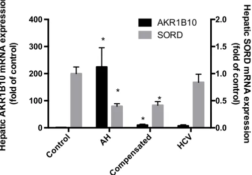
AKR1B10 and SORD mRNA Expression in Patients with Alcoholic Hepatitis A previous study using microarray analysis identified AKR1B10 as the most highly
up-regulated gene in patients with alcoholic hepatitis (AH) (1)
. That study also found that
SORD was down-regulated in AH patients (1)
. Therefore, using real-time qPCR analysis,
we analyzed both hepatic AKR1B10 and SORD mRNA expression in a cohort of patients
diagnosed with AH, non-alcoholic mediated cirrhosis, and hepatitis C virus. These results
indicate that there is significant AKR1B10 overexpression and SORD under-expression
in patients with alcoholic hepatitis, compared to controls and patients with other liver
diseases, which included hepatitis-c and non-alcohol mediated cirrhosis. Patients with
AH had a 220-fold increase when compared to controls and SORD had a 0.6 fold
decrease (Figure 1).
Figure 1. Hepatic AKR1B10 and SORD mRNA expression in patients with liver diseases (*p<0.05 compared to all other groups
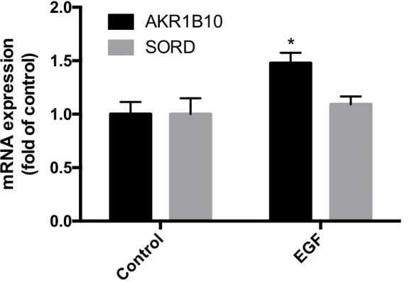
AKR1B10 and SORD mRNA Expression in HepG2 Cells
AKR1B10 and SORD mRNA was analyzed in HepG2 cells, considering HepG2 cells
originate from a hepatocellular carcinoma cell line and are widely used for the study of
liver diseases (11)
. Cells were exposed to EGF, as EGF has shown to induce AKR1B10
mRNA expression in other studies (5)
. EGF exposure caused a significant increase in
AKR1B10 mRNA expression as it was increased by 1.5 fold in HepG2 cells (Figure 3).
There was no significant change in SORD mRNA expression.
Figure 2. The effect of EGF exposure on AKR1B10 and SORD mRNA expression in HepG2 cells (*p<0.05 compared to all other groups)
Control
EGF
0.0
0.5
1.0
1.5
2.0
mRNA
exp
ressi
o
n
(fo
ld
o
f c
o
n
tr
o
l)
AKR1B10
*
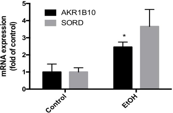
AKR1B10 and SORD mRNA Expression in VL-17A Cells
AKR1B10 and SORD mRNA expression were analyzed in VL-17A cells after exposure
to 100 mM of ethanol for 24 hours. VL-17A cells are HepG2 cells that have been altered
to express enzymes that play a key role in alcohol metabolism, ADH and CYP2E1(13) .
AKR1B10 mRNA expression was significantly increased 2.5 fold after exposure to 100
mM ethanol for 24 hours. Ethanol exposure did not cause a significant change in SORD
mRNA expression (figure 4).
Figure 3. The effect of 24 hour 100 mM ethanol exposure on AKR1B10 and SORD mRNA expression in VL-17A cells (*p<0.05 compared to all other groups)
Control
EtOH
0
1
2
3
4
5
mRNA
exp
ressi
on
(fo
ld
o
f c
on
tr
ol
)
AKR1B10
SORD
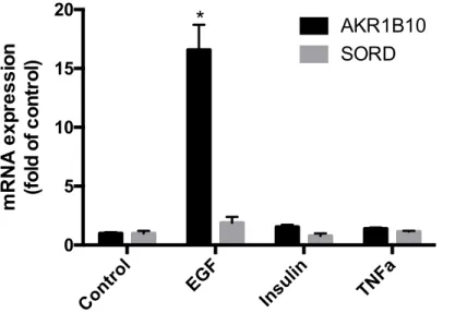
AKR1B10 and SORD mRNA Expression in Human Hepatocytes
AKR1B10 and SORD mRNA expression were determined in cryopreserved primary
human hepatocytes, as human hepatocytes maintain some metabolic functions of
hepatocytes that are lost in the immortalized hepatocellular carcinoma cell lines
(i.e-HepG2) (13)
. The human hepatocytes were exposed to EGF or insulin, since previous
published manuscripts indicate that EGF and insulin can induce AKR1B10 in HepG2
cells (5)
. Cells were exposed to TNFα, considering TNFα is highly involved in severe
liver diseases, including alcoholic hepatitis. Results indicate that AKR1B10 is
up-regulated when exposed to EGF, but that there is no major change when exposed to
insulin and TNFα. AKR1B10 had a 16-fold increase when exposed to EGF in
comparison to other groups. There was also no significant change in SORD mRNA
expression (figure 2).
Figure 4. The Effect of EGF, insulin, TNFα on AKR1B10 and SORD mRNA
expression in primary human hepatocytes (*p<0.05 compared to all other groups)
Control EGF Insulin TNFa
AKR1B10 and SORD mRNA Expression in Animal Models of Alcoholic Hepatitis Mice were fed an ethanol-containing diet or were pair-fed control diet for four weeks,
according to the Lieber-DeCarli method (8)
. AKR1B10 mRNA expression was not
significantly changed by ethanol exposure (Figure 5). SORD mRNA expression was
significantly up-regulated in mice exposed to alcohol by 1.7 fold.
Figure 5. The effect of Ethanol Exposure on AKR1B10 and SORD mRNA Expression in Mice Models (*p<0.05 compared to all other groups)
Control
EtOH
0.0
0.5
1.0
1.5
2.0
2.5
mRNA
exp
ressi
on
(fo
ld
o
f c
on
tr
ol
)
AKR1B10
V. DISCUSSION
Alcohol over-consumption is a prevalent problem in today’s society. As alcohol is
primarily metabolized in the liver, over-consumption can lead to a number of adverse
effects, resulting in hepatic cell damage(1)
. Alcoholic hepatitis is an example of a rare
condition that leads to the inflammation of the liver, usually after alcoholic liver disease
is already present (1)
. Unfortunately, not much is known on the pathogenesis of alcoholic
hepatitis. Thus, a major goal of this study was to explore a potential mechanism
underlying alcoholic hepatitis and find specific targets for therapy.
Liver tissue from patients with AH was previously deep-sequenced by our
research group. This study identified that AKR1B10 was the most up-regulated gene and
that SORD was significantly under-expressed. In order to confirm these results and
identify how AKR1B10 and SORD mRNA expression are altered in patients with liver
conditions, seventeen liver samples from patients who presented with alcoholic hepatitis,
non-alcohol mediated cirrhosis, and hepatitis-C induced liver diseases were collected and
qPCR was performed in our study. AKR1B10 was significantly up regulated and SORD
was significantly down-regulated in patients with alcoholic hepatitis (Figure 1). AH
patients presented with a 220-fold increase in terms of AKR1B10 mRNA expression, in
comparison to the control, and a 0.6 fold decrease for SORD mRNA expression (Figure
1). These findings were consistent with the previously performed experiments that
involved the deep sequencing of liver tissue from patients with AH. They also indicate
that AKR1B10 and SORD are likely involved in the progression of AH and should be
As aforementioned, besides being up-regulated in those with alcoholic hepatitis,
AKR1B10 is also highly expressed in patients with hepatocellular carcinoma and lung
non-small-cell carcinoma, and may play a major role in tumor progression (3)
. AKR1B10
is also involved in the sorbitol polyol pathway, as it converts glucose to sorbitol (9) .
The SORD gene codes for a multitude of enzymes that play an integral role in
converting polyols and their corresponding ketoses (7)
. Like ARK1B10, SORD, along
with aldose reductase, is involved in the sorbitol polyol pathway, as it converts sorbitol to
fructose (10) .
Considering both of these genes are involved in the sorbitol polyol pathway, it is
possible that this pathway may play a role in the progression of alcoholic hepatitis. Since
AKR1B10 is over-expression and SORD is under-expressed, there may be increased
sorbitol accumulation in the hepatocytes. Since there is no known transporter in
hepatocytes that can remove sorbitol, increased sorbitol production and a decrease in
sorbitol degradation may lead to osmotic stress13 .
By using in vivo and in vitro models, we explored AKR1B10 and SORD mRNA
expression in primary human hepatocytes and HepG2 cells, and determine what happens
to expression when exposed to ethanol in VL-17A cells and mice. We hypothesized that
TNFα, insulin, and EGF could induce expression and that ethanol could cause AKR1B10
mRNA expression to be up-regulated and SORD to be down-regulated. This would be
consistent with the idea that sorbitol may be accumulating in the cells, which is a
potential mechanism for hepatocellular damage.
After identifying that both ARK1B10 and SORD had altered mRNA expression
explore AKR1B10 and SORD mRNA expression in other hepatocytes, such as HepG2
cells (Figure 2). HepG2 cells are widely used for the study of liver diseases, and originate
from a hepatocellular carcinoma cell line. Results indicate that, when exposed to EGF,
AKR1B10 mRNA expression is increased by 1.5 fold in HepG2 cells. However, there
seemed to be no significant change in SORD mRNA expression (Figure 3). These results
also confirm studies that state AKR1B10 can be induced in the presence of EGF and
demonstrated that AKR1B10 mRNA expression can be stimulated in various cell types.
However, HepG2 cells are not the most similar to actual human liver cells and doing
experiments with a more accurate cell model may present with different results.
Since AKR1B10 was found to be inducible in the presence of EGF, it was
possible that other cell models could be used to see if ethanol had any effect on
AKR1B10 or SORD mRNA expression, since ethanol exposure is the a contributing
factor to the development of alcoholic hepatitis. Thus, these genes were analyzed in
VL-17A cells, which are HepG2 cells that have been altered to express alcohol-metabolizing
genes, ADH and CYP2E1 (11). Cells were exposed to 100 mM of ethanol for 24 hours, and
results indicated a 2.5 fold increase for AKR1B10 mRNA expression, but no significant
change for SORD mRNA expression (Figure 3). Unlike HepG2 cells, Vl-17A cells are
more representative of actual human liver cells since they can metabolize alcohol, but are
still not an ideal model for the study of hepatocytes. AKR1B10 up-regulation due to
ethanol exposure in VL-17A cells indicate that ethanol may contribute to the
over-expression of AKR1B10 in AH.
Similarly, AKR1B10 and SORD mRNA expression were studied in primary
maintain many of the metabolic functions of hepatocytes that are lost in other
immortalized hepatocellular carcinoma cell lines10
. The primary human hepatocytes were
exposed to TNFα, EGF, or insulin. According to previous research, AKR1B10 mRNA
expression could be induced in HepG2 cells in the presence of either insulin or EGF. In
addition, cells were exposed to TNFα, as TNFα is highly involved in the progression of
various liver diseases. When examining AKR1B10 and SORD mRNA expression in
human hepatocytes, it is clear that there was significant change in AKR1B10 mRNA
expression when exposed to EGF. AKR1B10 has a 16-fold increase in comparison to
other groups (Figure 4). These results confirmed previous studies that stated that
AKR1B10 is induced in the presence of EGF (5)
and were consistent with our hypothesis.
They also demonstrated that EGF could be a potential regulator of AKR1B10
up-regulation in patients with AH. Considering EGF is present in humans, it is possible that
patients with higher levels of EGF are more susceptible to developing AH, which is
important to experiment on further. SORD was unaffected, suggesting that EGF may not
be the cause of SORD under-expression in patients with AH. In addition, results indicate
that insulin and TNFα do not affect significantly affect the expression of AKR1B10 or
SORD.
Considering AKR1B10 mRNA expression in increased in patients with AH, an in
vivo model of ethanol exposure was created to explore AKR1B10 and SORD mRNA expression. This is because in vivo and in vitro models are two complimentary methods
of investigating what may cause AKR1B10 up-regulation and what effect it may have on
hepatocytes. Eight mice were pair-fed ethanol for four weeks, according to the
Lieber-DeCarli method (8)
was no significant change in AKR1B10 mRNA expression (Figure 5). These results are
not consistent with any of the hypotheses in our study. This demonstrates that moderate
alcohol exposure alone does not induce AKR1B10 expression or cause SORD
under-expression in the model studied. However, it is important to note that mice metabolize
alcohol differently from humans and that human drinking patters should be applied to the
mice models in order to create a more effective model.
Overall, results from the experiments conducted, with the exception of the in vivo
model, demonstrate no significant change in SORD expression. This could be due to the
fact that the cell models that we utilized are not perfect models of actual hepatocytes, the
experimental environment was not representative of the human body, and because there
could be other factors that regulate SORD dysregulation besides EGF, insulin, ethanol, or
TNFα. In addition, it is important to note that no experiments that we conducted
demonstrated both an up-regulation of AKR1B10 and down-regulation of SORD, which
does not seem consistent with the idea of sorbitol accumulation. This shows that there are
other underlying multifactorial mechanisms that may lead to sorbitol accumulation.
However, with the exception of the in vivo model of ethanol exposure, experiments
showed a significant up-regulation of AKR1B10, which does indicate that AKR1B10
VI. CONCLUSION
In all, it is evident from the experiments conducted that AKR1B10 is inducible by
EGF in HepG2 cells and in primary human hepatocytes, as by ethanol in VL-17A cells.
These suggest that that EGF and ethanol may be involved in the over-expression of
AKR1B10 in patients with AH. However, SORD regulation was not altered as expected
in any of the results. Thus, for our future studies, it is important to identify additional
stimuli that could dysregulate ARK1B10 or SORD expression, such as LPS. In addition,
to mimic the human body environment, a co-culture can be performed with our particular
cell model and Kupffer or stellate cells, which are both native to the liver. A logical next
step is to perform addition in vitro ethanol exposure experiments, particularly in primary
human hepatocytes, as they are very similar to actual human liver cells. Our in vivo
model could be improved by creating an ethanol exposure model that is more similar to
human drinking patterns. This could include exposing the mice to a greater concentration
of ethanol or exposing them for a longer period of time. To further investigate whether
sorbitol accumulation can cause negative effects in patients with AH, sorbitol levels
References
1.Affo S, Dominguez M, Lozano JJ, Sancho-Bru P, Rodrigo-Torres D, Morales-Ibanez O, Moreno M, et al. Transcriptome analysis identifies TNF superfamily receptors as potential therapeutic targets in alcoholic hepatitis. Gut 2013;62:452-460.
2.Cards G. AKR1B10 Gene. In; 2016.
3.Matkowskyj KA, Bai H, Liao J, Zhang W, Li H, Rao S, Omary R, et al.
Aldoketoreductase family 1B10 (AKR1B10) as a biomarker to distinguish hepatocellular carcinoma from benign liver lesions. Hum Pathol 2014;45:834-843.
4.Petrash JM. All in the family: aldose reductase and closely related aldo-keto reductases. Cell Mol Life Sci 2004;61:737-749.
5.Cao D, Fan ST, Chung SS. Identification and characterization of a novel human aldose reductase-like gene. J Biol Chem 1998;273:11429-11435.
6.Pastel E, Pointud JC, Volat F, Martinez A, Lefrancois-Martinez AM. Aldo-Keto Reductases 1B in Endocrinology and Metabolism. Front Pharmacol 2012;3:148.
7.El-Kabbani O, Darmanin C, Chung RP. Sorbitol dehydrogenase: structure, function and ligand design. Curr Med Chem 2004;11:465-476.
8.Massey VL, Poole LG, Siow DL, Torres E, Warner NL, Schmidt RH, Ritzenthaler JD, et al. Chronic Alcohol Exposure Enhances Lipopolysaccharide-Induced Lung Injury in Mice: Potential Role of Systemic Tumor Necrosis Factor-Alpha. Alcohol Clin Exp Res 2015;39:1978-1988.
9.Lorenzi M. The polyol pathway as a mechanism for diabetic retinopathy: attractive, elusive, and resilient. Exp Diabetes Res 2007;2007:61038.
10.Kracke GR, Preston GG, Stanley TH. Identification of a sorbitol permease in human erythrocytes. Am J Physiol 1994;266:C343-350.
11.Donohue TM, Osna NA, Clemens DL. Recombinant Hep G2 cells that express alcohol dehydrogenase and cytochrome P450 2E1 as a model of ethanol-elicited cytotoxicity. Int J Biochem Cell Biol 2006;38:92-101.
12.Labs TR. Cryopreserved Hepatocytes In; 2016.