16I CATARACT & REFRACTIVE SURGERY TODAY EUROPE IOCTOBER 2009
K
eratoconus is one of the most challenging corneal pathologies faced by ophthalmologists. The onset of the disease during puberty and its often pro-gressive course can have a potentially devastating impact on the patient’s vision and overall quality of life.Keratoconus treatment options were, until recently, limited to spectacles, contact lenses, and corneal transplantation. However, over the past few years, new
diagnostic and therapeutic tools have greatly expanded our options to address keratoconus and other corneal ectatic disorders. Advanced contact lens materials and designs, ultraviolet (UV) collagen crosslinking (CXL), intrastromal corneal ring seg-ments (ICRS), phakic IOLs, conductive keratoplasty, and modern wavefront-or topography-guided photoablative procedures have all been proposed for treatment of keratoconus.
Techniques that allow us to reshape the cornea for therapeutic purposes may collectively be called
corneoplastics, a term coined by Arun C. Gulani, MD. Corneoplastics is spreading rapidly and becoming a new subspecialty allied to refractive and corneal surgery. But with so many treatment options now available, which is best? I believe there is not one best solution for all cases; our job is to select the best corneoplastic treatment or combination of treatments for each patient. Herein, I present my pre-ferred option, Keraring ICRS (Mediphacos, Belo Horizonte, Brazil). Two articles to follow present other intrastromal corneal ring options.
A N I M P O RTA N T O P T I O N
In my personal practice at the Clinique de la Vision in Paris, femtosecond–laser-assisted implantation of the Keraring has been an important corneoplastic option for
most of our keratoconic patients since 2004. The Keraring is an implantable device designed to correct corneal surface irregularities, improve UCVA and BCVA, and reduce refrac-tive errors associated with keratoconus and other corneal ectatic disorders (Figure 1).
Our main indications for Keraring implantation are kera-toconus with reduced BCVA and contact lens intolerance,
progressing keratoconus, pellucid marginal degeneration, post-LASIK ectasia, and high amounts of regular or irregular astigma-tism after penetrating keratoplasty.
In my experience with many eyes treated, post-LASIK ectasia is an excellent indication for Keraring implantation and probably one of the best options as it not only treats the ectatic cornea and reduces higher-order aberrations but also corrects refractive error in patients who desire a refractive proce-dure. I exclude patients presenting with ker-atometry readings greater than 60.00 D, extensive central corneal opacity, severe atopic disease, or unrealistic expectations regarding refractive result. I believe that it is extremely important to take time to properly educate patients about keratoconus and the objectives of the Keraring procedure so that they have realistic expectations.
V E R S AT I L E S Y S T E M
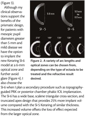
The Keraring system is quite versatile, offering 40 combi-nations of ring segment thicknesses, arc lengths, and optical zones (Figure 2). The segments are available with thicknesses from 150 to 350 µm in 50 µm steps; arc lengths from 90º to 210º in 30º steps; and optical zones of 5, 5.5, and 6 mm. Depending on the case, one or two Keraring segments of the same or different sizes may be implanted. The implant is delivered with one ring segment per box so the surgeon can mix and match the sizes and number of segments implant-ed as desirimplant-ed.
Corneal Remodeling
Using the Keraring
A variety of thicknesses, arc lengths, and optical zone sizes
allows tailoring of the procedure to the individual patient.
BY DOMINIQUE PIETRINI, MD; AND TONY GUEDJ
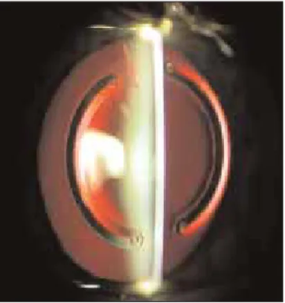
Figure 1. A pair of 160° Kerarings, as seen in the eye.
Each Keraring size can be compared with a unique struc-tural building block with specific properties of action on the corneal dome. By increasing the thickness we can increase the segment’s effect; by varying the arc length we can inde-pendently modulate the sphere and cylinder correction; and by using different optical zones we can address variations in corneal pachymetry, pupil diameter, and stage of the dis-ease. With an understanding of these properties, we can design a truly customized approach to corneal remodeling and refractive effect according to the individual case.
The refractive component of Keraring implantation improves UCVA. This is crucial for keratoconic patients, who are usually contraindicated for corneal refractive treat-ments and often intolerant of any optical correction.
Unlike other ICRS originally developed for myopic correc-tion, which are mostly astigmatically neutral, the Keraring was designed specifically for keratoconus to intentionally induce high degrees of astigmatism. This mechanism allows us to position the implant in such a way that it corrects pre-existing astigmatism by producing cylinder at the comple-mentary axis. I find this ability to correct cylinder especially helpful in addressing advanced keratoconus and high astig-matism after penetrating keratoplasty. Asymmetric implan-tation allows us to treat irregular astigmatism and to decrease large amounts of higher-order aberrations, espe-cially coma.
In my experience, the design of the Keraring and its vari-able optical zone of 5 to 6 mm produce greater effect than other ICRS that are implanted more peripherally. This is espe-cially important to allow us to more effectively treat the severe corneal surface irregularities and high refractive errors that often accompany keratoconus.1A smaller optical zone
also prevents corneal neovascularization, which can some-times be observed with other ICRS implanted closer to the limbus.
The incidence of glare and halos, which one would expect with the smaller optical zone, is reduced due to the Keraring’s design. Its triangular cross-sectional shape produces a pris-matic effect and mitigates visual disturbances in low light
(Figure 3). Although my clinical observa-tions support the benefits of the prismatic design, for patients with mesopic pupil diameters greater than 5 mm and mild disease we have the option to implant the new Keraring SI-6 model at a 6-mm optical zone and further avoid glare (Figure 4). I also choose the
SI-6 when I plan a secondary procedure such as topography-guided PRK or posterior chamber phakic IOL implantation. The SI-6 has a wide base, scalene triangular cross-section, and truncated apex design that provides 25% more implant vol-ume compared with the SI-5 Keraring of similar thickness. This increased volume offsets the loss of effect expected from the larger optical zone.
S U R G I C A L T E C H N I Q U E
Good surgical outcomes depend largely on careful patient selection, choice of implant size and location, and surgical technique. As a starting point for my surgical plans, I use the Keraring calculation guidelines and nomograms available from the manufacturer. These nomograms assist me in selecting the implant based on shape, extent, and dis-tribution of the corneal ectasia; visual acuity; and aberro-metric and manifest refraction values. However, as every ectatic cornea is unique, often I extrapolate the nomograms and design a more customized surgical plan. The technical help desk service offered by the manufacturer provides advice for surgical plan customization that I find useful for complex cases.
The surgical technique begins with construction of an intrastromal tunnel at a depth equivalent to 75% of the corneal thickness followed by implantation of the Keraring segments. The implantation tunnel can be dissected mechanically using special surgical instrumentation or cre-ated with a femtosecond laser such as the IntraLase2,3
(Abbott Medical Optics Inc., Santa Ana, California). We prefer to use a femtosecond–laser-assisted technique as we find it to be faster, less invasive, and more consistent than the mechanical technique. The IntraLase allows safe dissec-tion of the stromal tunnel with the exact dimensions
Figure 2. A variety of arc lengths and optical zones can be chosen from, depending on the type of ectasia to be treated and the refractive result desired.
Figure 3. The Keraring’s triangular cross-sectional shape produces a prismatic effect and mitigates visual disturbances in low light.
18I CATARACT & REFRACTIVE SURGERY TODAY EUROPE IOCTOBER 2009
desired and consistent centration on the visual axis. The entire procedure takes a few minutes and is per-formed under topical anesthesia on an outpatient basis. The technique is minimally invasive and sutureless. Patients receive a bandage contact lens for 24 hours and a prescrip-tion regimen with antibiotics and antiinflammatory drugs as in other corneal procedures. Most patients notice visual improvement by a few days after the procedure.
Topographic and refraction changes can fluctuate during the first 3 months, and then they tend to stabilize.
S Y N E R G I S T I C P R O C E D U R E S
Although the corneoplastic effects of implantation have been shown to be stable for several years in most cases, we can in cases of unstable cornea and progressive disease syn-ergistically combine the Keraring with UV CXL to further sta-bilize the cornea and lock in the achieved result (Figure 5).
We prefer to first reshape the cornea with the Keraring and then perform CXL once a good result is obtained, usu-ally 3 months or less after the first procedure. However, we prefer not to perform CXL unless we document progression of the disease or fluctuation of the refractive results.
Other interesting and potentially synergistic
combina-tions include Keraring implantation followed by phakic IOL implantation or PRK plus UV CXL, when indicated. These techniques can often enhance the corneoplastic result and correct residual refractive error, further improving the over-all result and patient satisfaction.
ICRS procedures are also reversible and adjustable. Topographic and refractive results may be adjusted by implant exchange or by changing the implant location. If necessary, explantation allows the cornea to revert to its preoperative shape with no sequelae. If the implant fails to provide acceptable results, or in the event of major complications, penetrating or lamellar kerato-plasty can be performed without being affected by the implant’s presence. The implant is removed with the host graft (Figure 6).
CO M P L I C AT I O N S
Complications are relatively rare and mostly related to poor surgical technique or wrong implant selection. Implant extrusion has been reported in rare cases and is often related to superficial implantation, excessive implant thickness, or implant position too close to the incision. Extrusion usually requires explantation with possible reimplantation after 90 days.4Migration of the
segment, usually caused by frequent eye rubbing, has also been occasionally described.4It requires surgical
repositioning of the implant and treatment of the asso-ciated atopic condition.
Infection is fortunately rare (0.2%), most often caused by poor patient compliance to the prescription drug regimen or an implant left too close to the incision. Depending on the severity of the infection, explantation or corneal trans-plantation may be required.
We have observed loss of BCVA in 2% of cases, attrib-utable to poor surgical planning or technique. In such cases the implant size or position can be adjusted to improve the outcome. Persistent glare and halos were
Figure 4. The SI-6 Keraring has a wide base,scalene triangular cross-section,and truncated apex design providing 25% more implant volume compared with the SI-5 Keraring of similar thickness.
Figure 5. In patients with an unstable cornea and progressive disease, the Keraring can be synergistically combined with UV CXL to further stabilize the cornea.
Figure 6. Deep anterior lamellar keratoplasty is performed on a cornea previously implanted with the Keraring (arrows).
reported by 3% of patients, often related to poor cen-tration of the implant or to patient susceptibility. Over time, most patients adjust to such symptoms, and for extreme cases miotic drops at dusk or even explanta-tion are possible alternatives.
C A S E E X A M P L E S
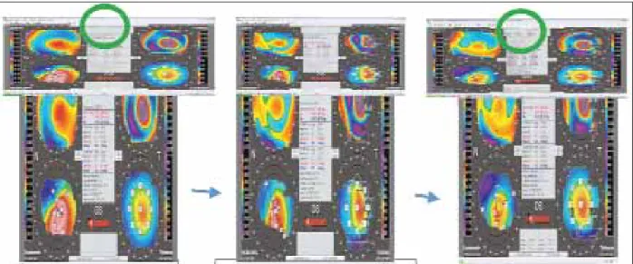
Case 1 (Figure 7) illustrates the evolution of the corneal topographic pattern over time. The initial effect of Keraring implantation is not complete at day 1. Stabilization of the cornea is achieved at 3 months after Keraring implantation. Preoperatively, this ectatic cornea had a maximal kerato-metric value of 53.00 D and BCVA of 20/32. At 3 months postop, UCVA was 20/20, even with the small amount of residual irregular astigmatism.
Case 2 (Figure 8) illustrates the effect induced by implan-tation of a pair of Keraring segments on the Orbscan (Bausch & Lomb, Rochester, New York) differential map. The effect on the cornea is clearly an induced cylindrical bowtie pattern when implantation is symmetrical.
Case 3 (Figures 9 and 10) is an incidence of post-LASIK ectasia after initial microkeratome LASIK probably per-formed on a cornea with pellucid marginal degeneration. BCVA is still 20/20, but the patient did not tolerate the opti-cal correction and complained of poor quality of vision. After uneventful femtosecond–laser-assisted Keraring implantation, UCVA improved to 20/20, restoring the initial postoperative result. This patient was crosslinked in a sec-ondary procedure, as ectasia showed recent progression.
Case 4 (Figure 11) illustrates how combining treatments
Figure 7. The initial effect of Keraring implantation is not complete at day 1. Stabilization of the cornea is achieved 3 months after implantation.
Figure 8. Differential corneal topography map (large image, top left) showing the symmetrical cylindrical effect of a pair of Kerarings of the same thickness and arc length in an eye with an advanced stage of keratoconus. Anterior chamber optical coherence tomography (bottom) shows the position
of the Kerarings in the stroma. • The Keraring is designed to correct corneal surface irregularities, improve UCVA and BCVA, and reduce refractive errors.
• This ICRS offers 40 combinations of thickness, arc lengths, and optical zones.
• Complications are rare.
22I CATARACT & REFRACTIVE SURGERY TODAY EUROPE IOCTOBER 2009
can improve results. The Keraring always provides the main improvement in vision, and its effect can be enhanced by a secondary procedure such as topography-guided PRK, in this case allowing a central ablation of 36 µm and a gain of three lines of BCVA.
Modern corneoplastic techniques are dramatically chang-ing the way we treat corneal ectatic disorders. Implantation of the Keraring either as a standalone or combined proce-dure can enhance our ability to deliver a customized approach to corneal remodeling and improve outcomes, potentially avoiding the need for corneal transplantation and improving patients’ quality of life5in many cases. ■
Dominique Pietrini, MD, practices at the Clinique de la Vision, Paris. He did not disclose financial interest. He may be reached at tel: +33 1 58 05 2000; fax: +33 1 58 05 2001; e-mail: [email protected].
Tony Guedj is an optometrist at the Clinique de la Vision, Paris. He did not disclose financial interest. He may be reached at e-mail: [email protected].
1. Uceda-Montanes A, Toms JD, Ali JL. Correction of severe ectasia after LASIK with intra-corneal ring segments. J Refract Surg. 2008;24(4):408-411.
2. Shabayek MH, Ali JL. Intrastromal corneal ring segment implantation by femtosecond laser for keratoconus correction. Ophthalmology. 2007;114(9):1643-1652.
3. Coskunseven E, Kymionis GD, Tsiklis NS, et al. One-year results of intrastromal corneal ring segment implantation (KeraRing) using femtosecond laser in patients with keratoconus. Am J Ophthalmol. 2008;145(5):775-779.
4. Coskunseven E, Kymionis GD, Tsiklis NS, et al. Complications of intrastromal corneal ring segment implantation using a femtosecond laser for channel creation: a survey of 850 eyes with keratoconus. Acta Ophthalmol. 2009 Aug 14 [Epub ahead of print].
5. Paranhos JF, Avila MP, Paranhos A Jr, Schor P. Evaluation of the impact of intracorneal ring segments implantation on the quality of life of patients with keratoconus using the NEI-RQL (National Eye Institute Refractive Error Quality of life) instrument. Br J Ophthalmol. 2009 Sep 11 [Epub ahead of print].
Figure 9. Post-LASIK ectasia after initial microkeratome LASIK probably performed on a cornea with pellucid marginal degeneration. After femtosecond–laser-assisted Keraring implantation, UCVA improved to 20/20.
Figure 10. Anterior chamber OCT shows the cornea before and after Keraring implantation.(Same eye as Figure 9.)
Figure 11. Combined Keraring implantation and topography-guided PRK. The Keraring provides the main improvement in vision; its effect is enhanced by topography-guided PRK, allowing a central ablation of 36 µm and a gain of three lines of BCVA.