R E V I E W
Open Access
Daptomycin-induced eosinophilic
pneumonia - a systematic review
Priyasha Uppal
1, Kerry L. LaPlante
1,2,3, Melissa M. Gaitanis
1,3, Matthew D. Jankowich
1,3and Kristina E. Ward
2*Abstract
Purpose:
Eosinophilic pneumonia comprises a group of lung diseases in which eosinophils appear in increased
numbers in the lungs and sometimes in the bloodstream. Several case reports link daptomycin use to this
phenomenon.
Summary:
We performed a systematic literature review to identify cases of eosinophilic pneumonia associated with
daptomycin use. Relevant studies were identified by searching Pubmed/Medline, EMBASE, Google Scholar, Cochrane
Database of Systematic Reviews, and Clin-Alert from inception to May 2016, and manual searches of reference lists. All
case reports that include information regarding patient age, indication, clinical and objective findings, treatment and
outcome were evaluated. Abstracts from conference proceedings as well as case reports not in English were excluded.
Descriptive statistics were used to analyze the data. Thirty-five patient-cases were included in the final analysis. Patients
most likely to be identified with daptomycin-induced eosinophilic pneumonia were male (83%) and elderly (mean age
65.4 ± 15 years). The dose for daptomycin ranged from 4 to 10 mg/kg/day, but included a large number of patients
with renal dysfunction. The average duration of daptomycin therapy upon onset of EP symptoms was 2.8 ± 1.6 weeks.
Majority of patients presented with dyspnea (94%), fever (57%) and were also found to have peripheral eosinophilia
(77%) and infiltrates/opacities of CT/CXR (86%). Symptom improvement was seen after daptomycin discontinuation
(24 h to 1 week). The majority of patients were also prescribed treatment with corticosteroids (66%).
Conclusion:
Clinicians should be aware of daptomycin-induced eosinophilic pneumonia and its symptoms along with
its presentation and treatment.
Keywords:
Daptomycin, Eosinophilia, Pneumonia
Background
Eosinophilic pneumonia is a rare, but serious respiratory
syndrome that occurs when eosinophils accumulate in the
lungs [1, 2]. It has been associated with several
medica-tions and chemicals, with antibiotics and nonsteroidal
anti-inflammatory drugs among the most common [3, 4].
The pathophysiology of acute eosinophilic pneumonia is
thought to be caused by detection of an antigen by
alveo-lar macrophages which leads to recruitment of T-helper 2
lymphocytes and subsequent release of interleukin 5.
Interleukin 5 promotes eosinophil production and
migra-tion to the lung. Addimigra-tionally, eotaxin (a potent eosinophil
chemoattractant) production by alveolar macrophages,
pulmonary endothelial cells, airway smooth muscle cells,
and alveolar epithelial cells leads to further accumulation
of eosinophils in the lungs [5].
Daptomycin is a cyclic lipopeptide antibiotic derived from
the fermentation of
Streptomyces roseosporus
. Daptomycin
has activity against Gram-positive organisms including
methicillin-resistant
Staphylococcus aureus
(MRSA) and
vancomycin-resistant enterococci (VRE) [6]. In 2007,
pulmonary eosinophilia was added to the
“Adverse
Reac-tions, Post-Marketing Experience”
section of the product
label for daptomycin [2]. A review of the literature and the
US FDA Adverse Event Reporting System database in 2012
revealed 7 definite, 13 probable, and 38 possible cases of
daptomycin-induced eosinophilic pneumonia [1]. While
the mechanism of daptomycin’s pulmonary toxicity is not
known, the drug undergoes conformational change through
interaction with calcium which allows binding to the
cyto-plasmic membrane, increased membrane permeability, and
intracellular ion escape [7]. The antibacterial activity of
* Correspondence:[email protected]2Department of Pharmacy Practice, University of Rhode Island, 7 Greenhouse Rd, Suite 295 J, Kingston, RI 02881, USA
Full list of author information is available at the end of the article
© The Author(s). 2016Open AccessThis article is distributed under the terms of the Creative Commons Attribution 4.0 International License (http://creativecommons.org/licenses/by/4.0/), which permits unrestricted use, distribution, and reproduction in any medium, provided you give appropriate credit to the original author(s) and the source, provide a link to the Creative Commons license, and indicate if changes were made. The Creative Commons Public Domain Dedication waiver (http://creativecommons.org/publicdomain/zero/1.0/) applies to the data made available in this article, unless otherwise stated. Uppalet al. Antimicrobial Resistance and Infection Control (2016) 5:55
daptomycin is decreased because of its binding to
pulmon-ary surfactant. Some have speculated two potential
mecha-nisms for daptomycin’s pulmonary toxicity: 1) chronic
daptomycin administration results in drug accumulation
near the epithelial alveolar surface causing epithelial injury
and pneumonia and 2) the daptomycin-surfactant
inter-action could alter lipid integrity which may stimulate an
in-flammatory response [3, 7, 8].
Per the FDA guidance, eosinophilic pneumonia is
attrib-uted to daptomycin when the following criteria are met:
1) concurrent exposure to daptomycin, 2) fever, 3)
dys-pnea with increased oxygen requirement or requiring
mechanical ventilation, 4) new infiltrates on chest x-ray or
computed tomography (CT) scan, 5) bronchoalveolar
lav-age (BAL) with > 25% eosinophils, and 6) clinical
improve-ment following daptomycin withdrawal [2]. Solomon and
Schwartz [4] have also developed criteria for drug- or
toxin-induced eosinophilic pneumonia that is similar
and includes 1) presence of simple, acute, or chronic
eosinophilic pneumonia by diagnostic criteria which
includes excess of eosinophils either on lung biopsy
or BAL (usually
≥
25%) in the setting of parenchymal
infiltrates 2) presence of a potential candidate drug or
toxin in an appropriate time frame 3) no other cause
of eosinophilic pneumonia such as fungal or parasitic
infection 4) clinical improvement after cessation of
the drug or toxin, and 5) recurrence of eosinophilic
pneumonia with re-challenge to the drug or toxin.
However, re-challenge is often not recommended as it
can be dangerous [4, 5].
Although the mechanism of daptomycin-induced
eosino-philic pneumonia is unknown, some have speculated that
daptomycin may bind to human surfactant and accumulate
in the alveolar space causing injury to the epithelium with
resulting inflammation [7, 8]. The purpose of this review is
to systematically evaluate the published literature describing
daptomycin-induced eosinophilic pneumonia.
Methods
All relevant cases and studies were identified by
systematic-ally searching of the PubMed, EMBASE, Google Scholar,
Cochrane Database of Systematic Reviews, and Clin-Alert
databases by two reviewers from inception through May
2016. The truncated terms
“daptomycin”,
“eosinophil*”, and
“pneumon*”
were searched in each database. All case
re-ports that included information regarding patient age,
indi-cation, clinical and objective findings, treatment, and
outcome were evaluated. Reports not published in English
were excluded as well as abstracts from conference
pro-ceedings. Descriptive analysis was used to present pooled
demographic information and other data where applicable.
Results
No clinical or observational trials assess
daptomycin-induced eosinophilic pneumonia; only case reports and case
series are published. In 2012, Kim et al. identified 7 definite,
13 probable, and 38 possible cases of daptomycin-induced
eosinophilic pneumonia via review of literature and FDA
Adverse Event Reporting System Reports (AERS) as defined
in Table 1 [1]. Details regarding the 38 possible cases
reported through AERS were not described. Of the 20
cases that were identified as definite or probable, 9
have been published in the literature, the remaining
11 were summarized by Kim [1, 3, 9–13]. We also
identified 39 additional cases of eosinophilic
pneumo-nia attributed to daptomycin for a total of 59 cases
described in the literature [7, 14–24]. Of those, 21
were excluded from this systematic review because
they
were
abstract
presentations
at
international
meetings and did not go through the peer review
publishing process [25–41]. Another three were
ex-cluded because they were not published in English
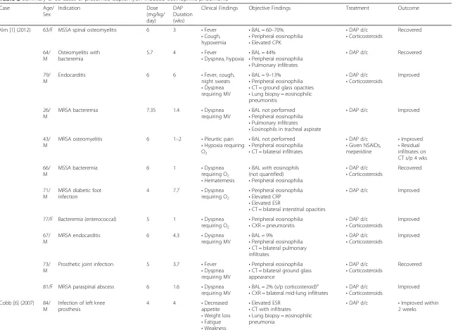
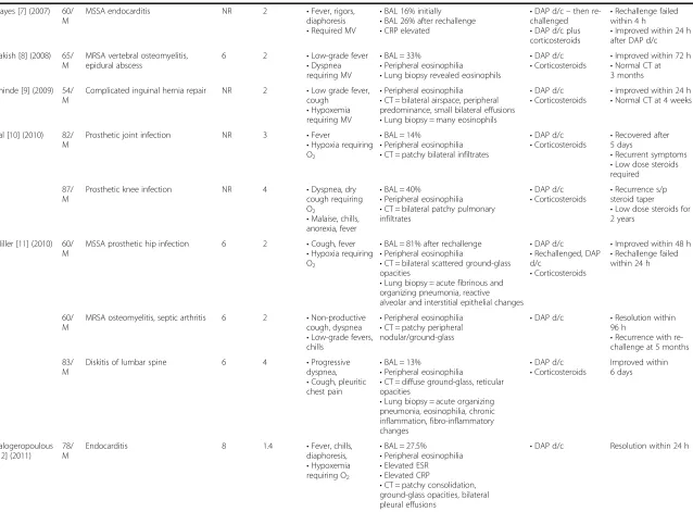
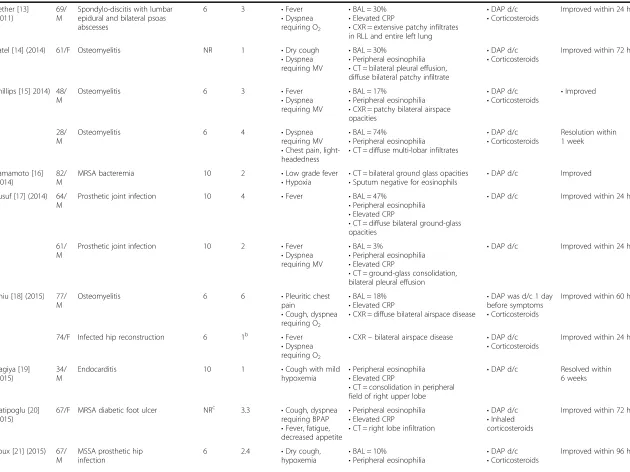
[42–44]. Currently available data on a total of 35
cases of daptomycin-induced eosinophilic pneumonia
is summarized in Table 2.
Table 1
Criteria for inclusion as definite, probably, possible, and unlikely cases of daptomycin-induced eosinophilic pneumonia [1, 2]
Definite Probable Possible Unlikely
Concurrent exposure to daptomycin Concurrent exposure to daptomycin Concurrent exposure to daptomycin All other cases that did not meet criteria
Dyspnea with increased oxygen requirement or requiring mechanical ventilation
Dyspnea with increased oxygen requirement or requiring mechanical ventilation
New infiltrates on CXR or CT
New infiltrates on CXR or CT New infiltrates on chest x-ray or CT Clinical improvement following daptomycin withdrawal OR the patient died
BAL with > 25% eosinophils BAL with≤25% eosinophils OR peripheral eosinophilia
Clinical improvement following daptomycin withdrawal
Clinical improvement following daptomycin withdrawal
Fever
Adapted from references 1 and 2
Abbreviation:BALbronchoalveolar lavage;CTcomputed tomography;CXRchest x-ray
Table 2
Summary of 35 cases of presumed daptomycin-induced eosinophilic pneumonia
Case Age/
Sex
Indication Dose
(mg/kg/ day)
DAP Duration (wks)
Clinical Findings Objective Findings Treatment Outcome
Kim [1] (2012) 63/F MSSA spinal osteomyelitis 6 3 •Fever
•Cough, hypoxemia
•BAL = 60–70%
•Peripheral eosinophilia
•Elevated CPK
•DAP d/c
•Corticosteroids
Recovered
64/ M
Osteomyelitis with bacteremia
5.7 4 •Fever
•Dyspnea, hypoxia
•BAL = 44%
•Peripheral eosinophilia
•Pulmonary infiltrates
•DAP d/c Recovered
79/ M
Endocarditis 6 6 •Fever, cough,
night sweats
•Dyspnea requiring MV
•BAL = 9–13%
•Peripheral eosinophilia
•CT = ground glass opacities
•Lung biopsy = eosinophilic pneumonitis
•DAP d/c
•Corticosteroids
Improved
26/ M
MRSA bacteremia 7.35 1.4 •Dyspnea
requiring MV
•BAL not performed
•Peripheral eosinophilia
•Pulmonary infiltrates
•Eosinophils in tracheal aspirate
•DAP d/c Improved
43/ M
MRSA osteomyelitis 6 1–2 •Pleuritic pain
•Hypoxia requiring O2
•BAL not performed
•Peripheral eosinophilia
•CT = bilateral infiltrates
•DAP d/c
•Given NSAIDs, meperidine
•Improved
•Residual infiltrates on CT s/p 4 wks
66/ M
MSSA bacteremia 6 1 •Dyspnea
requiring O2
•Hematemesis
•BAL with eosinophils (not quantified)
•Peripheral eosinophilia
•DAP d/c
•Corticosteroids
Recovered
71/ M
MRSA diabetic foot infection
4 7.7 •Dyspnea
requiring O2
•Peripheral eosinophilia
•Elevated CRP
•Elevated ESR
•CT = bilateral interstitial opacities
•DAP d/c Improved
77/F Bacteremia (enterococcal) 5 1 •Dyspnea
requiring O2 •
Peripheral eosinophilia
•CXR = pneumonitis •
DAP d/c
•Corticosteroids
Improved
67/ M
MRSA endocarditis 6 4.3 •Dyspnea
requiring MV
•BAL = 9%
•Peripheral eosinophilia
•CT = bilateral pulmonary infiltrates
•DAP d/c
•Corticosteroids
Improved
73/ M
Prosthetic joint infection 5 3.7 •Fever
•Dyspnea requiring MV
•Peripheral eosinophilia
•CT = bilateral ground glass appearance
•DAP d/c
•Corticosteroids
Recovered
81/F MRSA paraspinal abscess 6 1.6 •Dyspnea
requiring MV •
BAL = 2% (s/p corticosteroid)a
•CXR = bilateral mid-lung infiltrates • DAP d/c
•Corticosteroids
Improved
Cobb [6] (2007) 84/ M
Infection of left knee prosthesis
4 4 •Decreased
appetite
•Weight loss
•Fatigue
•Weakness
•Elevated ESR
•CT with infiltrates
•Lung biopsy = eosinophilic pneumonia
•DAP d/c •Improved within 2 weeks
Uppal
et
al.
Antimicrob
ial
Resistance
and
Infection
Control
(2016) 5:55
Page
3
of
Table 2
Summary of 35 cases of presumed daptomycin-induced eosinophilic pneumonia
(Continued)
Hayes [7] (2007) 60/ M
MSSA endocarditis NR 2 •Fever, rigors,
diaphoresis
•Required MV
•BAL 16% initially
•BAL 26% after rechallenge
•CRP elevated
•DAP d/c–then re-challenged
•DAP d/c plus corticosteroids
•Rechallenge failed within 4 h
•Improved within 24 h after DAP d/c
Kakish [8] (2008) 65/ M
MRSA vertebral osteomyelitis, epidural abscess
6 2 •Low-grade fever
•Dyspnea requiring MV
•BAL = 33%
•Peripheral eosinophilia
•Lung biopsy revealed eosinophils
•DAP d/c
•Corticosteroids •
Improved within 72 h
•Normal CT at 3 months
Shinde [9] (2009) 54/ M
Complicated inguinal hernia repair NR 2 •Low grade fever, cough
•Hypoxemia requiring MV
•Peripheral eosinophilia
•CT = bilateral airspace, peripheral predominance, small bilateral effusions
•Lung biopsy = many eosinophils
•DAP d/c
•Corticosteroids •
Improved within 24 h
•Normal CT at 4 weeks
Lal [10] (2010) 82/ M
Prosthetic joint infection NR 3 •Fever
•Hypoxia requiring O2
•BAL = 14%
•Peripheral eosinophilia
•CT = patchy bilateral infiltrates
•DAP d/c
•Corticosteroids
•Recovered after 5 days
•Recurrent symptoms
•Low dose steroids required
87/ M
Prosthetic knee infection NR 4 •Dyspnea, dry cough requiring O2
•Malaise, chills, anorexia, fever
•BAL = 40%
•Peripheral eosinophilia
•CT = bilateral patchy pulmonary infiltrates
•DAP d/c
•Corticosteroids
•Recurrence s/p steroid taper
•Low dose steroids for 2 years
Miller [11] (2010) 60/ M
MSSA prosthetic hip infection 6 2 •Cough, fever
•Hypoxia requiring O2
•BAL = 81% after rechallenge
•Peripheral eosinophilia
•CT = bilateral scattered ground-glass opacities
•Lung biopsy = acute fibrinous and organizing pneumonia, reactive alveolar and interstitial epithelial changes
•DAP d/c
•Rechallenged, DAP d/c
•Corticosteroids
•Improved within 48 h
•Rechallenge failed within 24 h
60/ M
MRSA osteomyelitis, septic arthritis 6 2 •Non-productive cough, dyspnea
•Low-grade fevers, chills
•Peripheral eosinophilia
•CT = patchy peripheral nodular/ground-glass
•DAP d/c •Resolution within 96 h
•Recurrence with re-challenge at 5 months
83/ M
Diskitis of lumbar spine 6 4 •Progressive
dyspnea,
•Cough, pleuritic chest pain
•BAL = 13%
•Peripheral eosinophilia
•CT = diffuse ground-glass, reticular opacities
•Lung biopsy = acute organizing pneumonia, eosinophilia, chronic inflammation, fibro-inflammatory changes
•DAP d/c
•Corticosteroids
Improved within 6 days
Kalogeropoulous [12] (2011)
78/ M
Endocarditis 8 1.4 •Fever, chills,
diaphoresis,
•Hypoxemia requiring O2
•BAL = 27.5%
•Peripheral eosinophilia
•Elevated ESR
•Elevated CRP
•CT = patchy consolidation, ground-glass opacities, bilateral pleural effusions
•DAP d/c Resolution within 24 h
Uppal
et
al.
Antimicrob
ial
Resistance
and
Infection
Control
(2016) 5:55
Page
4
of
Table 2
Summary of 35 cases of presumed daptomycin-induced eosinophilic pneumonia
(Continued)
Rether [13] (2011)
69/ M
Spondylo-discitis with lumbar epidural and bilateral psoas abscesses
6 3 •Fever
•Dyspnea requiring O2
•BAL = 30%
•Elevated CRP
•CXR = extensive patchy infiltrates in RLL and entire left lung
•DAP d/c
•Corticosteroids
Improved within 24 h
Patel [14] (2014) 61/F Osteomyelitis NR 1 •Dry cough
•Dyspnea requiring MV
•BAL = 30%
•Peripheral eosinophilia
•CT = bilateral pleural effusion, diffuse bilateral patchy infiltrate
•DAP d/c
•Corticosteroids
Improved within 72 h
Phillips [15] 2014) 48/ M
Osteomyelitis 6 3 •Fever
•Dyspnea requiring MV
•BAL = 17%
•Peripheral eosinophilia
•CXR = patchy bilateral airspace opacities
•DAP d/c
•Corticosteroids
•Improved
28/ M
Osteomyelitis 6 4 •Dyspnea
requiring MV
•Chest pain, light-headedness
•BAL = 74%
•Peripheral eosinophilia
•CT = diffuse multi-lobar infiltrates
•DAP d/c
•Corticosteroids
Resolution within 1 week
Yamamoto [16] (2014)
82/ M
MRSA bacteremia 10 2 •Low grade fever
•Hypoxia
•CT = bilateral ground glass opacities
•Sputum negative for eosinophils
•DAP d/c Improved
Yusuf [17] (2014) 64/ M
Prosthetic joint infection 10 4 •Fever •BAL = 47%
•Peripheral eosinophilia
•Elevated CRP
•CT = diffuse bilateral ground-glass opacities
•DAP d/c Improved within 24 h
61/ M
Prosthetic joint infection 10 2 •Fever
•Dyspnea requiring MV
•BAL = 3%
•Peripheral eosinophilia
•Elevated CRP
•CT = ground-glass consolidation, bilateral pleural effusion
•DAP d/c Improved within 24 h
Chiu [18] (2015) 77/ M
Osteomyelitis 6 6 •Pleuritic chest
pain
•Cough, dyspnea requiring O2
•BAL = 18%
•Elevated CRP
•CXR = diffuse bilateral airspace disease
•DAP was d/c 1 day before symptoms
•Corticosteroids
Improved within 60 h
74/F Infected hip reconstruction 6 1b •Fever
•Dyspnea requiring O2
•CXR–bilateral airspace disease •DAP d/c
•Corticosteroids
Improved within 24 h
Hagiya [19] (2015)
34/ M
Endocarditis 10 1 •Cough with mild
hypoxemia
•Peripheral eosinophilia
•Elevated CRP
•CT = consolidation in peripheral field of right upper lobe
•DAP d/c Resolved within 6 weeks
Hatipoglu [20] (2015)
67/F MRSA diabetic foot ulcer NRc 3.3 •Cough, dyspnea requiring BPAP
•Fever, fatigue, decreased appetite
•Peripheral eosinophilia
•Elevated CRP
•CT = right lobe infiltration
•DAP d/c
•Inhaled corticosteroids
Improved within 72 h
Roux [21] (2015) 67/ M
MSSA prosthetic hip infection
6 2.4 •Dry cough,
hypoxemia
•BAL = 10%
•Peripheral eosinophilia
•DAP d/c
•Corticosteroids
Improved within 96 h
Uppal
et
al.
Antimicrob
ial
Resistance
and
Infection
Control
(2016) 5:55
Page
5
of
Table 2
Summary of 35 cases of presumed daptomycin-induced eosinophilic pneumonia
(Continued)
•CT = diffuse alveolar and interstitial opacities
Wojtaszczyk [22] (2015)
76/ M
Septic arthritis and pacemaker vegetation
NR 2 •Dyspnea
requiring O2, cough
•Fever, fatigue
•BAL = 58%
•Elevated CRP
•CT = bilateral ground glass opacity, patchy consolidation
•DAP d/c
•Corticosteroids
Resolved within 72 h
Akcaer [23] (2016)
60/ M
MSSA post-amputation abscess
5 3.4 •Tachypnea,
hypoxia requiring O2
•Peripheral eosinophilia
•Elevated CRP
•Elevated ESR
•HRCT = right pleural effusion, bilateral tree-in-bud pattern, bilateral scattered ground-glass opacities
•DAP d/c Resolved within 72 h
KEY:BALbronchoalveolar lavage,BPAPbilevel positive airway,CPKcreatine phosphokinase,CRPC-reactive protein,CTcomputed tomography scan,CXRchest x-ray,DAPdaptomycin,d/cdiscontinued,ESRerythrocyte sedimentation rate,Ffemale,HRCThigh resolution computed tomography,Mmale,MRSAmethicillin resistantStaphylococcus aureus,MSSAmethicillin susceptibleStaphylococcus aureus, MVmechanical ventilation,NR not reported,NSAIDnonsteroidal anti-inflammatory drug, O2oxygen,RLLright lower lobe;s/pstatus post,wksweeks
a
= DAP given for 1 week, then held for 2 weeks, and restarted. Symptom onset in 72 h after restarting,b
= not included in analysis,c
= DAP 500 mg/day given (dose mg/kg unknown)
Uppal
et
al.
Antimicrob
ial
Resistance
and
Infection
Control
(2016) 5:55
Page
6
of
Analysis of the 35 cases shows eosinophilic pneumonia
resulting from daptomycin use is most likely to be
re-ported in males with a mean age of 65.4 ± 15 years and a
mean length of therapy of 2.8 ± 1.6 weeks at symptom
onset. The most common indication for daptomycin use
was osteomyelitis and/or diabetic foot infection closely
followed by prosthetic joint infection. Daptomycin dose
ranged from 4 to 10 mg/kg/day depending on renal
function; therefore, the adverse effect does not appear to
be dose dependent, but time dependent exposure. The
most common symptoms of eosinophilic pneumonia
included fever and dyspnea often requiring either oxygen
supplementation or mechanical ventilation. Other clinical
findings included malaise, elevated ESR (4/35 cases), or
el-evated C-reactive protein (11/35 cases). Peripheral
eosino-philia was also present in approximately 77% (27/35 cases)
of patients. Many cases also had computed tomography
scans or chest x-rays which revealed opacities (12/35
cases) and bilateral infiltrates (13/35 cases). Symptom
improvement was seen within 24 h through one week
after daptomycin discontinuation. The majority of patients
were also prescribed treatment with corticosteroids (23/35
cases); however, all patients were reported to recover
(See Table 3).
Discussion
While the criteria developed by Solomon and Schwartz
differ from the FDA guidance, they are largely similar in
that an offending agent (here, daptomycin) must be
present, >25% eosinophils are present, and that clinical
improvement is seen after discontinuation of the drug.
The FDA guidance also includes some measures of
symptomatology such as fever and dyspnea [2, 4].
Overall, dyspnea was the most common documented
symptom
associated
with
eosinophilic
pneumonia
followed by the presence of either pulmonary infiltrates
or opacities on chest x-ray or CT. A total of 10 cases
specifically mentioned the characteristic finding of
ground glass opacities on CT. A potential limitation of
this review is that some of the daptomycin-induced
eo-sinophilic pneumonia used lung biopsy in place of BAL
as part of the diagnostic criteria which is not part of the
FDA guidance, but is included in the Solomon and
Schwartz criteria. In addition, since some patients had
BAL < 25% eosinophils but lung biopsy revealed
eosino-philia [1, 13], a 25% cut off may be too strict in certain
situations.
Corticosteroids are believed to be beneficial at halting
clinical manifestations of daptomycin-induced
eosino-philic pneumonia and were used in the majority of
re-ported cases. Steroids exert action through eosinophilic
apoptosis and through accelerating intracellular signaling
involved in eosinophil death [45]. No dose or length of
corticosteroid treatment is established in guidelines for
eosinophilic pneumonia; however, a commonly employed
regimen is intravenous methylprednisolone 60–125 mg
every 6 h, with conversion to prednisone 40
–
60 mg oral
daily and taper over 2
–
6 weeks. Use of a 2- or 4-week
course appears to have similar time to resolution of
clin-ical symptoms and radiologclin-ical abnormalities [46].
Conclusion
As use of daptomycin continues to increase, it is
import-ant for clinicians to recognize and appropriately manage
daptomycin-induced eosinophilic pneumonia. Although
symptoms may resolve upon discontinuation of
dapto-mycin, use of corticosteroid may be beneficial for
recov-ery. Further research is needed to determine the exact
mechanism of daptomycin-induced eosinophilic
pneu-monia and identify the optimal treatment course.
Table 3
Compilation of available data on 35 cases of
daptomycin-induced eosinophilic pneumonia
Sex, n (%)Male 29 (83)
Female 6 (17)
Age(years), mean ± SD 65.4 ± 15
Daptomycin indication, n (%)
Osteomyelitis/diabetic foot infection 11 (31)
Prosthetic joint infection 9 (26)
Endocarditis 5 (14)
Bacteremia 4 (11)
Abscess 3 (9)
Other 3 (9)
Daptomycin dose (mg/kg/day), mean ± SD
6.4 ± 1.6
Treatment duration at symptom onset (weeks), mean ± SD
2.8 ± 1.6
Clinical findings, n (%)
Dyspnea 33 (94)
Fever 20 (57)
Cough 13 (37)
Requiring oxygen 15 (43)
Requiring mechanical ventilation 12 (34)
Infiltrates/opacities of CT/CXR, n (%) 30 (86)
BAL eosinophils %, mean ± SD 32 ± 22.4
Peripheral eosinophilia, n (%) 27 (77)
Lung biopsy consistent with AEP, n (%) 6 (17)
Treatment, n (%)
Daptomycin discontinued only 12 (34)
Daptomycin discontinued plus corticosteroid 23 (66)
AEPacute eosinophilic pneumonia,BALbronchoalveolar lavage,CTcomputed tomography,CXRchest x-ray,SDstandard deviation
Abbreviations
AERS:Aderverse Event Reporting System; BAL: Bronchoalveolar lavage; CT: Computed tomography; CXR: Chest x-ray; EP: Eosinophilic pneumonia; ESR: Erythrocyte sedimentation rate; FDA: Food and Drug Administration; MRSA: Methicillin resistant Staphylococcus aureus; VRE: Vancomycin resistant enterococcus
Acknowledgements
Not applicable.
Funding
The views expressed are those of the authors and do not necessarily reflect the position or policy of the United States Department of Veterans Affairs. This material is based upon work supported, in part, by the Office of Research and Development, Department of Veterans Affairs.
Availability of data and materials
Data sharing not applicable to this article as no datasets were generated or analyzed during the current study.
Authors’contributions
PU initiated the project, participated in review of literature, and was a major contributor in writing of the manuscript. KL provided analysis of intellectual content, writing of the manuscript, and critical revision. MG and MJ provided analysis of intellectual content and writing the manuscript. KW participated in review of literature, writing of the manuscript, and critical revision. All authors read and approved the final manuscript.
Competing interests
The views expressed are those of the authors and do not necessarily reflect the position or policy of the United States Department of Veterans Affairs. Kerry L. LaPlante has received research funding or acted as an advisor, or consultant for Cubist, Davol, Forest, and Pfizer Inc. Kristina Ward has received research funding from Pfizer Inc. Melissa Gaitanis, Matthew Jankowich, Priyasha Uppal report no conflicts.
Consent for publication
Not applicable
Ethics approval and consent to participate
Not applicable—systematic review of published literature.
Author details
1Providence Veterans Affairs Medical Center, Providence, RI, USA.
2Department of Pharmacy Practice, University of Rhode Island, 7 Greenhouse Rd, Suite 295 J, Kingston, RI 02881, USA.3Warren Alpert Medical School of Brown University, Providence, RI, USA.
Received: 12 August 2016 Accepted: 29 November 2016
References
1. Kim PW, Sorbello AF, Wassel RT, et al. Eosinophilic pneumonia in patients treated with daptomycin: review of the literature and US FDA adverse event reporting system records. Drug Saf. 2012;35:447–57.
2. US Food and Drug Administration. FDA drug safety communication: eosinophilic pneumonia associated with the use of Cubicin (daptomycin). http://www.fda.gov/Drugs/DrugSafety/
PostmarketDrugSafetyInformationforPatientsandProviders/ucm220273.htm. (Accessed 30 Nov 2016).
3. Allen JN. Drug-induced eosinophilic lung disease. Clin Chest Med. 2004;25:77–88. 4. Cubicin (daptomycin) package insert. Lexington, MA: Cubist
Pharmaceuticals; 2006 May.
5. Silverman JA, Mortin LI, Vanpraagh AD, et al. Inhibition of daptomycin by pulmonary surfactant: in vitro modeling and clinical impact. J Infect Dis. 2005;191:2149–52.
6. Cobb E, Kimbrough RC, Nugent KM, Phy MP. Organizing pneumonia and pulmonary eosinophilic infiltration associated with daptomycin. Ann Pharmacother. 2007;41:696–701.
7. Hayes Jr D, Anstead MI, Kuhn RJ. Eosinophilic pneumonia induced by daptomycin. J Infect. 2007;54:e211–3.
8. Kakish E, Wiesner AM, Winstead PS, Bensadoun ES. Acute respiratory failure due to daptomycin induced eosinophilic pneumonia. Respir Med. 2008;1:235–7.
9. Shinde A, Seifi A, DelRe S, et al. Daptomycin-induced pulmonary infiltrates with eosinophiiia. J Infect. 2009;58:173–4.
10. Lal Y, Assimacopoulos AP. Two cases of daptomycin induced eosinophilic pneumonia and chronic pneumonitis. Clin Infect Dis. 2010;50:737–40. 11. Miller BA, Gray A, Leblanc TW, Sexton DJ, et al. Acute eosinophilic
pneumonia secondary to daptomycin: a report of three cases. Clin Infect Dis. 2010;50:e63–8.
12. Kalogeropoulos AS, Tsiodras S, Loverdos D, et al. Eosinophilic pneumonia associated with daptomycin: a case report and a review of the literature. J Med Case Rep. 2011;5:13. doi:10.1186/1752-1947-5-13.
13. Rether C, Conen A, Grossenbacher M, Albrich WC. A rare cause of pulmonary infiltrates one should be aware of: a case of daptomycin-induced acute eosinophilic pneumonia. Infection. 2011;39:583–5. 14. Patel JJ, Atony A, Herrera M, Lipchik RJ. Daptomycin-induced acute
eosinophilic pneumonia. WMJ. 2014;113:199–201.
15. Phillips J, Cardile AP, Oatterson TF, Lewis II JS. Daptomycin-induced acute eosinophilic pneumonia: Analysis of the current data and illustrative case reports. Scand J Infect Dis. 2013;45:804–8.
16. Yamamoto K, Hayakawa K, Ohmagari N. Daptomycin-induced pneumonitis in a patient with chronic obstructive pulmonary disease (COPD). Intern Med. 2014;53:2559–60. doi:10.2169/internalmedicine.53.3058.
17. Yusuf E, Perrottet N, Orasch C, et al. Daptomycin-associated eosinophilic pneumonia in Two patients with prosthetic joint infection. Surg Infect. 2014; 15:834–7.
18. Chiu SY, Faust AC, Dand HM. Daptomycin-induced eosinophilic pneumonia treated with intravenous corticosteroids. J Pharm Pract. 2015;28:275–9. 19. Hagiya H, Hasegawa K, Asano K, et al. Myopathy and eosinophilic
pneumonia coincidentally induced by treatment with daptomycin. Intern Med. 2015;54:525–9. doi:10.2169/internalmedicine.54.3397.
20. Hatipoglu M, Memis A, Turhan V et al. Possible daptomycin-induced acute eosinophilic pneumonia in a patient with diabetic foot infection. Int J Antimicrob Agents. 2016;47:414–5. doi: 10.1016/j.ijantimicag.2016.02.014 21. Roux S, Ferry T, Chidiac C, Valour F. Daptomycin-induced eosinophilic
pneumonia. Int J Infect Dis. 2015;37:95–6.
22. Wojtaszczyk A, Jankowich M. Dyspnea on daptomycin: eosinophilic pneumonia. RI Med J. 2015;98:41–3.
23. Akcaer M, Karakas A, Tok D et al. Eosinophilic pneumonia: daptomycin-induced lung complication. Med Mal Infect. 2016 http://dx.doi.org/10.1016/j. medmal.2016.01.006
24. Prahl JD, Tripp MS, Stafford CM. Organizing pneumonia and pneumothorax associated with daptomycin use. Chest. 2010; 138 (4) Suppl: 77A1-2. American College of Chest Physicians Annual Meeting October 30 - November 4, 2010; Vancouver, British Columbia, Canada. doi: 10.1378/chest.9330
25. Brixey AG, Willers ED, Ferrel P, Light RW. Daptomycin-induced eosinophilic pneumonia. Am J Resp Crit Care Med. 2011; 183(1): A3876. American Thoracic Society 2011 International Conference, May 13–18, 2011; Denver, CO doi: 10.1164/ajrccm-conference.2011.183.1_MeetingAbstracts.A3876 26. Chow L, Gorga J, Zein J. Development of new pulmonary infiltrates in a
patient treated with daptomyinc. Chest. 2011:140(4_Meeting Abstracts): 159A American College of Chest Physicians Annual Meeting October 22–26, 2011; Honolulu, Hawaii. doi:10.1378/chest.1117499.
27. Gaines J, Mills A. Daptomycin-induced eosinophilic pneumonia. J Hosp Med. 2011;6 (4 Suppl 2): S181-182. Society of Hospital Medicine 2011 Abstracts Research, Innovations, Clinical Vignettes Competition, Hospital Medicine, May 10–13, 2011; Grapevine, Texas. doi: 10.1002/jhm.920 28. O’Brien C. Acute eosinophilic pneumonia secondary to daptomycin: A
potentially fatal adverse drug reaction. J Hosp Med. 2011;6 (4 Suppl 2): S224-225. Society of Hospital Medicine 2011 Abstracts Research, Innovations, Clinical Vignettes Competition, Hospital Medicine, May 10–13, 2011; Grapevine, Texas. doi: 10.1002/jhm.920
29. Mehta P, Wong W, Ramalingam S et al. Chest. 2012:142(4_Meeting Abstracts):1042A. American College of Chest Physicians Annual Meeting October 20–25, 2012; Atlanta, Georgia. doi: 10.1378/chest.1380652. 30. Safadi B, Carlos W, Bosslet G. Daptomycin-induced eosinophilic pneumonia.
Chest. 2012:142(4_Meeting Abstracts):485A. American College of Chest Physicians Annual Meeting October 20–25, 2012; Atlanta, Georgia. doi:10. 1378/chest.1388121.
31. Tolle LB, Hyzy RC. A 73-year-old man with acute eosinophilic pneumonia owing to daptomycin. Am J Resp Crit Care Med. 2012;185:A4620. American Thoracic Society 2012 International Conference, May 18–23, 2012; San Francisco, California. doi: 10.1164/ajrccm-conference.2012.185.1_ MeetingAbstracts.A4620
32. Edukulla J, Choudhary S, Glick A, DeSouza D. Eosinophilic pneumonia, rare but fatal side effect of daptomycin: A case report. J Gen Intern Med. 2013;28 (Suppl 1):S324. 36th Annual Meeting of the Society of General Internal Medicine April 24–27, 2013; Denver, Colorado. doi: 10. 1007/s11606-013-2436-y.
33. Azam M, Asghar S, Aggen D et al. Acute eosinophilic pneumonia: A rare side effect of daptomycin. Chest. 2014; 146 (4_Meeting Abstracts):179A. American College of Chest Physicians Annual Meeting October 25–30, 2014; Austin, Texas. doi:10.1378/chest.1989619.
34. Folkard C, Munoz R. Daptomycin-induced eosinophilic pneumonia. J Gen Intern Med. 2014;29 (Suppl 1): S341-342. 37th Annual Meeting of the Society of General Internal Medicine, April 23–26, 2014; San Diego, California. doi: 10.1007/s11606-014-2834-9.
35. Neyra K, Rahman A, Gupta, V, Cohen Z, Nafees. Acute eosinophilic pneumonia due to exposure to daptomycin leading to ventilator-dependent respiratory failure. Chest. 2014;148 (4_Meeting Abstracts):635A. American College of Chest Physicians Annual Meeting October 25–28, 2015; Montreal, Quebec, Canada. doi: 10.1378/chest.2247139
36. Rajagopal A, Mintz E, Reese L. Daptomycin-induced eosinophilic pneumonia without Peripheral eosinophilia. Chest. 2014;145(3_MeetingAbstracts):127A. CHEST World Congress 2014, March 21–24, 2014; Madrid, Spain. doi: 10. 1378/chest.1775453
37. Kuchlean D, Ie S, Foroozesh M. Daptomycin a pulmonary foe. Am J Respir Crit Care Med. 2015;191:A1661. American Thoracic Society 2015 International Conference, May 15–20, 2015; Denver, Colorado. doi: 10.1164/ ajrccm-conference.2015.191.1_MeetingAbstracts.A1661
38. Hilal T, Khosravi M. Daptomycin-induced acute eosinophilic pneumonia. Am J Respir Crit Care Med. 2015;191:A1533. American Thoracic Society 2015 International Conference, May 15–20, 2015; Denver, Colorado. doi: 10.1164/ ajrccm-conference.2015.191.1_MeetingAbstracts.A1533
39. Goyal P, Breen MJ, Hountras P et al. Daptomycin-induced acute eosinophilic pneumonia: Case report and review of literature. Am J Respir Crit Care Med. 2016; 193:A1658. American Thoracic Society 2016 International Conference, May 13–18, 2016; San Francisco, California. doi: 10.1164/ajrccm conference. 2016.193.1_MeetingAbstracts.A1658.
40. Hirai J, Kinjo T, Hagihara M et al. Eosinophilic pneumonia caused by daptomycin: Five case reports and review of the literature. Am J Respir Crit Care Med. 2016;193:A1586. American Thoracic Society 2016 International Conference, May 13–18, 2016; San Francisco, California. doi: 10.1164/ajrccm-conference.2016.193.1_MeetingAbstracts.A1586
41. Komur S, Ulu A, Kurtaran B et al. [Sudden respiratory failure and eosinophilic pneumonia in patients treated with daptomycin: a report of five cases].
Cukurova Med J. 2016; 41): 396–399. doi:10.17826/cutf.208261. Turkish 42. Montenegro O, Del Campo R, Del Rio JJ, Ambrós Checa A. [Acute
eosinophilic pneumonia secondary to daptomycin]. Enferm Infecc Microbiol Clin. 2015 Oct 31. doi: 10.1016/j.eimc.2015.08.010. Spanish.
43. Søndergaard TS, Schumacher H, Norup K. Possible daptomycin-induced organizing pneumonia. Ugeskr Laeger. 2010;172:2172–4. Danish. 44. Solomon J, Schwarz M. Drug-, toxin-, and radiation therapy induced
eosinophilic pneumonia. Semin Respir Crit Care Med. 2006;27:192–7. 45. Druilhe A, Létuvé S, Pretolani M. Glucocorticoid-induced apoptosis in
human eosinophils: mechanism of action. Apoptosis. 2003;8:481–95. 46. Rhee CK, Min KH, Yim NY, et al. Clinical characteristics and corticosteroid
treatment of acute eosinophilic pneumonia. Eur Respir J. 2013;41:402.
• We accept pre-submission inquiries
• Our selector tool helps you to find the most relevant journal
• We provide round the clock customer support
• Convenient online submission
• Thorough peer review
• Inclusion in PubMed and all major indexing services
• Maximum visibility for your research
Submit your manuscript at www.biomedcentral.com/submit
![Table 1 Criteria for inclusion as definite, probably, possible, and unlikely cases of daptomycin-induced eosinophilic pneumonia [1, 2]](https://thumb-us.123doks.com/thumbv2/123dok_us/8917513.1839867/2.595.62.539.552.719/criteria-inclusion-definite-probably-possible-daptomycin-eosinophilic-pneumonia.webp)